Ebola Virus Presentation: Symptoms, Transmission and Diagnosis
•Download as PPTX, PDF•
40 likes•16,474 views

EBOLA VIRUS ALERT

Recommended
More Related Content
What's hot
What's hot (20)
Similar to Ebola Virus Presentation: Symptoms, Transmission and Diagnosis
Similar to Ebola Virus Presentation: Symptoms, Transmission and Diagnosis (20)
More from Sachin Shekde
Recently uploaded
Recently uploaded (20)
Ebola Virus Presentation: Symptoms, Transmission and Diagnosis
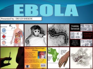
- 1. Presented by: DR S.D SHEKDE
- 3. EBOLA VIRUS • Presented by Dr.S.D.Shekde JR 2 • Guided by DR. V. M.HOLAMBE H.O.D. Assist. Professor Dept Of Comm. Medicine G.M.C. LATUR Date-30/08/14
- 4. INTRODUCTION Ebola first appeared in 1976 in 2 simultaneous outbreaks, in Nzara, Sudan, and in Yambuku, Democratic Republic of Congo. The latter was in a village situated near the Ebola River, from which the disease takes its name. Fruit bats of the Pteropodidae family are considered to be the natural host of the Ebola virus.
- 5. INTRODUCTION Ebola first appeared in 1976 in 2 simultaneous outbreaks, in Nzara, Sudan, and in Yambuku, Democratic Republic of Congo. The latter was in a village situated near the Ebola River, from which the disease takes its name. Fruit bats of the Pteropodidae family are considered to be the natural host of the Ebola virus.
- 6. VIRUS CLASSIFICATION Genus Ebolavirus is 1 of 3 members of the Filoviridae family ( filovirus), along with genus Marburg virus and genus Cueva virus. Group: Group V (-)ssRNA Order: Mononegavirales Family: Filoviridae Genus:Ebolavirus
- 7. SPECIES Genus Ebolavirus comprises 5 distinct species: Bundibugyo ebolavirus (BDBV) Zaire ebolavirus (EBOV) Sudan ebolavirus (SUDV) Reston ebolavirus (RESTV) Taï Forest ebolavirus (TAFV). Outbreaks of AFRICA BDBV, EBOV, and SUDV have been associated with large EVD outbreaks in Africa, whereas RESTV and TAFV have not. The RESTV species, found in Philippines and the People’s Republic of China, can infect humans, but no illness or death in humans from this species has been reported to date.
- 8. The Zaire ebolavirus is the most dangerous of the five species of Ebola viruses of the Ebolavirus genus which are the causative agents of Ebola virus disease.(EVD) The virus causes an extremely severe hemorrhagic fever in humans and other primate.
- 9. The name Zaire ebolavirus is derived from Zaire, the country (now the Democratic Republic of Congo) in which the Ebola virus was first discovered) and the taxonomic suffix ebola virus (which denotes an ebola virus species).
- 10. VIRION STRUCTURE
- 11. The EBOV genome is approximately 19 kb in length. It encodes seven structural proteins: nucleoprotein (NP) polymerase cofactor (VP35), (VP40) GP transcription activator (VP30), VP24 RNA polymerase (L)
- 12. The tubular Ebola virions are generally 80 nm in diameter and 800 nm long. In the center of the particle is the viral nucleocapsid which consists of the helical ssRNA genome wrapped about the NP, VP35, VP30 and L proteins. This structure is then surrounded by an outer viral envelope derived from the host cell membrane that is studded with 10 nm long viral glycoprotein (GP) spikes. Between the capsid and envelope are viral proteins VP40 and VP24 .
- 13. This envelope GP spike is expressed at the cell surface, and is incorporated into the virion to drive viral attachment and membrane fusion. It has also been shown as the crucial factor for Ebola virus pathogenicity . GP is actually post-translationally cleaved by the proprotein convertase furin to yield disulphide-linked GP1 and GP2 subunits.
- 14. GP1 allows for attachment to host cells, while GP2 mediates fusion of viral and host membranes. This protein assembles as a trimer of heterodimers on the viral envelope, and ultimately undergoes an irreversible conformation change to merge the two membranes
- 15. Pathophysiology
- 16. Endothelial cells, mononuclear phagocytes, and hepatocytes are the main targets of infection. After infection, a secreted glycoprotein (sGP) known as the Ebola virus glycoprotein (GP) is synthesized. Ebola replication overwhelms protein synthesis of infected cells and host immune defenses. The GP forms a trimeric complex, which binds the virus to the endothelial cells lining the interior surface of blood vessels.
- 17. The sGP forms a dimeric protein that interferes with the signaling of neutrophils, a type of white blood cell, which allows the virus to evade the immune system by inhibiting early steps of neutrophil activation. These white blood cells also serve as carriers to transport the virus throughout the entire body to places such as the lymph nodes, liver, lungs, and spleen.
- 19. EPIDEMIOLOGY
- 21. Current Ebola Outbreak in West Africa The current (2014) Ebola outbreak is occurring in the following West African countries: Guinea Liberia Sierra Leone Nigeria
- 26. • The virus is transmitted to people from wild animals and spreads in the human population through human-to-human transmission. • EVD outbreaks occur primarily in remote villages in Central and West Africa, near tropical rainforests. AlvinChew slideshare presentation alvinworks2006@yahoo.com
- 27. TRANSMISSION •The virus is spread through direct contact (through broken skin or mucous membranes) with i. sick person's blood or body fluids (urine, saliva, feces, vomit, sweat and semen) ii. objects (such as needles) that have been contaminated with infected body fluids. iii. Infected animals.
- 28. Exposure to ebola viruses can occur in healthcare settings where hospital staff are not wearing appropriate protective equipment, such as masks, gowns, and gloves. Proper cleaning and disposal of instruments, such as needles and syringes, is also important.
- 29. Transmission Animal to human • Consumption of raw meat • Contact wit fruit bat, pigs, apes-animal handlers • Animal products (blood, urine and feces.) Human to human • Close contact with infected patients • Care personnels of patient • Health care workers Prompt and safe burial of dead bodies. No to Autopsy Virus contained in dead body for a period 4 weeks.
- 30. Men who have recovered from the illness can still spread the virus to their partner through their semen for up to 7 weeks after recovery. Burial ceremonies in which mourners have direct contact with the body of the deceased person have played a role in the transmission of Ebola.
- 31. Who is most at risk? During an outbreak, those at higher risk of infection are: Health workers. Family members or others in close contact with infected people; and Mourners who have direct contact with the bodies of the deceased as part of burial ceremonies.
- 33. SUDDEN ONSET FEVER SORE THROAT INTENSE WEAKNESS MUSCLE PAIN, HEADACHE VOMITTING RASH DIARRHOEA IMPAIRED KIDNEY AND RENAL FUNCTION LAB •LOW WBC •LOW PLATELET •ELEVATED LIVER ENZYMES.
- 34. In some cases, both internal and external bleeding. People are infectious as long as their blood and secretions contain the virus. Ebola virus was isolated from semen 61 days after onset of illness in a man who was infected in a laboratory. The incubation period, that is, the time interval from infection with the virus to onset of symptoms, is 2 to 21 days.
- 35. The patient becomes contagious once they begin to show symptoms. They are not contagious during the incubation period. Ebola virus disease infections can only be confirmed through laboratory testing.
- 36. AlvinChew slideshare presentation alvinworks2006@yahoo.com
- 39. In some cases, both internal and external bleeding. People are infectious as long as their blood and secretions contain the virus. Ebola virus was isolated from semen 61 days after onset of illness in a man who was infected in a laboratory. The incubation period, that is, the time interval from infection with the virus to onset of symptoms, is 2 to 21 days.
- 41. Person Under Investigation (PUI) A person who has both consistent symptoms and risk factors as follows: 1) Clinical criteria, which includes fever of greater than 38.6 degrees Celsius or 101.5 degrees Fahrenheit, and additional symptoms such as severe headache, muscle pain, vomiting, diarrhea, abdominal pain, or unexplained hemorrhage.
- 42. 2) Epidemiologic risk factors within the past 21 days before the onset of symptoms, such as contact with blood or other body fluids or human remains of a patient known to have or suspected to have EVD; residence in—or travel to—an area where EVD transmission is active. or direct handling of bats, rodents, or primates from disease-endemic areas.
- 43. Probable Case A PUI who is a contact of an EVD case with either a high or low risk exposure. Confirmed Case A case with laboratory confirmed diagnostic evidence of ebola virus infection.
- 44. Contacts of an EVD Case (LEVELS OF EXPOSURE) High risk exposures Percutaneous, e.g. the needle stick, or mucous membrane exposure to body fluids of EVD patient. Direct care or exposure to body fluids of an EVD patient without appropriate personal protective equipment (PPE) Laboratory worker processing body fluids of confirmed EVD patients without standard biosafety precautions.
- 45. Low risk exposures Household member or other casual contactwith an EVD patient Providing patient care or casual contact without high-risk exposure with EVD patients in health care facilities in EVD outbreak affected countries No known exposure Persons with no known exposure in an EVD outbreak affected country in the past 21 days with no low risk or high risk exposures.
- 46. DIFFERENTIAL DIAGNOSIS Malaria Dengue Typhoid fever Shigellosis Cholera Leptospirosis Rickettsiosis Relapsing fever Other viral haemorrhagic fevers.
- 47. DIAGNOSIS Ebola virus infections can be diagnosed in a laboratory through several types of tests: Antibody-capture enzyme-linked immunosorbent assay (ELISA) -NP is one of the major viral structural components Antigen detection tests Serum neutralization test Reverse transcriptase polymerase chain reaction (RT-PCR) assay Electron microscopy Virus isolation by cell culture.
- 48. Samples from patients are an extreme biohazard risk; testing should be conducted under maximum biological containment conditions.
- 49. CDC- Diagnosis Timeline of Infection Diagnostic tests available Within a few days after symptoms begin •Antigen-capture enzyme-linked immunosorbent assay (ELISA) testing •IgM ELISA •Polymerase chain reaction (PCR) •Virus isolation Later in disease course or after recovery •IgM and IgG antibodies Retrospectively in deceased patients •Immunohistochemistry testing •PCR •Virus isolation
- 50. When Specimens Should Be Collected for Ebola Testing Ebola virus is detected in blood only after onset of symptoms, most notably fever. It may take up to 3 days post-onset of symptoms for the virus to reach detectable levels. Virus is generally detectable by real-time RT-PCR from 3-10 days post-onset of symptoms, but has been detected for several months in certain secretions.
- 51. Specimens ideally should be taken when a symptomatic patient reports to a healthcare facility and is suspected of having an EVD exposure However, if the onset of symptoms is <3 days, a subsequent specimen will be required to completely rule-out EVD.
- 52. From whom the samples are to be collected? The samples should be collected from any person ill or deceased who has or had fever with acute clinical symptoms and signs of hemorrhage, such as bleeding of the gums, nose-bleeds, conjunctival injection, red spots on the body, bloody stools and/or melaena (black liquid stools), or vomiting blood (haematemesis) with the history of travel to the affected area.
- 53. OR Any person (living or dead) having had contact with a clinical case of EBVD and with a history of acute fever. Anyone who has accidently come in contact with blood or body fluids should be kept under quarantine and observed for 30 days.
- 54. Preferred Specimens for Ebola Testing A minimum volume of 4mL whole blood / serum/ plasma preserved with EDTA, clot activator, sodium polyanethol sulfonate (SPS), or citrate in plastic collection tubes can be submitted for EVD testing. Postmortem: Tissue sample (liver, spleen, bone marrow, kidney, lung and brain)
- 55. Do not submit specimens preserved in heparin tubes. Specimens should be stored at 4°C or frozen. Before dispatching the sample disinfect the outer surface of container using 1:100 dilution of bleach or 5% Lysol solution.
- 56. Transporting Specimens within the Hospital / Institution Specimens should be placed in a durable, leak-proof secondary container for transport within a facility. To reduce the risk of breakage or leaks, do not use any pneumatic tube system for transporting suspected EVD specimens.
- 57. Samples should be sent to the following laboratories under cold chain with prior intimation: National Institute of Virology, Pune. National Centre for Disease Control, Delhi.
- 58. TREATMENT
- 59. No ebola virus Disease-specific treatment exists. Treatment is primarily supportive in nature and includes minimizing invasive procedures, balancing fluids and electrolytes to counter dehydration
- 60. Administration of anticoagulants early in infection to prevent or control disseminated intravascular coagulation, Administration of procoagulants late in infection to control bleeding. maintaining oxygen levels, pain management, and the use of medications to treat bacterial or fungal secondary infections.
- 61. Early treatment may increase the chance of survival. A number of experimental treatments are being studied
- 62. A hospital isolation ward in Gulu, Uganda, during the October 2000 outbreak
- 63. a high fatality rate for this disorder (80% to 90%) mortality from Ebola has ranged from 25% to 90% and recovery is slow in those who survive. Morbidity and mortality rates are very high, and they vary with the strain of Ebola
- 64. The most highly lethal Ebola subtype is EBO-Z, which has been reported to have a mortality rate as high as 88%. The EBO-S subtype has a reported mortality rate of 50%, similar to that of the Ebola outbreak in Gabon, where the mortality rate was 57-66%.
- 65. Vaccine: No vaccine is currently available for humans. The most promising candidates are DNA vaccines or vaccines derived from adenoviruses, vesicular stomatitis Indian virus (VSIV) or filovirus-like particles (VLPs)because these candidates could protect nonhuman primates from ebolavirus-induced disease. DNA vaccines, adenovirus-based vaccines, and VSIV-based vaccines have entered clinical trials. Vaccines have protected nonhuman primates.
- 66. PREVENTION:___
- 68. PREVENTION: A poster, among those being distributed by UNICEF, bears information and illustrations on the symptoms of Ebola virus disease (EVD)
- 69. Prevention: Prevention focuses on avoiding contact with the viruses. The following precautions can help prevent infection and spread of Ebola •Avoid areas of known outbreaks. •Wash your hands frequently. As with other infectious diseases, one of the most important preventive measures is frequent hand-washing. Use soap and water, or use alcohol-based hand rubs containing at least 60 percent alcohol when soap and water aren't available.
- 70. • Avoid wildlife /bush meat. In developing countries, avoid buying or eating the wild animals, including nonhuman primates, sold in local markets. • Avoid contact with infected people. In particular, caregivers should avoid contact with the person's body fluids and tissues, including blood, semen, vaginal secretions and saliva. People with Ebola are most contagious in the later stages of the disease.
- 71. •Follow infection-control procedures. If you're a health care worker, wear protective clothing, such as gloves, masks, gowns and eye shields. Keep infected people isolated from others. Dispose of needles and sterilize other instruments. •Don't handle remains. The bodies of people who have died of Ebola disease are still contagious. Specially organized and trained teams should bury the remains, using appropriate safety equipment.
- 72. Infection Control for Collecting and Handling Specimens This includes wearing appropriate personal protective equipment (PPE) and adhering to engineered safeguards, for all specimens regardless of whether they are identified as being infectious. Recommendations for specimen collection: full face shield or goggles, masks to cover all of nose and mouth, gloves, fluid resistant or impermeable gowns. Additional PPE may be required in certain situations.
- 73. This picture taken on April 9, 2005 shows five health workers, dressed in head-to-toe "Ebola suits." Doctors dressed up for Ebola in Bundibugyo
- 74. A researcher working with the Ebola virus while wearing a BSL-4 positive pressure suit to avoid infection
- 75. Proper Clothing
- 76. Recommendations for laboratory testing full face shield or goggles masks to cover all of nose and mouth gloves fluid resistant or impermeable gowns use of a certified class II Biosafety cabinet or plexiglass splash guard disinfectants routinely used to decontaminate the laboratory environment (benchtops and surfaces) and the laboratory instrumentation are sufficient to inactivate enveloped viruses, such as influenza, hepatitis C, and Ebola viruses.
- 77. PATIENT PLACEMENT Place the patient in Single room (containing a private bathroom) with the door closed. Maintain a log of persons entering the patient’s room. Allow access to only those necessary for the patient’s well-being and care, such as a child’s parent. Use of Personal Protective Equipment is essential.
- 78. All persons entering the patient room should wear at least: o Gloves o Gown (fluid resistant or impermeable)to cover clothing and exposed skin o Eye protection (goggles) to prevent splashes on eye. o Facemask to prevent splashes on nose and mouth. o Face shield, if used, will protect eye, nose and mouth. o Closed shoes
- 79. Waste Management
- 80. Waste should be segregated to enable appropriate and safe handling. Sharp objects (e.g. needles, syringes, glass articles) and tubing that has been in contact with the bloodstream should be placed inside puncture resistant containers. These should be located as close as practical to the area in which the items are used. Collect all solid, non-sharp, medical waste using leak-proof waste bags and covered bins.
- 81. Waste should be placed in a designated pit of appropriate depth (e.g. 2 m deep and filled to a depth of 1–1.5 m). After each waste load the waste should be covered with a layer of soil 10–15 cm deep. Placenta and anatomical samples should be buried in a separate pit or incinerated
- 82. An incinerator may be used to destroy solid waste. However, it is essential to ensure that total incineration has taken place. The area designated for the final treatment and disposal of waste should have controlled access to prevent entry by animals, untrained personnel or children.
- 83. Waste, such as faeces, urine and vomit, and liquid waste from washing, can be disposed of in the sanitary sewer or pit latrine. No further treatment is necessary. Wear gloves, gown, closed shoes and goggles/facial protection, when handling liquid infectious waste (e.g. any secretion or excretion with visible blood even if it originated from a normally sterile body cavity). Avoid splashing when disposing of liquid infectious waste
- 84. Quarantine: Quarantine, also known as enforced isolation, is usually effective in decreasing spread. Governments often quarantine areas where the disease is occurring or individuals who may be infected. In the United States the law allows quarantine of those infected with Ebola. The lack of roads and transportation may help slow the disease in Africa. During the 2014 outbreak Liberia closed schools.
- 85. AlvinChew slideshare presentation alvinworks2006@yahoo.com
- 86. Bioterrorism Locality of this virus has become less isolated as the threat of bioterrorism looms large. The Ebola virus is now on the “A” list for hopeful vaccination development. Experiments have even been formed to show how Ebola can be used as a bioterror agent.
- 87. CURRENT STATISTICS- INDIA The Union Health Ministry on Wednesday (20-08-14) said that there was no confirmed or even suspect case of Ebola virus disease in India as yet. In a statement, the Ministry said that a 30-year-old Nigerian national, who arrived at the Delhi Airport, was admitted to Dr. RML Hospital on Wednesday morning with fever. He has tested negative for Ebola at the National Centre for Disease Control, Delhi. Mandatory reporting of passengers from affected countries was in place. Since it began on August 10, about 3,089 passengers have been screened at the airports at Delhi, Mumbai, Kolkata, Bangalore, Chennai, Thiruvananthapuram and Kochi.
- 88. Conclusion EVD is very contagious. No aerosol route of transmission noted. Spreads through mucous membrane and contact with body fluids. Contact tracing and isolation very important. Early detection of cases to improve curative rate. Supportive treatment. To follow WHO/CDC methods of prevention.
- 89. References http://www.cdc.gov/ncidod/dvrd/spb/mnpages/dispages/ebotabl.htm http://www.who.int/mediacentre/factsheets/fs103/en/ http://mohfw.gov.in www.osha.gov Hampton, Tracy. Vaccines Against Ebola and Marburg Viruses Show Promise in Primates Studies. Maedical News and Perspectives. JAMA. Vol. 294 No. 2 July 2005. Jones, Steven. Live attenuated recombinant vaccine protects nonhuman primates against Ebola and Marburg viruses. Nature Medicine. Vol. 11 No. 7 July 2005.