Anatomy of brain sulcus and gyrus - Dr.Sajith MD RD
•Download as PPTX, PDF•
269 likes•128,846 views


Recommended
More Related Content
What's hot
What's hot (20)
Similar to Anatomy of brain sulcus and gyrus - Dr.Sajith MD RD
Similar to Anatomy of brain sulcus and gyrus - Dr.Sajith MD RD (20)
Recently uploaded
Recently uploaded (20)
Anatomy of brain sulcus and gyrus - Dr.Sajith MD RD
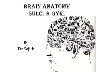
- 1. Brain Anatomy Sulci & Gyri By Dr.Sajith
- 2. • Sulcus : A sulcus is depression or fissure in the surface of the brain. ( valleys ) • Gyrus : A gyrus is a ridge on the cerebral cortex. It is generally surrounded by one or more sulci ( hills )
- 3. Typical continous fissure Interhemispheric fissure Sylvian fissure ( lateral sulcus ) Parieto-occipital fissure Collateral sulcus Central sulcus Calcarine Sulcus
- 4. Interhemispheric fissure Deep sulcus upto corpus collosum Divides brain into two hemisphere
- 5. Sylvian fissure deep, mostly horizontal insula is buried within it separates temporal lobe from parietal and frontal lobes
- 6. Parieto - occipital fissure very deep often Y-shaped from sagittal view X-shaped in horizontal and coronal views
- 7. Collateral sulcus Divides lingual and parahippocampal gyri from fusiform gyrus.
- 8. Central sulcus usually freestanding (no intersections) Seperates frontal and parietal lobe.
- 10. Lobes of brain Frontal lobe. Parietal lobe. Temporal lobe. Occipital lobe. Limbic lobe. Insular lobe.
- 11. • Frontal lobe: Anterior region of hemisphere; anterior to central sulcus, superior to sylvian fissure • Parietal lobe: Posterior region of hemisphere; posterior to central sulcus, anterior to parietooccipital sulcus • Occipital lobe: Posterior to parietooccipital sulcus • Temporal lobe: Inferior to sylvian fissure, anterior to angular gyrus
- 13. Lateral surface of brain
- 14. Medial surface of brain
- 15. Basal surface of brain
- 16. frontal lobe The frontal lobe constitutes the entire region in front of the central sulcus. Immediately in front of the central sulcus lies the precentral gyrus, which is the primary motor region of the cerebral cortex. In front of the precentral gyrus, the rest of the frontal lobe is divided into superior, middle and inferior frontal gyri.
- 17. Frontal lobe Sulci: Superior frontal sulcus. Inferior frontal sulcus. Precentral sulcus. Gyri: Superior frontal gyrus. Middle frontal gyrus. Inferior frontal gyrus. Precentral gyrus
- 18. Frontal lobe gyrus Lateral Surface Precentral Gyrus --- primary motor area Superior Frontal Gyrus Middle Frontal Gyrus Inferior Frontal Gyrus Pars Opercularis Pars Triangularis Pars Orbitalis Medial Surface Medial Frontal Gyrus Paracentral Lobule Basal Surface Rectus Gyrus Orbital Gyrus ( superior, medial ,lateral ,inferior )
- 23. Parietal lobe Behind the central sulcus, and above the lateral fissure, lies the parietal lobe. Its most anterior part is the postcentral gyrus, which is the site of the primary somatosensory cortex. Behind the postcentral gyrus, on the lateral surface of the hemisphere, the intraparietal sulcus divides the rest of the parietal lobe into superior and inferior parietal lobules.
- 24. Parietal lobe Sulci: Post-central sulcus. Intraparietal sulcus. Gyri: Post-central gyrus. Superior parietal gyrus. Inferior parietal gyrus.
- 25. Parietal lobe gyrus Lateral Surface Postcentral Gyrus ---- primary somatic area Superior Parietal Lobule Inferior Parietal Lobule ---- Wernicke’s area Supramarginal Gyrus Angular Gyrus Medial Surface Paracentral Lobule Precuneus
- 30. Temporal lobe The temporal lobe lies beneath the lateral fissure. On its lateral surface the temporal lobe is divided into three principal gyri that run roughly parallel to the lateral fissure: the superior, middle and inferior temporal gyri. The superior temporal gyrus includes the primary auditory cortex.
- 31. Temporal lobe Sulci: Superior temporal sulcus (parallel sulcus). Inferior temporal sulcus. Gyri: Superior temporal gyrus. Middle temporal gyrus. Inferior temporal gyrus.
- 32. Temporal lobe gyrus Lateral Surface Superior Temporal Gyrus Middle Temporal Gyrus Inferior Temporal Gyrus Basal Surface Occipitotemporal Gyrus Medial Occipitotemporal Gyrus Lateral Occipitotemporal Gyrus
- 36. Occipital lobe The boundary between the parietal lobe and occipital lobe is not coincident with a single sulcus on the lateral surface of the hemisphere; however, it is clearly marked by the deep parieto-occipital sulcus on the medial surface. The occipital lobe does not bear any important landmarks on its lateral surface but on the medial surface, the prominent calcarine sulcus indicates the location of the primary visual cortex.
- 37. Occipital lobe Sulci: • Superior occipital sulcus (extension of intraparietal sulcus). • Inferior occipital sulcus (extension of inferior temporal sulcus). Gyri: • Superior occipital gyrus. • Middle occipital gyrus. • Inferior occipital gyrus
- 38. Occipital lobe gyrus Lateral Surface Lateral Occipital Gyrus Superior Occipital Gyrus Inferior Occipital Gyrus Medial Surface Cuneus Lingual Gyrus Basal Surface Lateral occipito-temporal gyrus (fusiform gyrus). Medial occipito-temporal gyrus (lingual gyrus).
- 43. Limbic lobe On the medial surface of the hemisphere, certain portions of the frontal, parietal and temporal lobes also constitute components of the limbic system. Curving around the corpus callosum, and running parallel to it, lies the cingulate gyrus , separated from the rest of the hemisphere by the cingulate sulcus. The cingulate gyrus passes posteriorly and inferiorly round the posterior portion of the corpus callosum to become continuous with the parahippocampal gyrus of the temporal lobe. Deep to the parahippocampal gyrus, within the temporal lobe lies the hippocampus. The cingulate gyrus, parahippocampal gyrus and hippocampus are referred to as the limbic lobe.
- 44. Limbic lobe gyrus Outer Ring Cingulate Gyrus Isthmus of Cingulate Gyrus Parahippocampal Gyrus Uncus, Uncinate Gyrus Inner Ring Hippocampal Formation Hippocampus, Ammon's Horn Dentate Gyrus Fasciolar Gyrus Supracallosal Gyrus Subcallosal Area (Parolfactory Area)
- 48. Insular lobe • Lies deep in floor of sylvian fissure, overlapped by frontal, temporal and parietal. • The insular lobe is thought to be involved in sensory and motor visceral functions as well as taste perception.
- 49. Insular lobe Sulci: Central sulcus. Short insular sulcus. Precentral sulcus. Gyri: Anterior insular lobule. Posterior insular lobule.
- 50. Insular lobe Anterior insular lobule (short insular gyri): Anterior short insular gyrus. Middle short insular gyrus. Posterior short insular gyrus. Posterior insular lobule (long insular gyri): Anterior long insular gyrus. Posterior long insular gyrus.
- 55. Signs to identify sulcus in mri • The midline sulcus sign: The longest sulcus running horizontally and entering the interhemispheric fissure is the central sulcus . • The M sign: The typical upper case M shape of the inferior frontal gyrus is formed by its parts ( orbitalis, triangularis, and opercularis ).
- 56. signs • The bifid post cg sign: Toward the interhemispheric fissure, the postcentral gyrus is medially fissured, enclosing the pars marginalis of the cingulated sulcus with both legs and thereby forming the characteristic bifid post cg sign. • The thin post cg sign: On the surface, the precentral gyrus has a thicker anteroposterior diameter compared with the postcentral gyrus, which is thinner.
- 57. signs • The handknob: defined the "handknob " as 1 single -shaped dorsally convex bulge of the precentral gyrus. • The upper T sign: The intersection of the well-identifiable superior frontal sulcus with the precentral sulcus has the shape of an upper case T. The pre cg can be found immediately posterior to this T shape.
- 58. signs • The L sign: The sfg terminates in the pre cg, which runs laterally from the posterior end of the sfg, therefore together forming an upper case L. • The lower T sign: The inferior frontal sulcus posteriorly terminates in the pre sc, also in the shape of an upper case T that is dorsally bordered by the pre cg.
- 59. signs • Inverted omega sign : the central sulcus more likely to have the shape of inverted omega. • The bracket sign: The bihemispheric symmetric pars marginalis of the cingulate sulcus forms an anteriorly opened bracket.
- 63. Sagittal
- 64. Superior frontal gyrus Cingulate sulcus Marginal ramus of Cingulate sulcus Cingulate gyrus Paracentral lobule Superior parietal lobule Parietooccipital sulcus Cuneus Calcarine sulcus Lingual gyrus Subcallosal gyrus Gyrus rectus NP/MGH Fastigium, fourth ventricle
- 65. Superior frontal gyrus Cingulate sulcus Precentral gyrus Cingulate gyrus Central sulcus Marginal ramus of Cingulate sulcus Superior parietal lobule Precuneus Parietooccipital sulcus Cuneus Calcarine sulcus Frontomarginal gyrus Gyrus rectus Caudothallamic groove NP/MGH Lingual gyrus
- 66. Precentral sulcus Superior frontal gyrus Central sulcus Marginal ramus of Cingulate sulcus Corona radiata Superior parietal lobule Precuneus Parietooccipital sulcus Calcarine sulcus NP/MGH Lingual gyrus Inferior occipital gyrus
- 67. Central sulcus Superior parietal lobule Parietooccipital sulcus Frontopolar gyrus Frontomarginal gyrus Superior occipital gyrus Middle occipital gyrus Medial orbital gyrus Posterior orbital gyrus Inferior temporal gyrusNP/MGH Temporal horn, lateral ventricle Lingual gyrus Inferior occipital gyrus
- 68. Central sulcus uperior Temporal gyrus Middle Temporal gyrus Inferior Temporal gyrus NP/MGH
- 69. Central sulcus Inferior frontal gyrus Superior parietal gyrus Frontomarginal gyrus Anterior orbital gyrus Superior occipital gyrus Middle occipital gyrus Posterior orbital gyrus Superior Temporal gyrus Inferior occipital gyrus Middle Temporal gyrus Inferior Temporal gyrus NP/MGH Lingual gyrus
- 70. Precentral sulcus Superior frontal sulcus Central sulcus Postcentral sulcus Lateral fissure, posterior segment Inferior frontal gyrus, pars triangularis Angular gyrus Inferior frontal gyrus, pars orbitalis Superior Temporal gyrus Superior Temporal sulcus Middle occipital gyrus Middle Temporal gyrus Anterior occipital sulcus Inferior Temporal gyrus Inferior occipital gyrus NP/MGH
- 71. axial
- 72. Fusiform gyrus Superior Temporal gyrus Middle Temporal gyrus Inferior Temporal gyrus NP/MGH
- 73. Parahippocampal gyrus Superior Temporal gyrus Middle Temporal gyrus Inferior Temporal gyrus Hippocampal gyrus NP/MGH
- 74. Amygdala Hippocampus Superior Temporal gyrus Middle Temporal gyrus Temporo-occipital fissure Inferior occipital gyrus Gyrus descendens Inferior Temporal gyrus Lingual gyrus NP/MGH
- 75. Gyrus rectus Medial orbital gyrus Olfactory sulcus Subcallosal gyrus Posterior orbital gyrus Superior Temporal gyrus Amygdala Middle Temporal gyrus Hippocampus Inferior Temporal gyrus Temporo-occipital fissure Middle occipital gyrus Gyrus descendens NP/MGH Lingual gyrus
- 76. Superior frontal gyrus Middle frontal gyrus Inferior frontal gyrus, pars orbitalis Lateral fissure Inferior frontal gyrus, pars opercularis Inferior parietal gyrus Insula Lateral fissure Cingulate gyrus Superior temporal gyrus Parieto-occipital fissure Superior temporal sulcus Middle temporal gyrus Calcarine sulcus Middle occipital gyrus Superior occipital gyrus Intra-occipital sulcus NP/MGH Cuneus
- 77. Superior frontal gyrus Middle frontal gyrus Inferior frontal gyrus Central sulcus Postcentral gyrus Lateral fissure Inferior parietal gyrus Superior temporal gyrus Lateral fissure Superior temporal sulcus Middle occipital gyrus Intra-occipital sulcus Superior occipital gyrus NP/MGH Parieto-occipital sulcus
- 78. Precentral sulcus Precentral gyrus Central sulcus Middle occipital gyrus Central sulcus Cuneus NP/MGH Superior occipital gyrus Intra-occipital sulcus
- 79. Superior frontal gyrus Middle frontal gyrus Superior frontal sulcus Inferior frontal gyrus Centrum semiovale Central sulcus Central sulcus Postcentral sulcus Postcentral sulcus Supramarginal gyrus Intraparietal sulcus Angular gyrus Parietooccipital sulcus Superior parietal gyrus NP/MGH Precuneus
- 80. Superior frontal gyrus Middle frontal gyrus Superior frontal sulcus Central sulcus Central sulcus Supramarginal gyrus Postcentral sulcus Intraparietal sulcus Angular gyrus Pars marginalis Intraparietal sulcus Superior parietal gyrus NP/MGH
- 81. coronal
- 82. Interhemispheric Fissure Inferior Frontal gyrus Superior Frontal gyrus Middle Frontal gyrus Inferior Frontal gyrus Gyrus rectus Medial Orbital gyrus Olfactory bulb NP/MGH
- 83. Superior Frontal gyrus Forceps minor Superior Frontal sulcus Middle Frontal gyrus Inferior Frontal gyrus Lateral orbital sulcus Medial Orbital gyrus Lateral orbital gyrus Gyrus rectus Anterior Orbital gyrus Olfactory Sulcus NP/MGH
- 84. Circular insular sulcus Cingulate gyrus Superior Frontal gyrus short insular gyrus Middle Frontal gyrus Inferior Frontal sulcus Inferior Frontal gyrus pars opercularis Sylvian Fissure Posterior Orbital gyrus Middle Temporal gyrus Olfactory Sulcus Superior Temporal gyrus Inferior Temporal gyrus NP/MGH Gyrus rectus Medial Orbital gyrus
- 85. Superior Frontal sulcus Superior Frontal gyrus Cingulate sulcus Middle Frontal gyrus Precentral sulcus Precentral gyrus Sylvian Fissure Superior Temporal gyrus Superior Temporal Sulcus Middle Temporal gyrus Amygdala NP/MGH Anterior commissure Inferior Temporal gyrus
- 86. Superior Frontal gyrus Middle Frontal gyrus Central Sulcus Superior Temporal gyrus Hippocampus Middle Temporal gyrus Inferior Temporal gyrus Parahippocampal gyrus CA1, cornu ammonis NP/MGH Fusiform gyrus
- 87. Postcentral gyrus Central Sulcus Intraparietal sulcus Paracentral lobule Intraparietal sulcus Cingulate gyrus Supramarginal gyrus Superior Temporal gyrus Middle Temporal gyrus Inferior Temporal gyrus Fusiform gyrus Parahippocampal gyrus Collateral sulcus NP/MGH
- 88. Paracentral lobule Central sulcus Superior Temporal gyrus Middle Temporal gyrus Inferior temporal gyrus Fusiform gyrus NP/MGH
- 89. Paracentral lobule Postcentral gyrus Central sulcus Intraparietal sulcus Supramarginal gyrus Middle temporal gyrus Inferior temporal gyrus Fusiform gyrus Calcarine sulcus Lingual gyrus NP/MGH Cingulate gyrus
- 90. Thank u