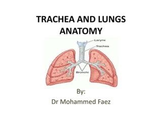
2. Trachea The trachea is a cartilaginous and membranous tube, extending from the cricoid cartilageof the larynx, on a level with C6 vertebra, to the level of the angle of Louis (T4/5) vertebra, where it divides into the two bronchi, one for each lung.
3. Trachea Structure: The trachea is a rigid fibroelastic structure. Incomplete rings of hyaline cartilage continuously maintain the patency of the lumen. The trachea is lined internally with ciliated columnar epithelium.
5. Trachea It measures about 11 cm. In length. Its diameter, from side to side, is from 2 to 2.5 cm. It has 18-22 cartilaginous rings It is greater in the male than in the female.
6. Trachea Relations: Anteriorly: Neck: The isthmus of the thyroid gland (2nd, 3rd and 4th tracheal rings ) The inferior thyroid veins The sternothyroid and sternohyoid muscles The cervical fascia Thorax: The manubriumsterni The remains of the thymus The left innominate vein The aortic arch Left common carotid arteries The deep cardiac plexus.
7. Trachea Relations: Posteriorly : the esophagus Laterally: Neck: Common carotid arteries Right and left lobes of the thyroid gland Inferior thyroid arteries The recurrent nerves
8. Trachea Relations: Laterally: Thorax: it lies in the superior mediastinum. Right The pleura Right vagus, The innominate artery Left Left recurrent nerve The aortic arch The left common carotid Subclavian arteries
9. Inferior thyroid a. Vagus n. Recurrent laryngeal n. Thoracic duct Sympathetic trunk Trachea
10. Trachea The trachea divides into two main bronchi : the left and the right bronchi, at the level of the sternal angle at the anatomical point known as the carina.
11. Bronchi The right bronchus: Wider, shorter, and more vertical in direction than the left. It is about 2.5 cm. Long, It enters the right lung nearly opposite the T5 vertebra.
12. Bronchi The Left Bronchus It is smaller in caliber but longer than the right It is about 5 cm. long. It enters the root of the left lung opposite the T6 vertebra.
13.
14. Wider, shorter, and more vertical than the left trachea Right Primary Bronchus Left primary bronchus
15. Bronchi The primary bronchi divide to form Secondary Bronchi (lobar bronchi). There is one secondary bronchus for each lobe of the lungs. There are 2 lobes on the left lung. There are 3 lobes on the right lung.
16. Bronchi The left main bronchus enters the hilum and divides into a superior and inferior lobar bronchus. The right main bronchus gives off the bronchus to the upper lobe prior to entering the hilum(superior lobar bronchus)and once into the hilumdivides into middle and inferiorlobar bronchi.
17. Bronchi Each lobar bronchus divides within the lobe into(Tertiary Bronchi) segmental bronchi. Each segmental bronchus enters a bronchopulmonary segment. Each bronchopulmonary segment is pyramidal in shape with its apex directed towards the hilum.
20. Trachea blood supply the trachea receives its blood supply from branches of the inferior thyroid and bronchial arteries.
21. Lungs The two lungs are organs of respiration and lie on either side of the mediastinum surrounded by the right and left pleural cavities. Right and left lungs.
22. Lungs The right lung is normally a little larger than the left lung because the middle mediastinum, containing the heart, bulges more to the left than to the right.
23. Lungs Each lung has a half-cone shape, with a base, apex, two surfaces and three borders: The base sits on the diaphragm. The apex projects above 1st rib and into the root of the neck.
24. Lungs The two surfaces-the costal surface lies immediately adjacent to the ribs and intercostal spaces of the thoracic wall. The mediastinal surface lies against the mediastinumanteriorly and the vertebral column posteriorly and contains the comma-shaped hilum of the lung through which structures enter and leave.
25. Lungs The three borders-the inferior border of the lung is sharp and separates the base from the costal surface. The anterior and posterior borders separate the costal surface from the medial surface. Unlike the anterior and inferior borders, which are sharp, the posterior border is smooth and rounded.
26. Lungs Root and hilum of lung: The root of each lung is a short tubular collection of structures that together attach the lung to structures in the mediastinum . The hilum, where structures enter and leave.
27. Lungs Structures within each root and located in the hilum: A pulmonary artery Two pulmonary veins A main bronchus Bronchial vessel Nerves Lymphatics
28. Lungs Structure the right lung: It is divided into upper, middle and lower lobes by oblique and horizontal fissures. The left lung: It has two lobes, upper and lower lobes. They are separated by the oblique fissure.
29. Lungs Read about right and left lungs relations and lungs impressions.
30.
31.
32. Bronchial Tree The trachea is divided into 2 bronchi The main bronchus divides within the lung into lobar bronchi. The lobar bronchi divide into segmental bronchi which supply bronchopulmonary segments. Each bronchopulmonary segment, the segmental bronchi divide into bronchioles, which further subdivide and supply the respiratory surfaces.
33. Bronchopulmonary Segment There are ten bronchopulmonary segments in each lung Each bronchopulmonary segment is shaped like an irregular cone with the apex at the origin of the segmental bronchus and the base projected peripherally onto the surface of the lung.
34. Bronchopulmonary Segment A bronchopulmonary segment is the smallest, functionally independent region of a lung and the smallest area of lung that can be isolated and removed without affecting adjacent regions.
35.
36.
37.
38. Blood supply of the lung The bronchi and parenchymal tissue of the lungs are supplied by bronchial arteries a branches of the descending thoracic aorta. Bronchial veins, which also communicate with pulmonary veins, drain into the azygos and hemiazygos.
39. Lymphatic drainage of the lungs lymph returns from the periphery towards the hilartracheobronchial groups of nodes and from here to mediastinallymph trunks.
40.
41. Nerve supply of the lungs A pulmonary plexus is located at the root of each lung. The plexus is composed of sympathetic fibres (from the sympathetic trunk) and parasympathetic fibres (from the vagus).