Intra-oral radiograph techniques guide
•
90 likes•15,840 views

1. There are several types of intraoral radiograph techniques including periapical, bite-wing, and occlusal radiographs. 2. Periapical radiographs show teeth and surrounding bone structures and are useful for detecting apical infections, impacted teeth, and evaluating implants. They can be taken using parallel or bisecting techniques. 3. Bite-wing radiographs show the crowns of teeth, interproximal areas, and crestal bone in one image. They are useful for detecting proximal caries and evaluating restorations. 4. Occlusal radiographs expose the entire maxilla or mandible and are useful for identifying supernumerary teeth, impacted teeth, foreign

Recommended
More Related Content
What's hot
What's hot (20)
Similar to Intra-oral radiograph techniques guide
Similar to Intra-oral radiograph techniques guide (20)
Recently uploaded
Recently uploaded (20)
Intra-oral radiograph techniques guide
- 2. Intra-oral radiograph techniques • What are the types of intra- oral radiograph????
- 3. 1. periapical • What are the benefits? I. Designed to show teeth and tissues around the apices. II. Show 2-4 teeth. III. Provide detail about the surrounding alveolar bone.
- 4. periapical • Indication???? I. Apical infection/cyst II. Impacted tooth III. Endo ttt IV. Root morphology before surgery V. Evaluate implants.
- 5. periapical • Our technique HOW WE DO IT??? • We have 2 technique: 1. Parallel tech; 2. Bisecting tech;
- 6. periapical A. Parallel tech; Concept “ film parallel to long axis of teeth” Soooo, the x-ray beam at right angle, That’s why it is called “right-angle Or long -cone tech”. • The goal: To reduce the geometric distortion True anatomic relation Increase sharpness, reduce magnification
- 8. periapical • Steps: 1. Film position; parallel to long axis and away from tooth. “object film distance” 1. Target –film distance “16 -inch” 2. Film holder.
- 9. periapical • Rules: I. Film placement “ cover target area” II. Film position “ parallellong axis” III. Vertical angulation “ perpendicular on both ilm and log axis” IV. Horizontal angulation “thr contact area of teeth” V. Film exposure “ avoid cone cut”
- 10. periapical • Adv: 1. Accurate image “ actual size, no distortion, max detail” 2. reproducible radiograph. 3. Vertical and horizontal angul. Automatically determined. 4. Relation of object-film-xray source maintained. • Dis adv: 1. Placement difficulties “inexperienced drs, child, small mouth or shallow palate” 2. Discomfort. 3. cost
- 11. periapical B. Bisecting technique: 1. Operator unable to do parallel tech. 2. Depend on rule of isometry “ 2 triangle with equal angulation. 3. Film placed close as possible to teeth. 4. Ptns hold the film 5. X-ray beam perpendicular to?????? The imaginary bisector
- 13. periapical 6. Horizontal angulation “ same parallel”. 7. Vertical angulation
- 14. periapical • Incorrect vertical angulation; A. Shortened image. B. Elongated image.
- 15. • Incorrect horizontal angulation: “overlapping”
- 17. periapical • Adv of bisecting: 1. Simple tech, quick 2. Comfortable 3. Less equipment “no film holder, no long cone” 4. Anatomy of ptns preclude the use of holder. • Dis adv: 1. Image distortion: using short PID cause divergence of x-ray. 2. Not reproducible. 3. Skill required. 4. Cone cut may occur
- 18. 2. Bite-Wing • Ptn bite on a small wing attached to film “ this explain name” • Include max. and mand. teeth, interproximal area , and crestal bone in one image
- 19. Bite-Wing • Indication: 1. Proximal caries. 2. Evaluation of restoration “overhang” 3. Evaluate alv. Bone crest level. 4. Evaluate pulp chamber
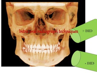
- 21. Bite-Wing • Principle: I. Film placed parallel to crowns II. Stabilized whn ptn bite on the tab/holder III. X-ray beam thr contact of the teeth wz +10 degree vertical andulation
- 22. Bite-Wing • Rules: 1. Film placement 2. Film position 3. Vertical angulation 4. Horizontal angulation 5. Film exposure
- 23. 3. oclussal • Expose all Maxilla Or Mandible.
- 26. oclussal • Upper (up. Standard or anterior)
- 27. oclussal • Upper
- 28. oclussal • Upper
- 29. oclussal • Indication: 1. Supernumerary teeth, impacted teeth 2. Foreign bodies in jaws and salivary gland. 3. Lesions in jaws “cyst ,malignancy,…..” 4. Examine max. sinus “anterior, medial, lateral wall” 5. Examine ptns wz trismus. 6. Fx in mand and max. 7. Cleft palate. 8. Size of the jaws