
Recommended
Recommended
More Related Content
What's hot
What's hot (20)
Viewers also liked
Viewers also liked (20)
Similar to Digital Radiography
Similar to Digital Radiography (20)
Recently uploaded
Recently uploaded (20)
Digital Radiography
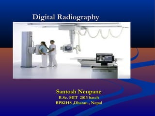
- 1. Digital RadiographyDigital Radiography 2121stst December 2016December 2016 Santosh NeupaneSantosh Neupane B.Sc. MIT 2013 batchB.Sc. MIT 2013 batch BPKIHS ,Dharan , NepalBPKIHS ,Dharan , Nepal
- 3. IntroductionIntroduction Today the world is moving in the guidance ofToday the world is moving in the guidance of computer and its application . Now we can call itcomputer and its application . Now we can call it digital world.digital world. Computer applications in radiology alsoComputer applications in radiology also continue to grow. Large scale radiologycontinue to grow. Large scale radiology applications are CT ,MRI, USG, direct digitalapplications are CT ,MRI, USG, direct digital radiography, indirect digital radiography, digitalradiography, indirect digital radiography, digital fluoroscopy, digital mammography ,computedfluoroscopy, digital mammography ,computed radiography and teleradiology.radiography and teleradiology.
- 4. Digital Vs Analog systemDigital Vs Analog system Analog system - a variable is measured on aAnalog system - a variable is measured on a continuous scale with infinite number ofcontinuous scale with infinite number of possible values .possible values . Digital system -measurments can have aDigital system -measurments can have a limited value .limited value .
- 6. Analog to digital conversionAnalog to digital conversion Conventional radiograph is produced by a seriesConventional radiograph is produced by a series of analog signals, from x-ray formation andof analog signals, from x-ray formation and interaction with the patient, to capture of x-raysinteraction with the patient, to capture of x-rays by the intensifying screen, which converts toby the intensifying screen, which converts to light which produces image on the film.light which produces image on the film. Digital x-ray converts the analog signal of theDigital x-ray converts the analog signal of the light to a digital signal. Converts the waves to alight to a digital signal. Converts the waves to a digital signal.digital signal.
- 8. Binary number systemBinary number system Computer operate on the simplest numberComputer operate on the simplest number system of all –the binary number system.It hassystem of all –the binary number system.It has only two digit 0 and 1.only two digit 0 and 1. Counting in the binary number system stars withCounting in the binary number system stars with 0 and ends in 1 and then counts over again.0 and ends in 1 and then counts over again. The computer performs all operation byThe computer performs all operation by converting alphabetic characters, decimal valuesconverting alphabetic characters, decimal values and logis functions to binary values,and logis functions to binary values,
- 10. Digital images are made of discrete pictureDigital images are made of discrete picture elements, pixels arranged in a matrix. The sizeelements, pixels arranged in a matrix. The size of image is described in the binary numberof image is described in the binary number system by power of 2 equivalents.system by power of 2 equivalents. Most images measure 256* 256(2^8) toMost images measure 256* 256(2^8) to 1024*1024(2^10) in CT and MRI.1024*1024(2^10) in CT and MRI. Digital fluoroscopy-1024*1024Digital fluoroscopy-1024*1024 Digital radiography-2048*2048 or 4096*4096Digital radiography-2048*2048 or 4096*4096
- 11. Pixel- picture element; the cell of the digitalPixel- picture element; the cell of the digital image matrix or X-ray beam is converted into aimage matrix or X-ray beam is converted into a electronic form that is digitized and converted toelectronic form that is digitized and converted to squares of information known as pixels.squares of information known as pixels. Matrix-Rows and columns of pixels displayed onMatrix-Rows and columns of pixels displayed on a digital image.a digital image. Smaller the pixel size,higher the imageSmaller the pixel size,higher the image resolution.resolution.
- 13. Digital radiographic systemDigital radiographic system The main difference between digital radiographicThe main difference between digital radiographic system and conventional radiographic system is thesystem and conventional radiographic system is the use of detector we use.use of detector we use. Conventional CR Cassette DR detectorConventional CR Cassette DR detector
- 14. Primary difference is that digital radiographs arePrimary difference is that digital radiographs are electronically captured, recorded and viewed at aelectronically captured, recorded and viewed at a computer terminal which replaces the radiographic filmcomputer terminal which replaces the radiographic film and the view box.and the view box. Screen and cassette is replaced by a reusable imageScreen and cassette is replaced by a reusable image receptor or detector.receptor or detector. Receptor receives x-rays and exposes a digital plate thatReceptor receives x-rays and exposes a digital plate that transforms light to an electrical image.transforms light to an electrical image. Hardware is essentially same (X-ray tube, stand, etc).Hardware is essentially same (X-ray tube, stand, etc).
- 15. Once digital radiographic image is made, it isOnce digital radiographic image is made, it is transferred to a dedicated digital radiographytransferred to a dedicated digital radiography computer for “image processing”.computer for “image processing”. Image can then be adjusted, rotated, etc.Image can then be adjusted, rotated, etc.
- 16. Limitations of Conventional Screen-Limitations of Conventional Screen- film Radiographyfilm Radiography Limitations:Limitations: Require fairly narrow exposure factors to produce aRequire fairly narrow exposure factors to produce a diagnostic quality radiograph.diagnostic quality radiograph. Radiographic image can not be adjusted once it isRadiographic image can not be adjusted once it is made.made. Requires physical handling and storage.Requires physical handling and storage.
- 17. Types of digital radiographyTypes of digital radiography
- 18. Computed RadiographyComputed Radiography Screen/Film & dark room is replaced by photoScreen/Film & dark room is replaced by photo stimulable phosphor plate (PSP) made up ofstimulable phosphor plate (PSP) made up of BaFx; (x-Br, I) (Europium Activated)BaFx; (x-Br, I) (Europium Activated) It is based upon Photostimulated LuminescenceIt is based upon Photostimulated Luminescence
- 19. MechanismMechanism X-rayX-ray tube primary beam patient secondary beamtube primary beam patient secondary beam Scanned by laser beam scanner PSP plateScanned by laser beam scanner PSP plate Blue light emitted input of PMT ADCBlue light emitted input of PMT ADC Digital imageDigital image
- 20. Components of CRComponents of CR Different size of cassetteDifferent size of cassette !0*12 square inch!0*12 square inch 12*15 square inch12*15 square inch !4*17 square inch!4*17 square inch ReaderReader Display monitorDisplay monitor PrinterPrinter
- 21. Advantage and disadvantage of CRAdvantage and disadvantage of CR Compatible with the conventional X-ray imagingCompatible with the conventional X-ray imaging system.system. Image produced are of constant image density.Image produced are of constant image density. Due to wide dynamic range of PSP plate, over andDue to wide dynamic range of PSP plate, over and under exposure can be manipulated, so no repeat ofunder exposure can be manipulated, so no repeat of radiograph.radiograph. Therefore patient dose is reduced.Therefore patient dose is reduced. Post-processing is possible.Post-processing is possible.
- 22. DisadvantageDisadvantage Error done in the selection of over exposureError done in the selection of over exposure cant be judged. So, risk of high patient dose.cant be judged. So, risk of high patient dose. Processing time 2 to 4 minute, so more timeProcessing time 2 to 4 minute, so more time consumption than conventional system.consumption than conventional system.
- 23. Picture of optics and dynamic rangePicture of optics and dynamic range
- 24. CR in our departmentCR in our department Kodak(USA)- Carestream classic CR system(Room 1).Kodak(USA)- Carestream classic CR system(Room 1). Kodak(USA)-Carestream Dry View Vita CR systemKodak(USA)-Carestream Dry View Vita CR system (room 3 and 4).(room 3 and 4). Konica Minolta CR system(Japan)-EmergencyKonica Minolta CR system(Japan)-Emergency
- 25. Scanned Projection RadiographyScanned Projection Radiography It is based on CT–Technology and is possibleIt is based on CT–Technology and is possible after introduction of 3after introduction of 3rdrd generation CT.generation CT. This system is based on the well collimatedThis system is based on the well collimated narrow fan beam coressponds to the detectornarrow fan beam coressponds to the detector array.array. SPR use the CT-Gantry & Computer toSPR use the CT-Gantry & Computer to produce an image that looks like Conventionalproduce an image that looks like Conventional Radiography.Radiography.
- 26. Pic of sprPic of spr
- 27. Components of SPRComponents of SPR Gantry- X-ray tubeGantry- X-ray tube Detector (NaI/CsI)Detector (NaI/CsI) Patient couchPatient couch Display monitorDisplay monitor
- 28. AdvantagesAdvantages 1.1. High Image Contrast.High Image Contrast. 2.2. wide Dynamic Range.wide Dynamic Range. 3.3. Post processing.Post processing. DisadvantagesDisadvantages 1.1. Exposure time is long.Exposure time is long. 2.2. Poor Spatial Resolution &Poor Spatial Resolution & 3.3. Radiation Dose is more.Radiation Dose is more.
- 29. Indirect DRIndirect DR Indirect digital radiography works on followingIndirect digital radiography works on following system.system. 1.1. Scintillator(CsI) +Optical coupling + CCDScintillator(CsI) +Optical coupling + CCD 2.2. Scintillator(CsI/GdOS)+photodiode(a:Silicon)+TF TScintillator(CsI/GdOS)+photodiode(a:Silicon)+TF T
- 31. CCD(charge coupled device)CCD(charge coupled device) CCD is a Silicon based semi conductor and it has threeCCD is a Silicon based semi conductor and it has three principle advantage.principle advantage. Sensitivity-High QDE and CESensitivity-High QDE and CE Dynamic range- Respond to wide range of light intensityDynamic range- Respond to wide range of light intensity Size- very smallSize- very small It converts light photon into electronic signal.It converts light photon into electronic signal. Cesium- 55Cesium- 55 Iodide- 53Iodide- 53 High QDE and CE.High QDE and CE.
- 32. Advantage of CsIAdvantage of CsI 1.1. It is manufactured in columnar shape(pipelineIt is manufactured in columnar shape(pipeline Structure) and spreading of light is minimum. Due toStructure) and spreading of light is minimum. Due to which image resolution is increased.which image resolution is increased. 2.2. Packing density is very high.Packing density is very high. 3.3. High QDE and CE.High QDE and CE.
- 33. MechanismMechanism X-RayX-Ray Scintillator (CsI)Scintillator (CsI) Light signalLight signal CCDCCD Electronic signalElectronic signal
- 35. Direct DRDirect DR The most commonly used element is Selenium (Z=34)The most commonly used element is Selenium (Z=34) which converts x-ray signal into electronic signal.which converts x-ray signal into electronic signal.
- 37. Wireless (Carestream) DDRWireless (Carestream) DDR system in our emergency.system in our emergency.
- 38. Indirect DR system(PhilipsIndirect DR system(Philips Diagnose) in room No.1 in ourDiagnose) in room No.1 in our departmentdepartment
- 39. Other modality of digital radiography are:Other modality of digital radiography are: CT-scanCT-scan OPGOPG CephalometryCephalometry CBCTCBCT Digital mammographyDigital mammography Digital fluoroscopyDigital fluoroscopy DEXA scanDEXA scan Gamma camera.Gamma camera.
- 40. Advantage of DR over CRAdvantage of DR over CR 1.1. Reduced scanning time, image is displayed immidately.Reduced scanning time, image is displayed immidately. 2.2. Slightly better resolution.Slightly better resolution. 3.3. Decreased exposure, reduced patient dose.Decreased exposure, reduced patient dose. Advantage of DDR over IDRAdvantage of DDR over IDR 1.1. The main advantage is the resolution is better as thereThe main advantage is the resolution is better as there is less chance of loss of signal.is less chance of loss of signal.
- 41. Advantages of Digital RadiographyAdvantages of Digital Radiography The imageThe image 1.1. Image can be adjusted after exposure.Image can be adjusted after exposure. 2.2. Need for re-takes are minimized.Need for re-takes are minimized. 3.3. Computer manipulation of the digital image isComputer manipulation of the digital image is possible.possible. 4.4. Able to see soft tissue and bony detail in singleAble to see soft tissue and bony detail in single image.image.
- 42. TimeTime 1.1. Reduces time takes to produce image.Reduces time takes to produce image. 2.2. Reduces processing time.Reduces processing time. Image storage and TransportImage storage and Transport 1.1. Stored in computer and no need of other storage.Stored in computer and no need of other storage. 2.2. Quick access to digital image.Quick access to digital image. 3.3. Ease of transfer of image.Ease of transfer of image. 4.4. Digital radiograph improves quality of imaging studies,Digital radiograph improves quality of imaging studies, which leads to better diagnosis.which leads to better diagnosis. 5.5. Not worried about lost or misplaced radiographs.Not worried about lost or misplaced radiographs.
- 43. Image Management Software andImage Management Software and Image ProcessingImage Processing Some digital radiology machines have pre-setSome digital radiology machines have pre-set selections based on species, body part ofselections based on species, body part of interest, and radiographic view to furtherinterest, and radiographic view to further identify the study type.identify the study type. Images will be sent to workstation or printed onImages will be sent to workstation or printed on transparent film for viewing.transparent film for viewing.
- 44. Digital ComputersDigital Computers Digital waveform is represented numerically byDigital waveform is represented numerically by binary numbers.binary numbers. Computer memory and storage consist of bitsComputer memory and storage consist of bits (binary digits).(binary digits). Eight bits become one byte.Eight bits become one byte.
- 45. Viewing Digital ImagesViewing Digital Images Display MonitorsDisplay Monitors Consideration given to size and typeConsideration given to size and type FilmFilm Should be printed on high-quality, transparent laserShould be printed on high-quality, transparent laser film and can be viewed on viewbox.film and can be viewed on viewbox.
- 46. Pixel Size and Matrix SizePixel Size and Matrix Size The amount of resolution in an image is determinedThe amount of resolution in an image is determined by the size of the pixels and the spacing betweenby the size of the pixels and the spacing between them, or pixel pitch. Larger matrices combined withthem, or pixel pitch. Larger matrices combined with small pixel size will increase resolution, but it may notsmall pixel size will increase resolution, but it may not be practical to use large matrices. The larger thebe practical to use large matrices. The larger the matrix, the larger the size of the image, and the greatermatrix, the larger the size of the image, and the greater the space needed for network transmission and picturethe space needed for network transmission and picture archiving and communication system (PACS) storage.archiving and communication system (PACS) storage. Typically, 2000 pixels/row are adequate for mostTypically, 2000 pixels/row are adequate for most diagnostic examinationsdiagnostic examinations
- 47. PACSPACS And we cannot forget the role of PACS in digitalAnd we cannot forget the role of PACS in digital radiography.radiography. The Picture Archiving and Communication SystemThe Picture Archiving and Communication System (PACS) is an integral part of the digital radiology(PACS) is an integral part of the digital radiology imaging departmentimaging department PACS is an electronic network for communicationPACS is an electronic network for communication between the image acquisition modalities, displaybetween the image acquisition modalities, display stations, and storage. For these different systems tostations, and storage. For these different systems to communicate with each other, a common language iscommunicate with each other, a common language is necessary. The Digital Imaging and Communicationsnecessary. The Digital Imaging and Communications in Medicine (DICOM) standard was first formulatedin Medicine (DICOM) standard was first formulated in 1983 for this purpose.in 1983 for this purpose.
- 48. P—Picture: the digital medical image(s)P—Picture: the digital medical image(s) A—Archiving: the “electronic” storage of the imagesA—Archiving: the “electronic” storage of the images C—Communication: the routing (retrieval/sending)C—Communication: the routing (retrieval/sending) and displaying of the imagesand displaying of the images S—System: the specialized computer network thatS—System: the specialized computer network that manages the complete systemmanages the complete system