SlideShare a Scribd company logo
1 of 230
Image Challenge What is the diagnosis? 1. Coxsackievirus infection 2. Gorlin's syndrome  3. Herpes zoster 4. Orbital cellulitis 5. Superficial pyoderma Q:
Answer: Image Challenge What is the diagnosis?   Q: 3. Herpes zoster The image illustrates the classical appearance of acute herpes zoster involving the first division of the trigeminal nerve.
Image Challenge What is the diagnosis? 1. Psoriatic arthropathy 2. Reflex sympathetic dystrophy 3. Osteoarthritis 4. Gout 5. Rheumatoid arthritis Q:
Answer: Image Challenge What is the diagnosis?   Q: 3. Osteoarthritis Examination of this patient's right hand reveals typical changes of osteoarthritis, with both Heberden's and Bouchard's nodes in association with irregular deformities.  Read More:  New Engl J Med 346;10:e3
Image Challenge This 35-year-old pet shop worker developed progressively spreading nodular lesions. He was afebrile. What is the most likely causative organism? 1. Acinetobacter baumannii 2. Erysipelothrix rhusiopathiae 3. Mycobacterium marinum 4. Pasteurella multocida 5. Staphylococcus epidermidis Q:
Answer: Image Challenge This 35-year-old pet shop worker developed progressively spreading nodular lesions. He was afebrile. What is the most likely causative organism?   Q: 3. Mycobacterium marinum Needle aspiration of a nodule recovered thick, purulent fluid that was positive for acid-fast bacilli, identified on culture as M. marinum. The patient was treated successfully with a four-month regimen of rifampin and ethambutol. M. marinum infection is an occupational hazard for aquarium cleaners. Infection is usually limited to the skin, with the hands, elbows, and feet most commonly affected. The appearance is not typical of infection with the other listed organisms.  Read More:  New Engl J Med 350;9:e8
Image Challenge This patient had left knee pain. What is the diagnosis? 1. Acanthosis nigricans 2. Erythema ab igne 3. Lymphangitis  4. Mycosis fungoides 5. Livedo reticularis Q:
Answer: Image Challenge This patient had left knee pain. What is the diagnosis?   Q: 2. Erythema ab igne This reticular, reddish-brown, pruritic, nontender, macular, nonblanching discoloration around the medial aspect of the left knee, with a few superficial erosions, is most consistent with erythema ab igne. This patient had repeatedly applied a heating pad to his left knee in the preceding weeks to relieve discomfort from osteoarthritis.  Read More:  New Engl J Med 356;9:e8
Image Challenge What is the most likely diagnosis? 1. Amyloidosis 2. Celiac disease 3. Hypothyroidism 4. Kawasaki disease 5. Type 2 diabetes Q:
Answer: Image Challenge What is the most likely diagnosis?   Q: 2. Celiac disease Atrophic glossitis is a typical manifestation of celiac disease.  Read More:  New Engl J Med 356;24:2547
Image Challenge What is the diagnosis? 1. Ankylosing spondylitis 2. Castleman disease 3. Metastatic prostate cancer 4. Osteopetrosis  5. Primary hyperparathyroidism Q:
Answer: Image Challenge What is the diagnosis?   Q: 1. Ankylosing spondylitis The radiograph shows extensive calcification of the intervertebral ligaments, bilateral ossification of the outer layer of the annulus fibrosis (forming bony bridges called marginal syndesmophytes), and apophyseal joint ankyloses all gave the appearance of a bamboo spine. This is most consistent with a diagnosis of ankylosing spondylitis.  Read More:  New Engl J Med 359;4:404
Image Challenge What is the diagnosis? 1. Flail chest 2. Pectus arcuatum 3. Pectus carinatum 4. Pectus excavatum 5. Spondylocostal dysostosis Q:
Answer: Image Challenge What is the diagnosis?   Q: 4. Pectus excavatum This patient has pectus excavatum, the most common congenital deformity of the chest wall.  Read More:  New Engl J Med 359;2:e2
Image Challenge What is the most likely diagnosis? 1. Renal tubular acidosis 2. Primary hypoparathyroidism 3. Familial hypocalciuric hypercalcemia 4. Salicylate overdose 5. Paget's disease Q:
Answer: Image Challenge What is the most likely diagnosis?   Q: 1. Renal tubular acidosis The film reveals bilateral symmetric calcification of the renal parenchyma, sparing only the renal pelvis. This patient had been diagnosed with renal tubular acidosis at 9 years of age, but did not undergo medical follow-up for 20 years. The other listed choices are not common causes of nephrocalcinosis.  Read More:  New Engl J Med 359;1:e1
Image Challenge What is the diagnosis? 1. Osgood-Schlatter disease 2. Cystinosis 3. Systemic sclerosis 4. Multiple myeloma 5. Paget's disease Q:
Answer: Image Challenge What is the diagnosis?   Q: 3. Systemic sclerosis Unlike the other listed choices, acral osteolysis is associated with systemic sclerosis. This patient presented with Raynaud's phenomenon and cutaneous sclerosis; she was missing the tips of all ten fingers and all ten toes.  Read More:  New Engl J Med 358;26:2812
Image Challenge Which one of the following patterns of visual disturbance would be predicted to be demonstrable on examination of this patient? 1. Inferior hemifield loss 2. Temporal quadrantanopsia 3. Uniocular blindness 4. Macular sparing hemianopia 5. Peripheral ring scotoma Q:
Answer: Image Challenge Which one of the following patterns of visual disturbance would be predicted to be demonstrable on examination of this patient?   Q: 1. Inferior hemifield loss Dilated ophthalmoscopy of this left eye illustrates a nonrefractile plaque in the proximal superior retinal artery with retinal whitening in the superior macula signifying retinal ischemia. Superior retinal ischemia will result in a defect in the inferior visual field, as in this patient. A diagnosis of hemiretinal arterial occlusion was made. Read More:  New Engl J Med 358;25:2716
Image Challenge What is the diagnosis? 1. Ludwig's angina 2. Glossopharyngeal nerve palsy 3. Pharyngeal gonorrhea 4. Bilateral peritonsillar abscesses 5. Infectious mononucleosis Q:
Answer: Image Challenge What is the diagnosis?   Q: 4. Bilateral peritonsillar abscesses Bilateral swelling of the soft palate is visible with a midline uvula pushed anteriorly. This is most consistent with a diagnosis of bilateral peritonsillar abscesses.  Read More:  New Engl J Med 358;23:e27
Image Challenge What term is used to describe this finding? 1. Hyphema 2. Hypopyon 3. Iridocyclitis 4. Iridodonesis 5. Synechia Q:
Answer: Image Challenge What term is used to describe this finding?   Q: 1. Hyphema Layering of blood in the anterior segment is termed hyphema. Hypopyon refers to pus in the anterior segment. Iridocyclitis refers to inflammation of the iris. Iridodonesis is a quivering of the iris when the patient moves the eye. Synechia is an adhesion between the iris and the lens.  Read More:  New Engl J Med 358;21:2265
Image Challenge What is the diagnosis? 1. Dermatopathia pigmentosa reticularis 2. Lichen planus 3. Psoriasis 4. Rubella 5. Keratoderma blennorrhagicum Q:
Answer: Image Challenge What is the diagnosis?   Q: 5. Keratoderma blennorrhagicum These vesiculopustular waxy lesions are most consistent with keratoderma blennorrhagicum. This finding should prompt diagnostic testing for sexually transmitted or gastrointestinal pathogens.  Read More:  New Engl J Med 358;20:2160
Image Challenge Which structure is most dilated? 1. Aorta 2. Left atrium 3. Left ventricle 4. Right atrium 5. Right ventricle Q:
Answer: Image Challenge Which structure is most dilated?   Q: 2. Left atrium The chest radiograph reveals cardiomegaly, with splaying of the carina and an elevated left main bronchus. These findings are most suggestive of an enlarged left atrium.  Read More:  New Engl J Med 358;19:2050
Image Challenge What is the diagnosis? 1. Central retinal artery occlusion 2. Diabetic papillopathy 3. Ocular toxoplasmosis 4. Optic neuritis 5. Malignant hypertension Q:
Answer: Image Challenge What is the diagnosis?   Q: 5. Malignant hypertension The fundus photograph shows disk edema, cottonwool spots, a swollen optic nerve, and retinal hemorrhages. Together, these findings suggest a diagnosis of malignant hypertension.  Read More:  New Engl J Med 358;18:1951
Image Challenge What is the diagnosis? 1. Babesiosis 2. Iron deficiency anemia 3. Hereditary spherocytosis 4. Malaria 5. Sideroblastic anemia Q:
Answer: Image Challenge What is the diagnosis?   Q: 1. Babesiosis The peripheral-blood smear shows numerous intracellular organisms in red blood cells. Ring forms are seen, as well as rare tetrads. These so-called Maltese cross formations are essentially pathognomonic of babesiosis, since they are not seen in malaria, the primary consideration in the differential diagnosis. Read More:  New Engl J Med 358;17:e19
Image Challenge This rash appeared following treatment for leukemia. What is the diagnosis? 1. Cryoglobulinemia 2. Leukemia cutis 3. Herpes zoster 4. Graft-versus-host disease 5. Urticaria pigmentosa Q:
Answer: Image Challenge This rash appeared following treatment for leukemia. What is the diagnosis?   Q: 4. Graft-versus-host disease There is hyperpigmentation and hypopigmentation of the skin, cutaneous atrophy, telangiectasia, and ulcerations. This is most consistent with graft-versus-host disease of the skin.
Image Challenge A patient with this tomogram would be most likely to present with which one of the following signs? 1. Uniocular blindness 2. Hemiplegia 3. Alexia without agraphia 4. Hemiballismus 5. Internuclear ophthalmoplegia Q:
Answer: Image Challenge A patient with this tomogram would be most likely to present with which one of the following signs?   Q: 2. Hemiplegia The tomogram shows a calcified object in the proximal right middle cerebral artery. Occlusion of the middle cerebral artery would be most likely to be associated with contralateral hemiparesis, as in this case. The other listed choices represent stroke syndromes that most typically involve other vascular territories.
Image Challenge What is the diagnosis? 1. Behçet's syndrome 2. Lichen simplex chronicus 3. Condyloma acuminatum 4. Lichen sclerosus 5. Vestibular papillomatosis Q:
Answer: Image Challenge What is the diagnosis?   Q: 5. Vestibular papillomatosis These shiny, soft, linearly arrayed papules are the typical presentation of vestibular papillomatosis, a variant of vestibular mucosa commonly mistaken for genital warts.  Read More:  New Engl J Med 358;14:1495
Image Challenge What is the most likely diagnosis? 1. Paget's disease 2. Meningioma 3. Neurocysticercosis 4. Pneumocephalus 5. Hyperparathyroidism Q:
Answer: Image Challenge What is the most likely diagnosis?   Q: 4. Pneumocephalus The cranial radiograph shows air in the left temporal region without evidence of fracture. Pneumocephalus can occur after neurosurgical procedures, head and facial trauma, or ear infection and can even occur spontaneously. The finding is not typical of a brown tumor, Paget's disease, neurocysticercosis, or meningioma.  Read More:  New Engl J Med 358;12:e13
Image Challenge Treatment with which one of the following medications is associated with this clinical finding? 1. Erlotinib 2. Leflunomide 3. Methotrexate 4. Pegvisomant 5. Psoralen Q:
Answer: Image Challenge Treatment with which one of the following medications is associated with this clinical finding?   Q: 1. Erlotinib Erlotinib, a tyrosine kinase inhibitor of the epidermal growth factor receptor, induces characteristic hair alterations. Trichomegaly, curling, elongation, and trichorrhexis are typical; these reverse after discontinuation of therapy. The pictured changes are not typical of leflunomide, methotrexate, pegvisomant, or psoralen.  Read More:  New Engl J Med 358;11:1175
Image Challenge What is the most likely diagnosis? 1. Cisplatin overdose 2. Lesch-Nyhan syndrome 3. Rhabdomyolysis 4. Primary hyperparathyroidism 5. Ethylene glycol poisoning Q:
Answer: Image Challenge What is the most likely diagnosis?   Q: 5. Ethylene glycol poisoning This urine sediment contains calcium oxalate crystals of two types. The crystals shaped like envelopes with diagonally crossing lines are octahedrons of calcium oxalate dihydrate. The needle-shaped crystals are calcium oxalate monohydrate. Calcium oxalate monohydrate crystals are rarely seen in the urinary sediment but are typical of ethylene glycol ingestion, and therefore when seen, they strongly suggest the diagnosis. Read More:  New Engl J Med 354;10:1065
Image Challenge What is the most likely diagnosis? 1. Chronic venous insufficiency 2. Reiter syndrome  3. Gunshot wound 4. Chronic renal failure 5. Pseudohypoparathyroidism Q:
Answer: Image Challenge What is the most likely diagnosis?   Q: 1. Chronic venous insufficiency Chronic venous insufficiency may be accompanied by subcutaneous calcifications. These calcifications are often discovered by chance on plain radiographs. Read More:  New Engl J Med 358;9:e10
Image Challenge This patient presented with transient, painless visual obscuration in the left eye. What is the diagnosis? 1. Papilledema 2. Hypertensive retinopathy 3. Cholesterol emboli 4. Temporal arteritis 5. Diabetic retinopathy Q:
Answer: Image Challenge This patient presented with transient, painless visual obscuration in the left eye. What is the diagnosis?   Q: 3. Cholesterol emboli The retinal photograph demonstrates multiple, tiny refractile retinal arteriolar cholesterol emboli and a saddle embolus superior to the optic nerve.  Read More:  New Engl J Med 358;8:826
Image Challenge This 43-year-old patient presented with bilateral pain, swelling, and stiffness in the hands and feet. Her chest radiograph was abnormal. What is the most likely diagnosis? 1. Miliary tuberculosis 2. Psoriasis 3. Syphilis 4. Reiter syndrome 5. Sarcoidosis Q:
Answer: Image Challenge This 43-year-old patient presented with bilateral pain, swelling, and stiffness in the hands and feet. Her chest radiograph was abnormal. What is the most likely diagnosis?   Q: 5. Sarcoidosis The circumscribed, corticated lytic bone lesions on the radiograph are more consistent with sarcoid granuloma than with the other listed choices. Manifestations of sarcoidosis involving the bones and joints can occur early or late in the illness.  Read More:  New Engl J Med 358;7:e7
Image Challenge This patient presented with jaw pain and was found to have an elevated alkaline phosphatase and a normal serum creatinine. Which one of the following tests would confirm the diagnosis?  1. Bone scan 2. Insulin-like growth factor-1 level 3. Serum calcium 4. Abdominal ultrasound 5. Testing the function of the facial nerve Q:
Answer: Image Challenge This patient presented with jaw pain and was found to have an elevated alkaline phosphatase and a normal serum creatinine. Which one of the following tests would confirm the diagnosis?  Q: 1. Bone scan Paget's disease, acromegaly, and renal osteodystrophy are among the causes of jaw enlargement, visible in this image. An elevated alkaline phosphatase makes Paget's disease the most likely diagnosis; the diagnosis can be confirmed with a bone scan.  Read More:  New Engl J Med 358;6:625
Image Challenge This patient presented with fatigue, fever, anorexia, and weight loss. What is the most likely diagnosis? 1. Leukemia 2. Scurvy 3. Acquired immunodeficiency syndrome  4. Sarcoidosis 5. Pellagra Q:
Answer: Image Challenge This patient presented with fatigue, fever, anorexia, and weight loss. What is the most likely diagnosis?   Q: 1. Leukemia Gingival infiltration in a patient with fever, fatigue, and weight loss is most suggestive of acute leukemia, especially monocytic variants of acute myelogenous leukemia. This patient's gingival infiltration resolved after treatment for acute myelomonocytic leukemia.  Read More:  New Engl J Med 358;3:274
Image Challenge What diagnosis is suggested by the finding on the sole of this patient's foot? 1. Chemical burn 2. Pemphigus 3. Thrombotic vasculopathy 4. Radiation dermatitis 5. Frostbite Q:
Answer: Image Challenge What diagnosis is suggested by the finding on the sole of this patient's foot?   Q: 3. Thrombotic vasculopathy The purpuric skin eruption has a netlike arrangement referred to as retiform. Retiform purpura is an indication of an acute thrombotic vasculopathy. If it is acute and rapidly progressive in a febrile patient, it suggests purpura fulminans. Read More:  New Engl J Med 358;2:e1
Image Challenge What is the diagnosis? 1. Rhinophyma 2. Leishmaniasis 3. Lupus pernio 4. Wegener's granulomatosis 5. Basal cell carcinoma Q:
Answer: Image Challenge What is the diagnosis?   Q: 1. Rhinophyma In rhinophyma, enlargement and distortion of the nose occur, with prominent pores and thickened skin due to hyperplasia of the sebaceous glands and fibrosis of the connective tissue. There is follicular prominence and a distorted nodular appearance.  Read More:  New Engl J Med 352;8:793
Image Challenge Which cardiac valves have been replaced? 1. Aortic and mitral valves 2. Aortic and tricuspid valves 3. Mitral and tricuspid valves 4. Pulmonary and mitral valves 5. Pulmonary and tricuspid valves Q:
Answer: Image Challenge Which cardiac valves have been replaced?   Q: 1. Aortic and mitral valves This patient had her mitral and aortic valves replaced with Starr-Edwards devices for severe rheumatic aortic and mitral stenosis.  Read More:  New Engl J Med 357;26:2706
Image Challenge What is the most likely diagnosis in this 36-year-old woman with chronic renal failure? 1. Lipoma 2. Osteitis fibrosa 3. Osteosarcoma 4. Fistula 5. Calcinosis Q:
Answer: Image Challenge What is the most likely diagnosis in this 36-year-old woman with chronic renal failure?   Q: 5. Calcinosis Tumoral calcinosis is characterized by soft-tissue calcium and phosphate deposits which are most commonly due to chronic renal failure. These nodules resolved after parathyroidectomy. Read More:  New Engl J Med 357;25:2615
Image Challenge This patient is most likely to have presented with which one of the following findings? 1. Amnesia 2. Ataxia 3. Dysphagia 4. Hemianopia 5. Hemiparesis Q:
Answer: Image Challenge This patient is most likely to have presented with which one of the following findings?   Q: 5. Hemiparesis An acute occlusion of the middle cerebral artery, as illustrated here, is most likely to present with contralateral hemiparesis.  Read More:  New Engl J Med 357;24:2495
Image Challenge Which one of following is typically associated with this finding? 1. Bulimia  2. Recent endotracheal intubation 3. Acquired immunodeficiency syndrome  4. Treatment with a sulfonylurea 5. Candida albicans infection Q:
Answer: Image Challenge Which one of following is typically associated with this finding?   Q: 5. Candida albicans infection Black hairy tongue (lingua villosa nigra) may be associated with the presence of chromogenic organisms (e.g., C. albicans) and the use of certain medications (e.g., doxycycline and bismuth). The pathophysiology is thought to be due to proliferation of the filiform papillae of the tongue, which stain black with porphyrin-producing chromogenic bacteria or yeast. Read More:  New Engl J Med 357;23:2388
Image Challenge What diagnosis is suggested by the findings on this anteroposterior radiograph of the right tibia? 1. Ewing's sarcoma 2. Marrow hyperplasia 3. Osteomalacia 4. Polyostotic fibrous dysplasia 5. Osteopetrosis Q:
Answer: Image Challenge What diagnosis is suggested by the findings on this anteroposterior radiograph of the right tibia?   Q: 2. Marrow hyperplasia The radiograph of the right tibia and fibula show the characteristic coarsened trabecular pattern, cortical thinning, and a widened medullary cavity consistent with marrow hyperplasia. This patient was being treated for beta-thalassemia major.  Read More:  New Engl J Med 357;22:e24
Image Challenge What is the diagnosis? 1. Bezoar 2. Pericardial effusion 3. Pneumatosis coli 4. Gastric carcinoma 5. Pheochromocytoma Q:
Answer: Image Challenge What is the diagnosis?   Q: 1. Bezoar The computed tomography shows a large gastric mass that is separate from the gastric wall. Endoscopy revealed a large trichobezoar occluding nearly the entire stomach.  Read More:  New Engl J Med 357;21:e23
Image Challenge This 81-year-old woman presented with swelling and pain in the left foot a week after treatment for acute bronchitis. Which one of the following treatments is most likely to have been contributory?  1. Beta-adrenergic agonist  2. Cephalosporin antibiotic  3. Leukotriene receptor antagonist 4. Guaifenesin expectorant 5. Fluoroquinolone antibiotic  Q:
Answer: Image Challenge This 81-year-old woman presented with swelling and pain in the left foot a week after treatment for acute bronchitis. Which one of the following treatments is most likely to have been contributory?  Q: 5. Fluoroquinolone antibiotic  The magnetic resonance image of the ankle reveals a near-full-thickness rupture of the Achilles’ tendon. Treatment with fluoroquinolone antibiotics has been associated with an increased risk of Achilles' tendon rupture. Quinolones may facilitate the enhanced expression of matrix metalloproteinases in tissue, which in turn causes tendon injury. The other listed choices have not been consistently linked with an increased risk of tendinopathy.  Read More:  New Engl J Med 357;20:2067
Image Challenge Which one of the following drugs of abuse is most typically associated with the illustrated complication? 1. Ketamine  2. Heroin 3. Cocaine 4. Phencyclidine 5. Mescaline Q:
Answer: Image Challenge Which one of the following drugs of abuse is most typically associated with the illustrated complication?   Q: 3. Cocaine Perforation of the nasal septum and palate are well recognized complications of intranasal cocaine use.  Read More:  New Engl J Med 357;19:1956
Image Challenge This patient presented with a 10-day history of left foot discoloration that resolved with elevation. What is the most likely diagnosis? 1. Angioneurotic edema  2. Arterial insufficiency 3. Erysipelas  4. Peripheral microembolization 5. Phlegmasia cerulea dolens Q:
Answer: Image Challenge This patient presented with a 10-day history of left foot discoloration that resolved with elevation. What is the most likely diagnosis?   Q: 2. Arterial insufficiency Buerger's symptom refers to dependent rubor after elevation and is suggestive of arterial insufficiency. The foot is not edematous. Erysipelas and peripheral microembolization would not typically resolve with elevation. Phlegmasia cerulea dolens presents with a bluish discoloration following deep venous thrombosis.  Read More:  New Engl J Med 357;18:e19
Image Challenge What is the diagnosis? 1. Cholangiocarcinoma 2. Cirrhosis 3. Echinococcosis 4. Hepatocellular carcinoma 5. Tuberculosis Q:
Answer: Image Challenge What is the diagnosis?   Q: 2. Cirrhosis The magnetic resonance image shows a connection between the umbilical vein and the left portal vein, as well as esophageal varices. Together these features suggest a diagnosis of portal hypertension and cirrhosis.  Read More:  New Engl J Med 357;16:e17
Image Challenge What is the diagnosis? 1. Pancreatic pseudocyst 2. Duodenal torsion 3. Cholangiocarcinoma 4. Gallbladder lipomatosis 5. Emphysematous cholecystitis Q:
Answer: Image Challenge What is the diagnosis?   Q: 5. Emphysematous cholecystitis The computed tomogram shows an air-fluid level in the lumen of a dilated gallbladder and gas within the gallbladder wall. These findings suggest emphysematous cholecystitis. Read More:  New Engl J Med 348;23:2329
Image Challenge What is the diagnosis? 1. Scleroderma 2. Thromboangiitis obliterans 3. Calciphylaxis 4. Hypoparathyroidism 5. Osteomyelitis Q:
Answer: Image Challenge What is the diagnosis?   Q: 3. Calciphylaxis The radiograph demonstrates severely calcified arteries and the loss of soft tissue at the tips of the index and middle fingers. A skin biopsy was consistent with calciphylaxis. Read More:  New Engl J Med 357;13:1326
Image Challenge What is the diagnosis?  1. Herpetic glossitis 2. Aphthous ulceration 3. Pemphigoid 4. Scurvy 5. Oral candidiasis Q:
Answer: Image Challenge What is the diagnosis?  Q: 1. Herpetic glossitis These multiple well-defined white papules with a central punctum are most consistent with herpetic glossitis.  Read More:  New Engl J Med 357;12:e13
Image Challenge What is the diagnosis? 1. Cutis laxa 2. Turner's syndrome 3. Ehlers-Danlos syndrome 4. Pseudoxanthoma elasticum 5. Marfan syndrome Q:
Answer: Image Challenge What is the diagnosis?   Q: 3. Ehlers-Danlos syndrome Hyperextensibility and hypermobility are features of Ehlers-Danlos syndrome, and result from quantitative or qualitative abnormalities in collagen synthesis.  Read More:  New Engl J Med 357;11:e12
Image Challenge What is the diagnosis? 1. Cytomegalovirus retinitis 2. Lipemia retinalis 3. Central retinal vein occlusion 4. Diabetic retinopathy 5. Retinitis pigmentosa Q:
Answer: Image Challenge What is the diagnosis?   Q: 2. Lipemia retinalis The creamy appearance of the vessels in the posterior pole and in the peripheral area of the eye is consistent with lipemia retinalis. Typically the retinal findings do not occur until the triglyceride level reaches 2500 mg/dl.  Read More:  New Engl J Med 357;10:e11
Image Challenge Which one of the following is the most likely diagnosis? 1. Chronic renal failure 2. Iron deficiency 3. Graves' disease 4. Chemotherapy treatment 5. Psoriasis Q:
Answer: Image Challenge Which one of the following is the most likely diagnosis?   Q: 4. Chemotherapy treatment A diagnosis of Muehrcke's lines related to chemotherapy treatment was made. Muehrcke's lines are the two smooth white bands that run parallel to the lunula across the width of the nail. The lines are nonpalpable and, unlike Beau's lines, do not indent the nail itself. Muehrcke's lines are a nonspecific finding that may be associated with periods of metabolic stress, which transiently impairs protein synthesis. Muehrcke's lines are also caused by infections and trauma. Read More:  New Engl J Med 357;9:917
Image Challenge This 12-year-old boy presented with abdominal pain. What is the diagnosis? 1. Cowden syndrome  2. Cronkhite-Canada syndrome 3. Osler-Weber-Rendu syndrome 4. Peutz-Jeghers syndrome 5. VonWillebrand syndrome Q:
Answer: Image Challenge This 12-year-old boy presented with abdominal pain. What is the diagnosis?   Q: 4. Peutz-Jeghers syndrome The presence of mucocutaneous pigmented lip lesions suggests the diagnosis of Peutz-Jeghers syndrome, an autosomal dominant disorder characterized by development of multiple hamartomatous gastrointestinal polyps.  Read More:  New Engl J Med 357;8:e9
Image Challenge What is the diagnosis? 1. Molluscum contagiosum 2. Pearly penile papules 3. Secondary syphilis 4. Obstruction of smegma-producing gland 5. Condyloma accuminatum Q:
Answer: Image Challenge What is the diagnosis?   Q: 2. Pearly penile papules These multiple tiny, smooth, skin-colored papules distributed circumferentially along the coronal sulcus of the glans penis are typical of pearly penile papules. Read More:  New Engl J Med 357;7:691
Image Challenge This patient presented with chest pain. What is the diagnosis? 1. Acute pulmonary embolism 2. Hypertrophic cardiomyopathy 3. Ascending aortic aneurysm 4. Coarctation of the aorta 5. Lymphoma Q:
Answer: Image Challenge This patient presented with chest pain. What is the diagnosis?   Q: 3. Ascending aortic aneurysm A widened mediastinum is visible on chest radiography. The patient was diagnosed with a 9.5-cm ascending aortic aneurysm.  Read More:  New Engl J Med 357;4:e5
Image Challenge This patient developed jaundice and an enlarging neck mass. What is the diagnosis? 1. Medullary carcinoma of the thyroid 2. Peripancreatic malignancy 3. Scrofula 4. Hodgkin's lymphoma 5. Pancoast tumor Q:
Answer: Image Challenge This patient developed jaundice and an enlarging neck mass. What is the diagnosis?   Q: 2. Peripancreatic malignancy The patient had a fungating mass around the Vater's ampulla. Abdominal cancers can metastasize through the thoracic duct to the left supraclavicular fossa.  Read More:  New Engl J Med 357;3:282
Image Challenge This patient developed progressive thickening of the skin following failure of a transplanted kidney. What is the most likely diagnosis? 1. Eosinophilic fasciitis 2. Myxedema 3. Nephrogenic fibrosing dermopathy 4. Scleroderma 5. Vitiligo Q:
Answer: Image Challenge This patient developed progressive thickening of the skin following failure of a transplanted kidney. What is the most likely diagnosis?   Q: 3. Nephrogenic fibrosing dermopathy The patient was diagnosed with nephrogenic fibrosing dermopathy, an uncommon sclerosing skin condition that has been associated with the use of gadolinium in patients with renal failure.  Read More:  New Engl J Med 357;2:e2
Image Challenge This smoker presented with a four-year history of blanching of the fingers on exposure to cold. What is the diagnosis? 1. Thromboangiitis obliterans  2. Marantic endocarditis 3. Kawasaki disease 4. Brachial entrapment syndrome 5. Takayasu's arteritis Q:
Answer: Image Challenge This smoker presented with a four-year history of blanching of the fingers on exposure to cold. What is the diagnosis?   Q: 1. Thromboangiitis obliterans  Thromboangiitis obliterans is a vaso-occlusive disease that involves small- and medium-sized vessels of the upper and lower extremities. It is strongly associated with tobacco use. An angiogram of this patient's right hand demonstrated multiple chronic occlusions of the digital branches that were unresponsive to intraarterial vasodilators, supporting the diagnosis of thromboangiitis obliterans. Read More:  New Engl J Med 339;10:672
Image Challenge What is the diagnosis? 1. Graves' ophthalmopathy 2. Retinal detachment 3. Choroidal melanoma 4. Ocular implant 5. Angle closure glaucoma Q:
Answer: Image Challenge What is the diagnosis?   Q: 2. Retinal detachment The magnetic resonance imaging shows an unusual appearance of retinal detachment with symmetrical bulging in the right eye.  Read More:  New Engl J Med 357;17:e18
Image Challenge What is the diagnosis? 1. Hypertriglyceridemia 2. Hypertensive retinopathy 3. Optic atrophy 4. Central retinal artery occlusion 5. Cytomegalovirus retinitis Q:
Answer: Image Challenge What is the diagnosis?   Q: 1. Hypertriglyceridemia The creamy white vessels in the fundus resulted from extreme hypertriglyceridemia. Read More:  New Engl J Med 340;25:1969
Image Challenge These lesions appeared spontaneously in a patient with untreated multiple myeloma. Coagulation studies were normal and platelet count was 80,000 per cubic millimeter. What is the diagnosis? 1. Orbital fracture 2. Disseminated intravascular coagulation 3. Tuberous sclerosis  4. Amyloid purpura 5. Horner's syndrome Q:
Answer: Image Challenge These lesions appeared spontaneously in a patient with untreated multiple myeloma. Coagulation studies were normal and platelet count was 80,000 per cubic millimeter. What is the diagnosis?   Q: 4. Amyloid purpura These lesions were consistent with amyloid purpura.  Read More:  New Engl J Med 356;23:2406
Image Challenge What is the diagnosis? 1. Nocardiosis 2. Adenocarcinoma 3. Ventricular rupture 4. Alpha-1 antitrypsin deficiency 5. Pulmonary hemosiderosis Q:
Answer: Image Challenge What is the diagnosis?   Q: 2. Adenocarcinoma Biopsy of the pictured pulmonary adenocarcinoma resulted in a pneumothorax.  Read More:  New Engl J Med 356;22:2312
Image Challenge These lesions developed over 1 to 2 years. What is the diagnosis? 1. Melanocytic nevi 2. Seborrheic keratoses 3. Pemphigus erythematosus 4. Bowenoid papulosis 5. Guttate psoriasis Q:
Answer: Image Challenge These lesions developed over 1 to 2 years. What is the diagnosis?   Q: 2. Seborrheic keratoses Multiple eruptive seborrheic keratosis are visible. In this case, they were associated with renal cell carcinoma. Read More:  New Engl J Med 356;21:2184
Image Challenge What is the diagnosis? 1. Central retinal vein occlusion 2. Profilerative diabetic retinopathy 3. Hypertensive retinopathy 4. Chorioretinitis 5. Papilledema Q:
Answer: Image Challenge What is the diagnosis?   Q: 2. Profilerative diabetic retinopathy Severe bilateral proliferative diabetic retinopathy with significant optic-disk neovascularization is visible. Read More:  New Engl J Med 356;19:1979
Image Challenge What is the diagnosis? 1. Intracranial hemorrhage 2. Osteoma 3. Neurocysticercosis 4. Arachnoid cyst 5. Meningioma Q:
Answer: Image Challenge What is the diagnosis?   Q: 5. Meningioma This well-circumscribed and highly calcified extra-axial mass is most consistent with a meningioma. Read More:  New Engl J Med 356;16:e14
Image Challenge What is the diagnosis? 1. Subcutaneous metastases 2. Filariasis 3. Caput Medusae 4. Neurofibromatosis 5. Hepatocellular carcinoma Q:
Answer: Image Challenge What is the diagnosis?   Q: 3. Caput Medusae These enlarged veins on his abdomen are consistent with caput medusae. Read More:  New Engl J Med 341;6:419
Image Challenge What is the diagnosis? 1. Chalazion 2. Papilloma 3. Pterygium 4. Pinguecula 5. Coloboma Q:
Answer: Image Challenge What is the diagnosis?   Q: 2. Papilloma This 9-year-old boy was diagnosed with conjunctival viral papilloma. Read More:  New Engl J Med 356;13:1352
Image Challenge What is the diagnosis? 1. Carpal tunnel syndrome 2. Rheumatoid Arthritis 3. Scleroderma 4. Diabetic peripheral neuropathy 5. Dupuyten's contracture Q:
Answer: Image Challenge What is the diagnosis?   Q: 5. Dupuyten's contracture The pictured flexion contractures involving bilateral third digits and the right fifth digit are most consistent with Dupuytren's contracture. Read More:  New Engl J Med 356;12:e11
Image Challenge This patient presented with loss of vision. What is the diagnosis? 1. Central retinal artery occlusion 2. Diabetic retinopathy 3. Tay-Sach's disease 4. Hypertensive retinopathy 5. Papilledema Q:
Answer: Image Challenge This patient presented with loss of vision. What is the diagnosis?   Q: 1. Central retinal artery occlusion Diffuse retinal whitening, constriction of the arteriole and venule with segmentation and a cherry red spot in the macula are most consistent with central retinal artery occlusion.
Image Challenge What is the diagnosis? 1. Tinea barbae 2. Herpes simplex infection 3. Eczema 4. Mycosis fungoides 5. Impetigo Q:
Answer: Image Challenge What is the diagnosis?   Q: 1. Tinea barbae The combination of alopecia and pustules is most consistent with tinea barbae.
Image Challenge What is the diagnosis? 1. Diphtheria 2. Secondary syphilis 3. Oral leukoplakia 4. Candida 5. Ludwig's angina Q:
Answer: Image Challenge What is the diagnosis?   Q: 2. Secondary syphilis These pseudomembranous lesions and erosions of the tongue, the hard and soft palate, and tonsils are consistent with secondary syphilis. Read More:  New Engl J Med 347;21:1677
Image Challenge What is the diagnosis? 1. Cholesterol emboli 2. Acute arterial insufficiency 3. Deep venous thrombosis 4. Frost bite 5. Thromboangiitis obliterans Q:
Answer: Image Challenge What is the diagnosis?   Q: 3. Deep venous thrombosis This patient was diagnosed with phlegmasia cerulea dolens, an uncommon manifestation of deep vein thrombosis. Unilateral swelling with discoloration makes arterial insufficiency less likely. Read More:  New Engl J Med 356;3:e3
Image Challenge What is the diagnosis? 1. Pulmonary hydatid disease 2. Bullous emphysema 3. Pulmonary silicosis 4. Multiple bacterial abscesses 5. Aspergillosis Q:
Answer: Image Challenge What is the diagnosis?   Q: 4. Multiple bacterial abscesses The finding of bilateral multiple cavitary lesions with air fluid levels is most consistent with multiple pulmonary bacterial abscesses.
Image Challenge This 19-year-old man presented with a 10-month history of Raynaud's phenomenon, fever, abdominal pain, and hypertension. What diagnosis is suggested by the findings on his angiogram? 1. Takayasu's arteritis 2. Wegener's granulomatosis 3. Paraganglioma 4. Systemic lupus erythematosus 5. Polyarteritis nodosa Q:
Answer: Image Challenge This 19-year-old man presented with a 10-month history of Raynaud's phenomenon, fever, abdominal pain, and hypertension. What diagnosis is suggested by the findings on his angiogram?   Q: 5. Polyarteritis nodosa The angiogram reveals multiple microaneurysms involving the renal artery. A biopsy of a subcutaneous nodule was consistent with polyarteritis nodosa.
Image Challenge What is the diagnosis? 1. Fabry's disease 2. Hereditary hemorrhagic telangiectasia 3. Peutz-Jegher's syndrome 4. Roseola 5. Discoid lupus Q:
Answer: Image Challenge What is the diagnosis?   Q: 3. Peutz-Jegher's syndrome The mucocutaneous pigmentation is most consistent with Peutz-Jeghers syndrome.
Image Challenge This patient presented with severe jaw pain while being treated for osteoporosis. What is the diagnosis? 1. Submandibular abscess 2. Osteonecrosis 3. Behcet's disease 4. Accessory tooth 5. Calciphylaxis Q:
Answer: Image Challenge This patient presented with severe jaw pain while being treated for osteoporosis. What is the diagnosis?   Q: 2. Osteonecrosis A diagnosis of bisphosphonate-associated osteonecrosis of the jaw was made in this case. Read More:  New Engl J Med 355;22:2348
Image Challenge What is the diagnosis? 1. Pericardial hydatid cysts 2. Diaphragmatic rupture 3. Empyema thoracis 4. Cystic fibrosis 5. Pericardial metastases Q:
Answer: Image Challenge What is the diagnosis?   Q: 5. Pericardial metastases This computed tomogram of the chest revealed multiple pericardial masses with central necrosis consistent with metastatic disease. Read More:  New Engl J Med 355;21:e24
Image Challenge Treatment with what antibiotic is most likely to have resulted in this patient's skin changes? 1. Rifampin 2. Chloramphenicol 3. Nitrofurantoin 4. Minocycline 5. Trimethoprim Q:
Answer: Image Challenge Treatment with what antibiotic is most likely to have resulted in this patient's skin changes?   Q: 4. Minocycline Minocycline was the cause of this patient's hyperpigmentation. Read More:  New Engl J Med 355;20:e23
Image Challenge These lesions were neither pruritic nor painful. What is the diagnosis? 1. Pyoderma gangenosus 2. Phlegmasia cerulea dolens 3. Pretibial myxedema 4. Necrobiosis lipoidica diabeticorum 5. Erythema nodosum Q:
Answer: Image Challenge These lesions were neither pruritic nor painful. What is the diagnosis?   Q: 4. Necrobiosis lipoidica diabeticorum This patient was diagnosed with necrobiosis lipoidica diabeticorum. Read More:  New Engl J Med 355;18:e20
Image Challenge What is the diagnosis? 1. Rocky mountain spotted fever 2. Hand foot and mouth disease 3. Infective endocarditis 4. Psoriasis 5. Secondary syphilis Q:
Answer: Image Challenge What is the diagnosis?   Q: 5. Secondary syphilis This symmetric, dusky red, and polymorphic papulosquamous rash is consistent with secondary syphilis.
Image Challenge This patient presented with discoloration of his palms and soles. He reported a normal diet and had normal serum creatinine, thyroxine, and bilirubin concentrations. What is the diagnosis? 1. Diabetes mellitus 2. Vitamin B12 deficiency 3. Amiodarone exposure 4. Hyperlipidemia type III 5. Syphilis Q:
Answer: Image Challenge This patient presented with discoloration of his palms and soles. He reported a normal diet and had normal serum creatinine, thyroxine, and bilirubin concentrations. What is the diagnosis?   Q: 1. Diabetes mellitus Yellow discoloration of the skin may be associated with carotenemia, hypothyroidism, diabetes mellitus, liver disease, and renal disease. Read More:  New Engl J Med 355;14:1486
Image Challenge What is the diagnosis? 1. Cytomegalovirus retinitis 2. Roth spots 3. Central retinal vein occlusion 4. Hypertensive retinopathy 5. Papilledema Q:
Answer: Image Challenge What is the diagnosis?   Q: 5. Papilledema The fundoscopic image suggests florid papilledema. Read More:  New Engl J Med 355;12:1262
Image Challenge What is the diagnosis? 1. Cardiac tamponade 2. Tension hydrothorax 3. Diaphragmatic eventeration 4. Pulmonary hydatid disease 5. Lymphangiomyomatosis Q:
Answer: Image Challenge What is the diagnosis?   Q: 1. Cardiac tamponade Cardiac tamponade resolved after removal of 6 liters of pus from the right chest. Read More:  New Engl J Med 355;11:e10
Image Challenge This 38-year-old woman developed recurrent right-sided chest pain synchronously with her menses. What is the most likely diagnosis? 1. Recurrent pulmonary emboli 2. Alpha-1 antitrypsin deficiency 3. Meig's syndrome 4. Thoracic endometriosis 5. Lymphangiomyomatosis Q:
Answer: Image Challenge This 38-year-old woman developed recurrent right-sided chest pain synchronously with her menses. What is the most likely diagnosis?   Q: 4. Thoracic endometriosis Her chest radiography demonstrated pneumothorax; endometrial deposits were identified during thoracoscopy implicating catamenial pneumothorax as the cause of her chest pain. Read More:  New Engl J Med 355;10:e9
Image Challenge This lesion developed after the patient consumed raw meat from a sick goat. What is the most likely diagnosis? 1. Brucellosis 2. Anthrax 3. Herpes simplex 4. Orf 5. Pasteurella multocida Q:
Answer: Image Challenge This lesion developed after the patient consumed raw meat from a sick goat. What is the most likely diagnosis?   Q: 2. Anthrax This lesion was found to be anthrax of the upper lip. Read More:  New Engl J Med 355;9:940
Image Challenge This patient's appearance is a consequence of what surgery? 1. Gastric bypass 2. Adrenalectomy 3. Liposuction 4. Pancreatectomy 5. Small bowel transplant Q:
Answer: Image Challenge This patient's appearance is a consequence of what surgery?   Q: 1. Gastric bypass This patient had undergone open gastric bypass for morbid obesity two years previously. His appearance is a result of massive weight loss. Read More:  New Engl J Med 355;8:830
Image Challenge The pupils of this female smoker were unresponsive to light and accommodation. She had noted weight loss and postural hypotension and was found to have an irregular centimeter-sized nodule in the right middle lobe. A test for which one of the following would most likely lead to the diagnosis? 1. Calcium-receptor antibody 2. Parathyroid hormone related protein 3. Serum ceruloplasmin 4. Treponema pallidum haemagglutination assay 5. Anti-Hu antibody Q:
Answer: Image Challenge The pupils of this female smoker were unresponsive to light and accommodation. She had noted weight loss and postural hypotension and was found to have an irregular centimeter-sized nodule in the right middle lobe. A test for which one of the following would most likely lead to the diagnosis?   Q: 5. Anti-Hu antibody A screen for antineuronal antibodies identified anti-Hu; the patient was found to have a pulmonary neuroendocrine tumor. Anti-Hu antibodies are associated with irregular, asymmetric pupils that are unresponsive to light and accommodation. Read More:  New Engl J Med 355;5:e4
Image Challenge Treatment with which antihypertensive is most likely to cause this appearance? 1. Beta-blocker 2. Diuretic 3. Alpha-blocker 4. Angiotensin converting-enzyme inhibitor 5. Calcium-channel blocker Q:
Answer: Image Challenge Treatment with which antihypertensive is most likely to cause this appearance?   Q: 4. Angiotensin converting-enzyme inhibitor Angioedema of the tongue is most associated with treatment with an angiotensin converting-enzyme inhibitor. Read More:  New Engl J Med 355;3:295
Image Challenge What diagnosis is suggested? 1. Parotitis 2. Otitis externa 3. Herpes zoster 4. Frey's syndrome 5. Parotid adenoma Q:
Answer: Image Challenge What diagnosis is suggested?   Q: 4. Frey's syndrome Sweating over the site of a resected parotid can indicate Frey's syndrome. Read More:  New Engl J Med 355;1:66
Image Challenge A 55-year-old kidney-transplant recipient presented with headache and fever. The cerebrospinal fluid contained 84 percent neutrophils. What is the most likely diagnosis? 1. Nocardia asteroides infection 2. Cerebral toxoplasmosis 3. Listeria moncytogenes infection 4. Miliary tuberculosis 5. Cryptococcus neoformans infection Q:
Answer: Image Challenge A 55-year-old kidney-transplant recipient presented with headache and fever. The cerebrospinal fluid contained 84 percent neutrophils. What is the most likely diagnosis?   Q: 1. Nocardia asteroides infection Nocardia asteroides infection typically presents with an abscess or multiple enhancing lesions, with neutrophilic pleocytosis in the cerebrospinal fluid. Read More:  New Engl J Med 354;26:2802
Image Challenge This 68-year-old woman presented with hair growth and a sore furrowed tongue. What is the most likely diagnosis? 1. Cushing's syndrome 2. Prophyria cutanea tarda 3. Paraneoplastic disorder 4. Acromegaly 5. Polycystic ovarian syndrome Q:
Answer: Image Challenge This 68-year-old woman presented with hair growth and a sore furrowed tongue. What is the most likely diagnosis?   Q: 3. Paraneoplastic disorder Acquired hypertrichosis lanuginosa is a rare disorder of abnormal and excessive growth of fine lanugo hair, most often associated with an internal malignancy. Read More:  New Engl J Med 354;25:2696
Image Challenge A 70-year-old man presented with weight loss and hemoptysis. Multiple painless cutaneous nodules had developed over several weeks. What is the most likely diagnosis? 1. Neurofibromatosis 2. Acute myelogenous leukemia 3. Disseminated leishmaniasis 4. Acquired immunodeficiency syndrome 5. Small-cell lung cancer Q:
Answer: Image Challenge A 70-year-old man presented with weight loss and hemoptysis. Multiple painless cutaneous nodules had developed over several weeks. What is the most likely diagnosis?   Q: 5. Small-cell lung cancer An endobronchial tumor was recognized on flexible bronchoscopy. Cutaneous and bronchial biopsies were positive for small-cell lung cancer. Read More:  New Engl J Med 354;24:2583
Image Challenge What diagnosis is suggested by this radiograph taken following a barium swallow? 1. Achalasia 2. Esophageal carcinoma 3. Zenker's diverticulum 4. Esophageal stricture 5. Diffuse esophageal spasm Q:
Answer: Image Challenge What diagnosis is suggested by this radiograph taken following a barium swallow?   Q: 3. Zenker's diverticulum The barium swallow radiograph demonstrates a large Zenker's diverticulum. Read More:  New Engl J Med 354;23:e24
Image Challenge This 9-year-old boy presented with a two-day history of right shoulder pain after an upper respiratory tract infection. What is the cause of the abnormality demonstrated? 1. Palsy of the long thoracic nerve 2. Subcutaneous emphysema 3. Neuralgic amyotrophy 4. Pleural prolapse 5. Scapular subluxation Q:
Answer: Image Challenge This 9-year-old boy presented with a two-day history of right shoulder pain after an upper respiratory tract infection. What is the cause of the abnormality demonstrated?   Q: 1. Palsy of the long thoracic nerve Weakness or paralysis of the serratus anterior due to palsy of the long thoracic nerve is a common cause of a winged scapula.
Image Challenge What diagnosis explains the combination of findings on this lateral chest radiograph? 1. Syphilis 2. Dressler's syndrome 3. Turner's syndrome 4. Rheumatic heart disease 5. Tertiary hyperparathyroidism Q:
Answer: Image Challenge What diagnosis explains the combination of findings on this lateral chest radiograph?   Q: 4. Rheumatic heart disease The lateral chest radiograph shows complete left atrial calcification. In combination with the Starr Edwards mitral prosthesis, the most likely diagnosis is rheumatic heart disease. Read More:  New Engl J Med 354;21:2262
Image Challenge This 61-year-old man presented with abdominal pain. Basophilic stippling was evident on a blood smear. What is the most likely diagnosis? 1. Acute myelogenous leukemia 2. Chronic lead poisoning 3. Beta-thallasemia 4. Megaloblastic anemia 5. Sickle cell anemia Q:
Answer: Image Challenge This 61-year-old man presented with abdominal pain. Basophilic stippling was evident on a blood smear. What is the most likely diagnosis?   Q: 2. Chronic lead poisoning Chronic lead poisoning was the diagnosis in this case. Lead poisoning is associated with abdominal pain, blue discoloration of the gums, and basophilic stippling. Read More:  New Engl J Med 354;20:e21
Image Challenge A 39-year-old Zambian man who has tested positive for the human immunodeficiency virus presented with a three-week history of a draining neck mass. What is the most likely diagnosis? 1. Actinomycosis 2. Scrofula 3. Kaposi sarcoma 4. Burkitt lymphoma 5. Nocardiosis Q:
Answer: Image Challenge A 39-year-old Zambian man who has tested positive for the human immunodeficiency virus presented with a three-week history of a draining neck mass. What is the most likely diagnosis?   Q: 2. Scrofula The patient was found to have scrofula (tuberculous lymphadenitis in the cervical region). Scrofula presents as a chronic, nontender lymphadenopathy, which may fistulize and drain cutaneously. Read More:  New Engl J Med 354;18:e18
Image Challenge This 46-year-old woman developed pruritus and similar papular lesions over her axillae, groin, and buttocks. What is the most likely diagnosis? 1. Pilonidal sinus 2. Psoriasis 3. Impetigo 4. Dermatomyositis 5. Scabies Q:
Answer: Image Challenge This 46-year-old woman developed pruritus and similar papular lesions over her axillae, groin, and buttocks. What is the most likely diagnosis?   Q: 5. Scabies These lesions were caused by bird mite, and are similar to the lesions caused by scabies. Read More:  New Engl J Med 354;16:1728
Image Challenge This 63-year-old man with a 10-year history of progressive neck enlargement noted a sudden increase in the size of the swelling on the left side. What is the most likely diagnosis? 1. Superior vena cava syndrome 2. Scrofula 3. Bleeding into a thyroid nodule 4. Non-hodgkins lymphoma 5. Follicular thyroid carcinoma Q:
Answer: Image Challenge This 63-year-old man with a 10-year history of progressive neck enlargement noted a sudden increase in the size of the swelling on the left side. What is the most likely diagnosis? Q: 3. Bleeding into a thyroid nodule An acute hematoma into an established multinodular goiter caused the acute swelling. Read More:  New Engl J Med 354;17:1827
Image Challenge This 36-year-old man has undergone renal transplantation and parathyroidectomy. What is the most likely cause of the changes in his hands? 1. Pseudoclubbing 2. Albright's hereditary osteodystrophy 3. Hypertrophic osteoarthropathy 4. Yellow nail syndrome 5. Psoriasis Q:
Answer: Image Challenge This 36-year-old man has undergone renal transplantation and parathyroidectomy. What is the most likely cause of the changes in his hands?   Q: 1. Pseudoclubbing The history and preservation of the nail fold angle suggest pseudoclubbing as the diagnosis, a change that results from soft-tissue collapse due to severe bony erosions. Read More:  New Engl J Med 354;15:e14
Image Challenge This abnormality was found during auditory screening of a five-year-old girl at school. What is the most likely diagnosis? 1. Serous otitis media 2. Auditory polyp 3. Mastoiditis 4. Glomus jugalare 5. Cholesteatoma Q:
Answer: Image Challenge This abnormality was found during auditory screening of a five-year-old girl at school. What is the most likely diagnosis?   Q: 5. Cholesteatoma A cholesteatoma medial to an intact translucent tympanic membrane is shown in the left ear of this five-year-old girl.
Image Challenge Twelve hours after urgent coronary angiography, the appearance of this patient's feet had changed. What is the most likely explanation for the finding? 1. Endocarditis 2. Raynaud phenomenon 3. Heparin-induced thrombocytopenia 4. Contrast allergy 5. Cholesterol emboli Q:
Answer: Image Challenge Twelve hours after urgent coronary angiography, the appearance of this patient's feet had changed. What is the most likely explanation for the finding?   Q: 5. Cholesterol emboli The bluish discoloration of the toes with livedo reticularis that appeared after coronary angiography suggests a diagnosis of cholesterol emboli. Read More:  New Engl J Med 354;12:1294
Image Challenge What diagnosis explains this 15-year-old patient's chest pain? 1. Aortic coarctation 2. Metastatic carcinoma 3. Pneumomediastinum 4. Acute severe asthma 5. Pulmonary hypertension Q:
Answer: Image Challenge What diagnosis explains this 15-year-old patient's chest pain?   Q: 3. Pneumomediastinum The chest radiograph shows clear lungs, pneumomediastinum and pneumopericardium without evidence of rib fracture or pneumothorax. Read More:  New Engl J Med 354;11:1177
Image Challenge A 31-year-old woman presented with fever following a trip to Brazil. What diagnosis is suggested by the findings on her blood smear? 1. Paracoccidioides brasiliensis 2. Trypanozoma cruzi 3. Rickettsia typhi 4. Plasmodium vivax 5. Leishmania donovani Q:
Answer: Image Challenge A 31-year-old woman presented with fever following a trip to Brazil. What diagnosis is suggested by the findings on her blood smear?   Q: 4. Plasmodium vivax The presence of a ring form and basophilic stippling within the red cell accompanied by serpentine forms suggest Plasmodium vivax as the responsible organism. Read More:  New Engl J Med 354;10:1064
Image Challenge What nebulized medication is most likely to have caused this patient's anisocoria? 1. Racemic epinephrine 2. Ipatropium 3. Ribavirin 4. Salbutamol 5. Theophylline Q:
Answer: Image Challenge What nebulized medication is most likely to have caused this patient's anisocoria?   Q: 2. Ipatropium This patient's BiPAP face mask leaked to the right, exposing the eye to the anticholinergic agent, ipratropium. Read More:  New Engl J Med 354;8:e7
Image Challenge This 75-year-old man presented with cough and hoarseness. What is the most likely cause of his appearance? 1. Superior vena cava syndrome 2. Mitral stenosis 3. Chronic obstructive airways disease 4. Angioedema 5. Systemic lupus erythematosus Q:
Answer: Image Challenge This 75-year-old man presented with cough and hoarseness. What is the most likely cause of his appearance?   Q: 1. Superior vena cava syndrome The combination of facial plethora, swelling, and prominent spidery telangiectasia suggests obstruction of the superior vena cava. A malignancy would be the most likely underlying cause. Read More:  New Engl J Med 354;8:e7
Image Challenge This 16-year-old boy presented with malaise and fever. What is the diagnosis? 1. Scarlet fever 2. Infectious mononucleosis 3. Measles 4. Mumps 5. Coxsackie viral infection Q:
Answer: Image Challenge This 16-year-old boy presented with malaise and fever. What is the diagnosis?   Q: 3. Measles The evanescent white papules on the buccal mucosa, Koplik's spots, are diagnostic of measles. Read More:  New Engl J Med 354;7:740
Image Challenge This patient underwent computed tomography of the pelvis having presented with lower extremity edema. What diagnosis is suggested? 1. Uterine fibroid 2. Ovarian cyst 3. Urinary retention 4. Teratoma 5. Aortic aneurysm Q:
Answer: Image Challenge This patient underwent computed tomography of the pelvis having presented with lower extremity edema. What diagnosis is suggested?   Q: 3. Urinary retention Bladder outflow obstruction resulted in bladder distension and compression of the iliac veins. Read More:  N Engl J Med 2006;354:e5
Image Challenge This 6-year-old boy presented with fever and rash that did not improve despite treatment with cephalexin. What diagnosis is suggested? 1. Bullous pemphigoid 2. Staphylococcal scalded skin 3. Stevens-Johnson syndrome 4. Herpes simplex infection 5. Kawasaki disease Q:
Answer: Image Challenge This 6-year-old boy presented with fever and rash that did not improve despite treatment with cephalexin. What diagnosis is suggested?   Q: 2. Staphylococcal scalded skin Flaccid bullae, perioral crusting and fever suggest the staphylococcal scalded skin syndrome. The patient improved following treatment with intravenous nafcillin.
Image Challenge What is the most likely etiology of the findings on this computed tomogram of the chest? 1. Aspergillus fumigatus 2. Bronchiolitis obliterans 3. Amiodarone 4. Tobacco 5. Pneumoconiosis Q:
Answer: Image Challenge What is the most likely etiology of the findings on this computed tomogram of the chest?   Q: 4. Tobacco Cigarettes are present in the patient's pocket and are the most likely etiology of the pictured lung findings. Read More:  N Engl J Med 2006;354:397
Image Challenge What is most likely to account for the findings on this abdominal radiograph? 1. Schistosomiasis 2. Chronic laxative use 3. Hyperparathyroidism 4. Ischemic colitis 5. Ingestion of a heavy metal Q:
Answer: Image Challenge What is most likely to account for the findings on this abdominal radiograph?   Q: 5. Ingestion of a heavy metal The radiograph shows punctate opacities along the entire colon most consistent with ingestion of a heavy metal. Read More:  N Engl J Med 2006;354:e3
Image Challenge A 37-year-old woman with idiopathic T-cell deficiency underwent computed tomography of the abdomen. What diagnosis is most likely to account for the findings? 1. Histoplasmosis 2. Miliary tuberculosis 3. Amyloidosis 4. Lymphoma 5. Portal hypertension Q:
Answer: Image Challenge A 37-year-old woman with idiopathic T-cell deficiency underwent computed tomography of the abdomen. What diagnosis is most likely to account for the findings?   Q: 1. Histoplasmosis The image shows calcifications in the spleen and mesenteric lymph nodes, most consistent with prior infection with Histoplasma. Read More:  N Engl J Med 2006;354:179
Image Challenge What is the diagnosis? 1. Aspergillosis 2. Adrenal insufficiency 3. Oral leukoplakia 4. Pellagra 5. Lingua villosa nigra Q:
Answer: Image Challenge What is the diagnosis? Q: 5. Lingua villosa nigra Black hairy tongue (also known as lingua villosa nigra) is a painless benign disorder of unclear etiology. Read More:  N Engl J Med 2006;354:67
Image Challenge What endocrinopathy is most frequently associated with this sign? 1. Addison's disease 2. Insulin resistance 3. Growth hormone excess 4. Glucagonoma 5. Diabetes insipidus Q:
Answer: Image Challenge What endocrinopathy is most frequently associated with this sign?   Q: 2. Insulin resistance Acanthosis nigricans is most often associated with insulin resistance. Read More:  N Engl J Med 2005;353:2797
Image Challenge What is the diagnosis? 1. Pterygium 2. Retroorbital hematoma 3. Retained suture 4. Loa loa 5. Toxocariasis Q:
Answer: Image Challenge What is the diagnosis?   Q: 4. Loa loa The eyeworm illustrated is Loa loa, a filarial parasite endemic to west and central Africa. Read More:  N Engl J Med 2005;353:e22
Image Challenge What is the diagnosis? 1. Gout 2. Rheumatoid arthritis 3. Gonococcal arthritis 4. Leprosy 5. Psoriasis Q:
Answer: Image Challenge What is the diagnosis?   Q: 1. Gout The image illustrates severe deforming gout. Read More:  N Engl J Med 2005;353:e20
Image Challenge This 61-year-old man is receiving care for an arrhythmia. What is the cause of his appearance? 1. Procainemide 2. Bretylium 3. Amiodarone 4. Sotalol 5. Hydralazine Q:
Answer: Image Challenge This 61-year-old man is receiving care for an arrhythmia. What is the cause of his appearance?   Q: 3. Amiodarone The blue-grey appearance is a result of treatment with amiodarone. Read More:  N Engl J Med 1997;337:1813
Image Challenge What is the diagnosis? 1. Budd chiari syndrome 2. Cutaneous larva migrans 3. Umbilical hernia 4. Portal hypertension 5. Gastric carcinoma Q:
Answer: Image Challenge What is the diagnosis?   Q: 4. Portal hypertension The image illustrates dilated veins radiating from the umbilicus secondary to portal hypertension. Read More:  N Engl J Med 2005;353:e19
Image Challenge What is the most important diagnosis to exclude in this 81-year-old woman? 1. Lentigo maligna  2. Basal cell carcinoma 3. Varicella zoster 4. Systemic sclerosis 5. Actinic keratosis Q:
Answer: Image Challenge What is the most important diagnosis to exclude in this 81-year-old woman?   Q: 1. Lentigo maligna  The image shows a poorly circumscribed macule with variegated pigmentation. The histopathological diagnosis was lentigo maligna, a diagnosis that should be considered in any pigmented skin lesion. Read More:  N Engl J Med 2005;353:2176
Image Challenge The pictured patient suffered a traumatic head injury. What underlying diagnosis is suggested by the examination findings? 1. Skull fracture 2. Infratentorial hemorrhage 3. Intraorbital foreign body 4. Uncal herniation 5. Carotid dissection Q:
Answer: Image Challenge The pictured patient suffered a traumatic head injury. What underlying diagnosis is suggested by the examination findings?   Q: 1. Skull fracture Ptosis and mydriasis suggest a cranial nerve III palsy. The appearance of these signs after a crush injury suggests that a skull fracture is impinging on the nerve canal. Read More:  N Engl J Med 2005;353:1955
Image Challenge What process is illustrated in the radiograph? 1. Paget's disease 2. Osteopetrosis 3. Hyperparathyroidism 4. Bone marrow hyperplasia 5. Acromegaly Q:
Answer: Image Challenge What process is illustrated in the radiograph?   Q: 4. Bone marrow hyperplasia The "hair-on-end" appearance of the skull in the radiograph is consistent with massive bone marrow hyperplasia.
Image Challenge What is the diagnosis? 1. Left facial palsy 2. Cavernous sinus thrombosis 3. Orbital lymphoma 4. Herpes zoster ophthalmicus 5. Orbtial fracture Q:
Answer: Image Challenge What is the diagnosis?   Q: 4. Herpes zoster ophthalmicus The image illustrates a left VI nerve palsy from herpes zoster ophthalmicus.  Read More:  N Engl J Med 2005;353:e14

More Related Content

What's hot (20)

Medical student surgery osce
Medical student surgery osceMedical student surgery osce
Medical student surgery osce
 
Mrcp Part 2 Witten Exam
Mrcp Part 2 Witten ExamMrcp Part 2 Witten Exam
Mrcp Part 2 Witten Exam
 
Internal Medicine Board Review - Rheumatology Flashcards - by Knowmedge
Internal Medicine Board Review - Rheumatology Flashcards -  by KnowmedgeInternal Medicine Board Review - Rheumatology Flashcards -  by Knowmedge
Internal Medicine Board Review - Rheumatology Flashcards - by Knowmedge
 
Osce surgery
Osce surgeryOsce surgery
Osce surgery
 
DNB OSCE SGRH - 2
DNB OSCE SGRH - 2DNB OSCE SGRH - 2
DNB OSCE SGRH - 2
 
OSCE - Pune mock OSCE 2012
OSCE - Pune mock OSCE 2012OSCE - Pune mock OSCE 2012
OSCE - Pune mock OSCE 2012
 
OSCE Pediatrics KKCTH
OSCE Pediatrics KKCTHOSCE Pediatrics KKCTH
OSCE Pediatrics KKCTH
 
Golden book for Medicine OSCE: First View
Golden book for Medicine OSCE: First ViewGolden book for Medicine OSCE: First View
Golden book for Medicine OSCE: First View
 
I. Med OSCE STATIONS_044558.pptx
I. Med OSCE  STATIONS_044558.pptxI. Med OSCE  STATIONS_044558.pptx
I. Med OSCE STATIONS_044558.pptx
 
Hyper-IgE syndrome
Hyper-IgE syndromeHyper-IgE syndrome
Hyper-IgE syndrome
 
DNB Pediatrics OSCE CME (Command Hospital, Pune)
DNB Pediatrics OSCE CME (Command Hospital, Pune)DNB Pediatrics OSCE CME (Command Hospital, Pune)
DNB Pediatrics OSCE CME (Command Hospital, Pune)
 
OSCE in pediatric 1 By Omar Albesshrei
OSCE in pediatric 1 By Omar AlbesshreiOSCE in pediatric 1 By Omar Albesshrei
OSCE in pediatric 1 By Omar Albesshrei
 
Mock OSCE Pediatrics Apr 2013
Mock OSCE Pediatrics Apr 2013Mock OSCE Pediatrics Apr 2013
Mock OSCE Pediatrics Apr 2013
 
Surgery Osce Quiz 5
Surgery Osce Quiz 5Surgery Osce Quiz 5
Surgery Osce Quiz 5
 
Infectious diseases
Infectious diseasesInfectious diseases
Infectious diseases
 
Final year ospe
Final year ospeFinal year ospe
Final year ospe
 
Surgery Osce Quiz 4
Surgery Osce Quiz 4Surgery Osce Quiz 4
Surgery Osce Quiz 4
 
Picture test for MD Exam
Picture test for MD ExamPicture test for MD Exam
Picture test for MD Exam
 
Pathology of Hepatitis - Quiz
Pathology of Hepatitis - QuizPathology of Hepatitis - Quiz
Pathology of Hepatitis - Quiz
 
Vasculitis
VasculitisVasculitis
Vasculitis
 

Viewers also liked (14)

Infectious disease
Infectious diseaseInfectious disease
Infectious disease
 
Medical image analysis and big data evaluation infrastructures
Medical image analysis and big data evaluation infrastructuresMedical image analysis and big data evaluation infrastructures
Medical image analysis and big data evaluation infrastructures
 
Anti-GBM Diseases
Anti-GBM DiseasesAnti-GBM Diseases
Anti-GBM Diseases
 
Medical Quiz 10 05 08
Medical Quiz 10 05 08Medical Quiz 10 05 08
Medical Quiz 10 05 08
 
Medical image analysis
Medical image analysisMedical image analysis
Medical image analysis
 
Git pathology m scyear2011 12
Git pathology m scyear2011 12Git pathology m scyear2011 12
Git pathology m scyear2011 12
 
Dermatology for MRCP
Dermatology for MRCPDermatology for MRCP
Dermatology for MRCP
 
Gastrointestinal disorders 2
Gastrointestinal disorders 2Gastrointestinal disorders 2
Gastrointestinal disorders 2
 
MRCP Classical Diagnostics and Keywords.
MRCP Classical Diagnostics and Keywords.MRCP Classical Diagnostics and Keywords.
MRCP Classical Diagnostics and Keywords.
 
Mrcp Radiology
Mrcp RadiologyMrcp Radiology
Mrcp Radiology
 
Medical image analysis
Medical image analysisMedical image analysis
Medical image analysis
 
Basics of CT
Basics of CTBasics of CT
Basics of CT
 
Patrones radiologicos pulmonares
Patrones radiologicos pulmonaresPatrones radiologicos pulmonares
Patrones radiologicos pulmonares
 
Interpretacion de radiografia PA de torax o tele de torax
Interpretacion de radiografia PA de torax o tele de toraxInterpretacion de radiografia PA de torax o tele de torax
Interpretacion de radiografia PA de torax o tele de torax
 

Similar to Nejm Medical Image Challenge

Most common opthalmology slides with anwers in uhs ospe
Most common opthalmology slides with anwers in uhs ospeMost common opthalmology slides with anwers in uhs ospe
Most common opthalmology slides with anwers in uhs ospeVerdah Sabih
 
Dermatolody quizzes
Dermatolody quizzesDermatolody quizzes
Dermatolody quizzesFayzaRayes
 
23205056
2320505623205056
23205056radgirl
 
Serotoninsyndrome Ser (1) (1)
Serotoninsyndrome Ser (1) (1)Serotoninsyndrome Ser (1) (1)
Serotoninsyndrome Ser (1) (1)lenny6998
 
Serotoninsyndrome Ser
Serotoninsyndrome SerSerotoninsyndrome Ser
Serotoninsyndrome Serlenny6998
 
white_dot_syndromes.pdf
white_dot_syndromes.pdfwhite_dot_syndromes.pdf
white_dot_syndromes.pdfssuser0f453c
 
Premier Medillectuals - Mains
Premier Medillectuals - MainsPremier Medillectuals - Mains
Premier Medillectuals - MainsDhananjay Bansal
 
peripheral ulcerative keratopathy
peripheral ulcerative keratopathyperipheral ulcerative keratopathy
peripheral ulcerative keratopathyHossein Mirzaie
 
Eye and Beyond Quiz of the ophthalmology
Eye and Beyond Quiz of the ophthalmologyEye and Beyond Quiz of the ophthalmology
Eye and Beyond Quiz of the ophthalmologykrishnenduUnnikannan
 
Etiological surprises in a series of sterile central corneal melts as a rare ...
Etiological surprises in a series of sterile central corneal melts as a rare ...Etiological surprises in a series of sterile central corneal melts as a rare ...
Etiological surprises in a series of sterile central corneal melts as a rare ...iosrjce
 
Emergency Quiz Cases
Emergency Quiz CasesEmergency Quiz Cases
Emergency Quiz Casesejheffernan
 
Slides Review 01 FOR DOCTORS
Slides  Review 01 FOR DOCTORSSlides  Review 01 FOR DOCTORS
Slides Review 01 FOR DOCTORSinojustin
 
neetpg LRR PART2 (for march24).pdf
neetpg LRR PART2 (for march24).pdfneetpg LRR PART2 (for march24).pdf
neetpg LRR PART2 (for march24).pdfShubhamRoy947322
 

Similar to Nejm Medical Image Challenge (20)

Most common opthalmology slides with anwers in uhs ospe
Most common opthalmology slides with anwers in uhs ospeMost common opthalmology slides with anwers in uhs ospe
Most common opthalmology slides with anwers in uhs ospe
 
Dermatolody quizzes
Dermatolody quizzesDermatolody quizzes
Dermatolody quizzes
 
23205056
2320505623205056
23205056
 
Thyroid eye disease
Thyroid eye disease Thyroid eye disease
Thyroid eye disease
 
Ecgs at bedside
Ecgs at bedsideEcgs at bedside
Ecgs at bedside
 
Serotoninsyndrome Ser (1) (1)
Serotoninsyndrome Ser (1) (1)Serotoninsyndrome Ser (1) (1)
Serotoninsyndrome Ser (1) (1)
 
Serotoninsyndrome Ser
Serotoninsyndrome SerSerotoninsyndrome Ser
Serotoninsyndrome Ser
 
Opthalmology
OpthalmologyOpthalmology
Opthalmology
 
white_dot_syndromes.pdf
white_dot_syndromes.pdfwhite_dot_syndromes.pdf
white_dot_syndromes.pdf
 
Premier Medillectuals - Mains
Premier Medillectuals - MainsPremier Medillectuals - Mains
Premier Medillectuals - Mains
 
Premier Medillectuals :- Mains
Premier Medillectuals :- MainsPremier Medillectuals :- Mains
Premier Medillectuals :- Mains
 
peripheral ulcerative keratopathy
peripheral ulcerative keratopathyperipheral ulcerative keratopathy
peripheral ulcerative keratopathy
 
Eye and Beyond Quiz of the ophthalmology
Eye and Beyond Quiz of the ophthalmologyEye and Beyond Quiz of the ophthalmology
Eye and Beyond Quiz of the ophthalmology
 
Orbital defects1
Orbital defects1Orbital defects1
Orbital defects1
 
Etiological surprises in a series of sterile central corneal melts as a rare ...
Etiological surprises in a series of sterile central corneal melts as a rare ...Etiological surprises in a series of sterile central corneal melts as a rare ...
Etiological surprises in a series of sterile central corneal melts as a rare ...
 
Emergency Quiz Cases
Emergency Quiz CasesEmergency Quiz Cases
Emergency Quiz Cases
 
Heterochromia
HeterochromiaHeterochromia
Heterochromia
 
Slides Review 01 FOR DOCTORS
Slides  Review 01 FOR DOCTORSSlides  Review 01 FOR DOCTORS
Slides Review 01 FOR DOCTORS
 
Osce bat surgery
Osce bat surgeryOsce bat surgery
Osce bat surgery
 
neetpg LRR PART2 (for march24).pdf
neetpg LRR PART2 (for march24).pdfneetpg LRR PART2 (for march24).pdf
neetpg LRR PART2 (for march24).pdf
 

More from Dr Ahmed Sayeed

10 steps to case report-BMJ
10 steps to case report-BMJ10 steps to case report-BMJ
10 steps to case report-BMJDr Ahmed Sayeed
 
UPDATED 2019 novel coronavirus wuhan, china 2 feb 2020
 UPDATED 2019 novel coronavirus wuhan, china  2 feb 2020 UPDATED 2019 novel coronavirus wuhan, china  2 feb 2020
UPDATED 2019 novel coronavirus wuhan, china 2 feb 2020Dr Ahmed Sayeed
 
2019 novel corona virus wuhan, china
2019 novel corona virus wuhan, china2019 novel corona virus wuhan, china
2019 novel corona virus wuhan, chinaDr Ahmed Sayeed
 
Respiratory devices 4 december 2018(21 nov)
Respiratory  devices 4 december 2018(21 nov)Respiratory  devices 4 december 2018(21 nov)
Respiratory devices 4 december 2018(21 nov)Dr Ahmed Sayeed
 
Lun g transplant indications and complications
Lun g transplant indications and complicationsLun g transplant indications and complications
Lun g transplant indications and complicationsDr Ahmed Sayeed
 
Gateway to pu bmed 25 feb 2018
Gateway  to pu bmed 25 feb 2018Gateway  to pu bmed 25 feb 2018
Gateway to pu bmed 25 feb 2018Dr Ahmed Sayeed
 
Management of Diabetes in Ramadan 2010 ADA guidelines
Management of Diabetes in Ramadan 2010 ADA guidelinesManagement of Diabetes in Ramadan 2010 ADA guidelines
Management of Diabetes in Ramadan 2010 ADA guidelinesDr Ahmed Sayeed
 
middle east respiratory virus syndrome
middle east respiratory virus syndromemiddle east respiratory virus syndrome
middle east respiratory virus syndromeDr Ahmed Sayeed
 
right middle lobe syndrome
right middle lobe syndromeright middle lobe syndrome
right middle lobe syndromeDr Ahmed Sayeed
 

More from Dr Ahmed Sayeed (13)

Covid vaccine 2021
Covid vaccine 2021Covid vaccine 2021
Covid vaccine 2021
 
10 steps to case report-BMJ
10 steps to case report-BMJ10 steps to case report-BMJ
10 steps to case report-BMJ
 
Covid pt education
Covid  pt educationCovid  pt education
Covid pt education
 
Lung cancer screening
Lung cancer screening Lung cancer screening
Lung cancer screening
 
UPDATED 2019 novel coronavirus wuhan, china 2 feb 2020
 UPDATED 2019 novel coronavirus wuhan, china  2 feb 2020 UPDATED 2019 novel coronavirus wuhan, china  2 feb 2020
UPDATED 2019 novel coronavirus wuhan, china 2 feb 2020
 
2019 novel corona virus wuhan, china
2019 novel corona virus wuhan, china2019 novel corona virus wuhan, china
2019 novel corona virus wuhan, china
 
Respiratory devices 4 december 2018(21 nov)
Respiratory  devices 4 december 2018(21 nov)Respiratory  devices 4 december 2018(21 nov)
Respiratory devices 4 december 2018(21 nov)
 
Lun g transplant indications and complications
Lun g transplant indications and complicationsLun g transplant indications and complications
Lun g transplant indications and complications
 
Gateway to pu bmed 25 feb 2018
Gateway  to pu bmed 25 feb 2018Gateway  to pu bmed 25 feb 2018
Gateway to pu bmed 25 feb 2018
 
Management of Diabetes in Ramadan 2010 ADA guidelines
Management of Diabetes in Ramadan 2010 ADA guidelinesManagement of Diabetes in Ramadan 2010 ADA guidelines
Management of Diabetes in Ramadan 2010 ADA guidelines
 
THYROID CARCINOMA
THYROID CARCINOMATHYROID CARCINOMA
THYROID CARCINOMA
 
middle east respiratory virus syndrome
middle east respiratory virus syndromemiddle east respiratory virus syndrome
middle east respiratory virus syndrome
 
right middle lobe syndrome
right middle lobe syndromeright middle lobe syndrome
right middle lobe syndrome
 

Recently uploaded

Call Girls Service Jaipur Grishma WhatsApp ❤8445551418 VIP Call Girls Jaipur
Call Girls Service Jaipur Grishma WhatsApp ❤8445551418 VIP Call Girls JaipurCall Girls Service Jaipur Grishma WhatsApp ❤8445551418 VIP Call Girls Jaipur
Call Girls Service Jaipur Grishma WhatsApp ❤8445551418 VIP Call Girls Jaipurparulsinha
 
(Rocky) Jaipur Call Girl - 09521753030 Escorts Service 50% Off with Cash ON D...
(Rocky) Jaipur Call Girl - 09521753030 Escorts Service 50% Off with Cash ON D...(Rocky) Jaipur Call Girl - 09521753030 Escorts Service 50% Off with Cash ON D...
(Rocky) Jaipur Call Girl - 09521753030 Escorts Service 50% Off with Cash ON D...indiancallgirl4rent
 
💎VVIP Kolkata Call Girls Parganas🩱7001035870🩱Independent Girl ( Ac Rooms Avai...
💎VVIP Kolkata Call Girls Parganas🩱7001035870🩱Independent Girl ( Ac Rooms Avai...💎VVIP Kolkata Call Girls Parganas🩱7001035870🩱Independent Girl ( Ac Rooms Avai...
💎VVIP Kolkata Call Girls Parganas🩱7001035870🩱Independent Girl ( Ac Rooms Avai...Taniya Sharma
 
Vip Call Girls Anna Salai Chennai 👉 8250192130 ❣️💯 Top Class Girls Available
Vip Call Girls Anna Salai Chennai 👉 8250192130 ❣️💯 Top Class Girls AvailableVip Call Girls Anna Salai Chennai 👉 8250192130 ❣️💯 Top Class Girls Available
Vip Call Girls Anna Salai Chennai 👉 8250192130 ❣️💯 Top Class Girls AvailableNehru place Escorts
 
Call Girls Bhubaneswar Just Call 9907093804 Top Class Call Girl Service Avail...
Call Girls Bhubaneswar Just Call 9907093804 Top Class Call Girl Service Avail...Call Girls Bhubaneswar Just Call 9907093804 Top Class Call Girl Service Avail...
Call Girls Bhubaneswar Just Call 9907093804 Top Class Call Girl Service Avail...Dipal Arora
 
Low Rate Call Girls Kochi Anika 8250192130 Independent Escort Service Kochi
Low Rate Call Girls Kochi Anika 8250192130 Independent Escort Service KochiLow Rate Call Girls Kochi Anika 8250192130 Independent Escort Service Kochi
Low Rate Call Girls Kochi Anika 8250192130 Independent Escort Service KochiSuhani Kapoor
 
Call Girls Kochi Just Call 9907093804 Top Class Call Girl Service Available
Call Girls Kochi Just Call 9907093804 Top Class Call Girl Service AvailableCall Girls Kochi Just Call 9907093804 Top Class Call Girl Service Available
Call Girls Kochi Just Call 9907093804 Top Class Call Girl Service AvailableDipal Arora
 
Call Girls Jabalpur Just Call 9907093804 Top Class Call Girl Service Available
Call Girls Jabalpur Just Call 9907093804 Top Class Call Girl Service AvailableCall Girls Jabalpur Just Call 9907093804 Top Class Call Girl Service Available
Call Girls Jabalpur Just Call 9907093804 Top Class Call Girl Service AvailableDipal Arora
 
Bangalore Call Girls Nelamangala Number 7001035870 Meetin With Bangalore Esc...
Bangalore Call Girls Nelamangala Number 7001035870  Meetin With Bangalore Esc...Bangalore Call Girls Nelamangala Number 7001035870  Meetin With Bangalore Esc...
Bangalore Call Girls Nelamangala Number 7001035870 Meetin With Bangalore Esc...narwatsonia7
 
Top Rated Bangalore Call Girls Mg Road ⟟ 8250192130 ⟟ Call Me For Genuine Sex...
Top Rated Bangalore Call Girls Mg Road ⟟ 8250192130 ⟟ Call Me For Genuine Sex...Top Rated Bangalore Call Girls Mg Road ⟟ 8250192130 ⟟ Call Me For Genuine Sex...
Top Rated Bangalore Call Girls Mg Road ⟟ 8250192130 ⟟ Call Me For Genuine Sex...narwatsonia7
 
Premium Call Girls Cottonpet Whatsapp 7001035870 Independent Escort Service
Premium Call Girls Cottonpet Whatsapp 7001035870 Independent Escort ServicePremium Call Girls Cottonpet Whatsapp 7001035870 Independent Escort Service
Premium Call Girls Cottonpet Whatsapp 7001035870 Independent Escort Servicevidya singh
 
Call Girls Nagpur Just Call 9907093804 Top Class Call Girl Service Available
Call Girls Nagpur Just Call 9907093804 Top Class Call Girl Service AvailableCall Girls Nagpur Just Call 9907093804 Top Class Call Girl Service Available
Call Girls Nagpur Just Call 9907093804 Top Class Call Girl Service AvailableDipal Arora
 
Call Girls Siliguri Just Call 9907093804 Top Class Call Girl Service Available
Call Girls Siliguri Just Call 9907093804 Top Class Call Girl Service AvailableCall Girls Siliguri Just Call 9907093804 Top Class Call Girl Service Available
Call Girls Siliguri Just Call 9907093804 Top Class Call Girl Service AvailableDipal Arora
 
Russian Call Girls in Jaipur Riya WhatsApp ❤8445551418 VIP Call Girls Jaipur
Russian Call Girls in Jaipur Riya WhatsApp ❤8445551418 VIP Call Girls JaipurRussian Call Girls in Jaipur Riya WhatsApp ❤8445551418 VIP Call Girls Jaipur
Russian Call Girls in Jaipur Riya WhatsApp ❤8445551418 VIP Call Girls Jaipurparulsinha
 
Best Rate (Guwahati ) Call Girls Guwahati ⟟ 8617370543 ⟟ High Class Call Girl...
Best Rate (Guwahati ) Call Girls Guwahati ⟟ 8617370543 ⟟ High Class Call Girl...Best Rate (Guwahati ) Call Girls Guwahati ⟟ 8617370543 ⟟ High Class Call Girl...
Best Rate (Guwahati ) Call Girls Guwahati ⟟ 8617370543 ⟟ High Class Call Girl...Dipal Arora
 
Call Girls Bareilly Just Call 9907093804 Top Class Call Girl Service Available
Call Girls Bareilly Just Call 9907093804 Top Class Call Girl Service AvailableCall Girls Bareilly Just Call 9907093804 Top Class Call Girl Service Available
Call Girls Bareilly Just Call 9907093804 Top Class Call Girl Service AvailableDipal Arora
 
Top Rated Bangalore Call Girls Richmond Circle ⟟ 8250192130 ⟟ Call Me For Gen...
Top Rated Bangalore Call Girls Richmond Circle ⟟ 8250192130 ⟟ Call Me For Gen...Top Rated Bangalore Call Girls Richmond Circle ⟟ 8250192130 ⟟ Call Me For Gen...
Top Rated Bangalore Call Girls Richmond Circle ⟟ 8250192130 ⟟ Call Me For Gen...narwatsonia7
 
Call Girls Faridabad Just Call 9907093804 Top Class Call Girl Service Available
Call Girls Faridabad Just Call 9907093804 Top Class Call Girl Service AvailableCall Girls Faridabad Just Call 9907093804 Top Class Call Girl Service Available
Call Girls Faridabad Just Call 9907093804 Top Class Call Girl Service AvailableDipal Arora
 
Call Girls Bangalore Just Call 9907093804 Top Class Call Girl Service Available
Call Girls Bangalore Just Call 9907093804 Top Class Call Girl Service AvailableCall Girls Bangalore Just Call 9907093804 Top Class Call Girl Service Available
Call Girls Bangalore Just Call 9907093804 Top Class Call Girl Service AvailableDipal Arora
 

Recently uploaded (20)

Call Girls Service Jaipur Grishma WhatsApp ❤8445551418 VIP Call Girls Jaipur
Call Girls Service Jaipur Grishma WhatsApp ❤8445551418 VIP Call Girls JaipurCall Girls Service Jaipur Grishma WhatsApp ❤8445551418 VIP Call Girls Jaipur
Call Girls Service Jaipur Grishma WhatsApp ❤8445551418 VIP Call Girls Jaipur
 
(Rocky) Jaipur Call Girl - 09521753030 Escorts Service 50% Off with Cash ON D...
(Rocky) Jaipur Call Girl - 09521753030 Escorts Service 50% Off with Cash ON D...(Rocky) Jaipur Call Girl - 09521753030 Escorts Service 50% Off with Cash ON D...
(Rocky) Jaipur Call Girl - 09521753030 Escorts Service 50% Off with Cash ON D...
 
💎VVIP Kolkata Call Girls Parganas🩱7001035870🩱Independent Girl ( Ac Rooms Avai...
💎VVIP Kolkata Call Girls Parganas🩱7001035870🩱Independent Girl ( Ac Rooms Avai...💎VVIP Kolkata Call Girls Parganas🩱7001035870🩱Independent Girl ( Ac Rooms Avai...
💎VVIP Kolkata Call Girls Parganas🩱7001035870🩱Independent Girl ( Ac Rooms Avai...
 
Vip Call Girls Anna Salai Chennai 👉 8250192130 ❣️💯 Top Class Girls Available
Vip Call Girls Anna Salai Chennai 👉 8250192130 ❣️💯 Top Class Girls AvailableVip Call Girls Anna Salai Chennai 👉 8250192130 ❣️💯 Top Class Girls Available
Vip Call Girls Anna Salai Chennai 👉 8250192130 ❣️💯 Top Class Girls Available
 
Call Girls Bhubaneswar Just Call 9907093804 Top Class Call Girl Service Avail...
Call Girls Bhubaneswar Just Call 9907093804 Top Class Call Girl Service Avail...Call Girls Bhubaneswar Just Call 9907093804 Top Class Call Girl Service Avail...
Call Girls Bhubaneswar Just Call 9907093804 Top Class Call Girl Service Avail...
 
Low Rate Call Girls Kochi Anika 8250192130 Independent Escort Service Kochi
Low Rate Call Girls Kochi Anika 8250192130 Independent Escort Service KochiLow Rate Call Girls Kochi Anika 8250192130 Independent Escort Service Kochi
Low Rate Call Girls Kochi Anika 8250192130 Independent Escort Service Kochi
 
Call Girls Kochi Just Call 9907093804 Top Class Call Girl Service Available
Call Girls Kochi Just Call 9907093804 Top Class Call Girl Service AvailableCall Girls Kochi Just Call 9907093804 Top Class Call Girl Service Available
Call Girls Kochi Just Call 9907093804 Top Class Call Girl Service Available
 
Call Girls Jabalpur Just Call 9907093804 Top Class Call Girl Service Available
Call Girls Jabalpur Just Call 9907093804 Top Class Call Girl Service AvailableCall Girls Jabalpur Just Call 9907093804 Top Class Call Girl Service Available
Call Girls Jabalpur Just Call 9907093804 Top Class Call Girl Service Available
 
Bangalore Call Girls Nelamangala Number 7001035870 Meetin With Bangalore Esc...
Bangalore Call Girls Nelamangala Number 7001035870  Meetin With Bangalore Esc...Bangalore Call Girls Nelamangala Number 7001035870  Meetin With Bangalore Esc...
Bangalore Call Girls Nelamangala Number 7001035870 Meetin With Bangalore Esc...
 
Top Rated Bangalore Call Girls Mg Road ⟟ 8250192130 ⟟ Call Me For Genuine Sex...
Top Rated Bangalore Call Girls Mg Road ⟟ 8250192130 ⟟ Call Me For Genuine Sex...Top Rated Bangalore Call Girls Mg Road ⟟ 8250192130 ⟟ Call Me For Genuine Sex...
Top Rated Bangalore Call Girls Mg Road ⟟ 8250192130 ⟟ Call Me For Genuine Sex...
 
Premium Call Girls Cottonpet Whatsapp 7001035870 Independent Escort Service
Premium Call Girls Cottonpet Whatsapp 7001035870 Independent Escort ServicePremium Call Girls Cottonpet Whatsapp 7001035870 Independent Escort Service
Premium Call Girls Cottonpet Whatsapp 7001035870 Independent Escort Service
 
Call Girls Nagpur Just Call 9907093804 Top Class Call Girl Service Available
Call Girls Nagpur Just Call 9907093804 Top Class Call Girl Service AvailableCall Girls Nagpur Just Call 9907093804 Top Class Call Girl Service Available
Call Girls Nagpur Just Call 9907093804 Top Class Call Girl Service Available
 
Call Girls Siliguri Just Call 9907093804 Top Class Call Girl Service Available
Call Girls Siliguri Just Call 9907093804 Top Class Call Girl Service AvailableCall Girls Siliguri Just Call 9907093804 Top Class Call Girl Service Available
Call Girls Siliguri Just Call 9907093804 Top Class Call Girl Service Available
 
Russian Call Girls in Jaipur Riya WhatsApp ❤8445551418 VIP Call Girls Jaipur
Russian Call Girls in Jaipur Riya WhatsApp ❤8445551418 VIP Call Girls JaipurRussian Call Girls in Jaipur Riya WhatsApp ❤8445551418 VIP Call Girls Jaipur
Russian Call Girls in Jaipur Riya WhatsApp ❤8445551418 VIP Call Girls Jaipur
 
Best Rate (Guwahati ) Call Girls Guwahati ⟟ 8617370543 ⟟ High Class Call Girl...
Best Rate (Guwahati ) Call Girls Guwahati ⟟ 8617370543 ⟟ High Class Call Girl...Best Rate (Guwahati ) Call Girls Guwahati ⟟ 8617370543 ⟟ High Class Call Girl...
Best Rate (Guwahati ) Call Girls Guwahati ⟟ 8617370543 ⟟ High Class Call Girl...
 
Russian Call Girls in Delhi Tanvi ➡️ 9711199012 💋📞 Independent Escort Service...
Russian Call Girls in Delhi Tanvi ➡️ 9711199012 💋📞 Independent Escort Service...Russian Call Girls in Delhi Tanvi ➡️ 9711199012 💋📞 Independent Escort Service...
Russian Call Girls in Delhi Tanvi ➡️ 9711199012 💋📞 Independent Escort Service...
 
Call Girls Bareilly Just Call 9907093804 Top Class Call Girl Service Available
Call Girls Bareilly Just Call 9907093804 Top Class Call Girl Service AvailableCall Girls Bareilly Just Call 9907093804 Top Class Call Girl Service Available
Call Girls Bareilly Just Call 9907093804 Top Class Call Girl Service Available
 
Top Rated Bangalore Call Girls Richmond Circle ⟟ 8250192130 ⟟ Call Me For Gen...
Top Rated Bangalore Call Girls Richmond Circle ⟟ 8250192130 ⟟ Call Me For Gen...Top Rated Bangalore Call Girls Richmond Circle ⟟ 8250192130 ⟟ Call Me For Gen...
Top Rated Bangalore Call Girls Richmond Circle ⟟ 8250192130 ⟟ Call Me For Gen...
 
Call Girls Faridabad Just Call 9907093804 Top Class Call Girl Service Available
Call Girls Faridabad Just Call 9907093804 Top Class Call Girl Service AvailableCall Girls Faridabad Just Call 9907093804 Top Class Call Girl Service Available
Call Girls Faridabad Just Call 9907093804 Top Class Call Girl Service Available
 
Call Girls Bangalore Just Call 9907093804 Top Class Call Girl Service Available
Call Girls Bangalore Just Call 9907093804 Top Class Call Girl Service AvailableCall Girls Bangalore Just Call 9907093804 Top Class Call Girl Service Available
Call Girls Bangalore Just Call 9907093804 Top Class Call Girl Service Available
 

Nejm Medical Image Challenge

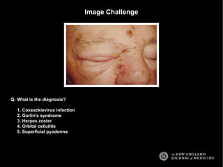
  • 1. Image Challenge What is the diagnosis? 1. Coxsackievirus infection 2. Gorlin's syndrome 3. Herpes zoster 4. Orbital cellulitis 5. Superficial pyoderma Q:
  • 2. Answer: Image Challenge What is the diagnosis? Q: 3. Herpes zoster The image illustrates the classical appearance of acute herpes zoster involving the first division of the trigeminal nerve.
  • 3. Image Challenge What is the diagnosis? 1. Psoriatic arthropathy 2. Reflex sympathetic dystrophy 3. Osteoarthritis 4. Gout 5. Rheumatoid arthritis Q:
  • 4. Answer: Image Challenge What is the diagnosis? Q: 3. Osteoarthritis Examination of this patient's right hand reveals typical changes of osteoarthritis, with both Heberden's and Bouchard's nodes in association with irregular deformities. Read More: New Engl J Med 346;10:e3
  • 5. Image Challenge This 35-year-old pet shop worker developed progressively spreading nodular lesions. He was afebrile. What is the most likely causative organism? 1. Acinetobacter baumannii 2. Erysipelothrix rhusiopathiae 3. Mycobacterium marinum 4. Pasteurella multocida 5. Staphylococcus epidermidis Q:
  • 6. Answer: Image Challenge This 35-year-old pet shop worker developed progressively spreading nodular lesions. He was afebrile. What is the most likely causative organism? Q: 3. Mycobacterium marinum Needle aspiration of a nodule recovered thick, purulent fluid that was positive for acid-fast bacilli, identified on culture as M. marinum. The patient was treated successfully with a four-month regimen of rifampin and ethambutol. M. marinum infection is an occupational hazard for aquarium cleaners. Infection is usually limited to the skin, with the hands, elbows, and feet most commonly affected. The appearance is not typical of infection with the other listed organisms. Read More: New Engl J Med 350;9:e8
  • 7. Image Challenge This patient had left knee pain. What is the diagnosis? 1. Acanthosis nigricans 2. Erythema ab igne 3. Lymphangitis 4. Mycosis fungoides 5. Livedo reticularis Q:
  • 8. Answer: Image Challenge This patient had left knee pain. What is the diagnosis? Q: 2. Erythema ab igne This reticular, reddish-brown, pruritic, nontender, macular, nonblanching discoloration around the medial aspect of the left knee, with a few superficial erosions, is most consistent with erythema ab igne. This patient had repeatedly applied a heating pad to his left knee in the preceding weeks to relieve discomfort from osteoarthritis. Read More: New Engl J Med 356;9:e8
  • 9. Image Challenge What is the most likely diagnosis? 1. Amyloidosis 2. Celiac disease 3. Hypothyroidism 4. Kawasaki disease 5. Type 2 diabetes Q:
  • 10. Answer: Image Challenge What is the most likely diagnosis? Q: 2. Celiac disease Atrophic glossitis is a typical manifestation of celiac disease. Read More: New Engl J Med 356;24:2547
  • 11. Image Challenge What is the diagnosis? 1. Ankylosing spondylitis 2. Castleman disease 3. Metastatic prostate cancer 4. Osteopetrosis 5. Primary hyperparathyroidism Q:
  • 12. Answer: Image Challenge What is the diagnosis? Q: 1. Ankylosing spondylitis The radiograph shows extensive calcification of the intervertebral ligaments, bilateral ossification of the outer layer of the annulus fibrosis (forming bony bridges called marginal syndesmophytes), and apophyseal joint ankyloses all gave the appearance of a bamboo spine. This is most consistent with a diagnosis of ankylosing spondylitis. Read More: New Engl J Med 359;4:404
  • 13. Image Challenge What is the diagnosis? 1. Flail chest 2. Pectus arcuatum 3. Pectus carinatum 4. Pectus excavatum 5. Spondylocostal dysostosis Q:
  • 14. Answer: Image Challenge What is the diagnosis? Q: 4. Pectus excavatum This patient has pectus excavatum, the most common congenital deformity of the chest wall. Read More: New Engl J Med 359;2:e2
  • 15. Image Challenge What is the most likely diagnosis? 1. Renal tubular acidosis 2. Primary hypoparathyroidism 3. Familial hypocalciuric hypercalcemia 4. Salicylate overdose 5. Paget's disease Q:
  • 16. Answer: Image Challenge What is the most likely diagnosis? Q: 1. Renal tubular acidosis The film reveals bilateral symmetric calcification of the renal parenchyma, sparing only the renal pelvis. This patient had been diagnosed with renal tubular acidosis at 9 years of age, but did not undergo medical follow-up for 20 years. The other listed choices are not common causes of nephrocalcinosis. Read More: New Engl J Med 359;1:e1
  • 17. Image Challenge What is the diagnosis? 1. Osgood-Schlatter disease 2. Cystinosis 3. Systemic sclerosis 4. Multiple myeloma 5. Paget's disease Q:
  • 18. Answer: Image Challenge What is the diagnosis? Q: 3. Systemic sclerosis Unlike the other listed choices, acral osteolysis is associated with systemic sclerosis. This patient presented with Raynaud's phenomenon and cutaneous sclerosis; she was missing the tips of all ten fingers and all ten toes. Read More: New Engl J Med 358;26:2812
  • 19. Image Challenge Which one of the following patterns of visual disturbance would be predicted to be demonstrable on examination of this patient? 1. Inferior hemifield loss 2. Temporal quadrantanopsia 3. Uniocular blindness 4. Macular sparing hemianopia 5. Peripheral ring scotoma Q:
  • 20. Answer: Image Challenge Which one of the following patterns of visual disturbance would be predicted to be demonstrable on examination of this patient? Q: 1. Inferior hemifield loss Dilated ophthalmoscopy of this left eye illustrates a nonrefractile plaque in the proximal superior retinal artery with retinal whitening in the superior macula signifying retinal ischemia. Superior retinal ischemia will result in a defect in the inferior visual field, as in this patient. A diagnosis of hemiretinal arterial occlusion was made. Read More: New Engl J Med 358;25:2716
  • 21. Image Challenge What is the diagnosis? 1. Ludwig's angina 2. Glossopharyngeal nerve palsy 3. Pharyngeal gonorrhea 4. Bilateral peritonsillar abscesses 5. Infectious mononucleosis Q:
  • 22. Answer: Image Challenge What is the diagnosis? Q: 4. Bilateral peritonsillar abscesses Bilateral swelling of the soft palate is visible with a midline uvula pushed anteriorly. This is most consistent with a diagnosis of bilateral peritonsillar abscesses. Read More: New Engl J Med 358;23:e27
  • 23. Image Challenge What term is used to describe this finding? 1. Hyphema 2. Hypopyon 3. Iridocyclitis 4. Iridodonesis 5. Synechia Q:
  • 24. Answer: Image Challenge What term is used to describe this finding? Q: 1. Hyphema Layering of blood in the anterior segment is termed hyphema. Hypopyon refers to pus in the anterior segment. Iridocyclitis refers to inflammation of the iris. Iridodonesis is a quivering of the iris when the patient moves the eye. Synechia is an adhesion between the iris and the lens. Read More: New Engl J Med 358;21:2265
  • 25. Image Challenge What is the diagnosis? 1. Dermatopathia pigmentosa reticularis 2. Lichen planus 3. Psoriasis 4. Rubella 5. Keratoderma blennorrhagicum Q:
  • 26. Answer: Image Challenge What is the diagnosis? Q: 5. Keratoderma blennorrhagicum These vesiculopustular waxy lesions are most consistent with keratoderma blennorrhagicum. This finding should prompt diagnostic testing for sexually transmitted or gastrointestinal pathogens. Read More: New Engl J Med 358;20:2160
  • 27. Image Challenge Which structure is most dilated? 1. Aorta 2. Left atrium 3. Left ventricle 4. Right atrium 5. Right ventricle Q:
  • 28. Answer: Image Challenge Which structure is most dilated? Q: 2. Left atrium The chest radiograph reveals cardiomegaly, with splaying of the carina and an elevated left main bronchus. These findings are most suggestive of an enlarged left atrium. Read More: New Engl J Med 358;19:2050
  • 29. Image Challenge What is the diagnosis? 1. Central retinal artery occlusion 2. Diabetic papillopathy 3. Ocular toxoplasmosis 4. Optic neuritis 5. Malignant hypertension Q:
  • 30. Answer: Image Challenge What is the diagnosis? Q: 5. Malignant hypertension The fundus photograph shows disk edema, cottonwool spots, a swollen optic nerve, and retinal hemorrhages. Together, these findings suggest a diagnosis of malignant hypertension. Read More: New Engl J Med 358;18:1951
  • 31. Image Challenge What is the diagnosis? 1. Babesiosis 2. Iron deficiency anemia 3. Hereditary spherocytosis 4. Malaria 5. Sideroblastic anemia Q:
  • 32. Answer: Image Challenge What is the diagnosis? Q: 1. Babesiosis The peripheral-blood smear shows numerous intracellular organisms in red blood cells. Ring forms are seen, as well as rare tetrads. These so-called Maltese cross formations are essentially pathognomonic of babesiosis, since they are not seen in malaria, the primary consideration in the differential diagnosis. Read More: New Engl J Med 358;17:e19
  • 33. Image Challenge This rash appeared following treatment for leukemia. What is the diagnosis? 1. Cryoglobulinemia 2. Leukemia cutis 3. Herpes zoster 4. Graft-versus-host disease 5. Urticaria pigmentosa Q:
  • 34. Answer: Image Challenge This rash appeared following treatment for leukemia. What is the diagnosis? Q: 4. Graft-versus-host disease There is hyperpigmentation and hypopigmentation of the skin, cutaneous atrophy, telangiectasia, and ulcerations. This is most consistent with graft-versus-host disease of the skin.
  • 35. Image Challenge A patient with this tomogram would be most likely to present with which one of the following signs? 1. Uniocular blindness 2. Hemiplegia 3. Alexia without agraphia 4. Hemiballismus 5. Internuclear ophthalmoplegia Q:
  • 36. Answer: Image Challenge A patient with this tomogram would be most likely to present with which one of the following signs? Q: 2. Hemiplegia The tomogram shows a calcified object in the proximal right middle cerebral artery. Occlusion of the middle cerebral artery would be most likely to be associated with contralateral hemiparesis, as in this case. The other listed choices represent stroke syndromes that most typically involve other vascular territories.
  • 37. Image Challenge What is the diagnosis? 1. Behçet's syndrome 2. Lichen simplex chronicus 3. Condyloma acuminatum 4. Lichen sclerosus 5. Vestibular papillomatosis Q:
  • 38. Answer: Image Challenge What is the diagnosis? Q: 5. Vestibular papillomatosis These shiny, soft, linearly arrayed papules are the typical presentation of vestibular papillomatosis, a variant of vestibular mucosa commonly mistaken for genital warts. Read More: New Engl J Med 358;14:1495
  • 39. Image Challenge What is the most likely diagnosis? 1. Paget's disease 2. Meningioma 3. Neurocysticercosis 4. Pneumocephalus 5. Hyperparathyroidism Q:
  • 40. Answer: Image Challenge What is the most likely diagnosis? Q: 4. Pneumocephalus The cranial radiograph shows air in the left temporal region without evidence of fracture. Pneumocephalus can occur after neurosurgical procedures, head and facial trauma, or ear infection and can even occur spontaneously. The finding is not typical of a brown tumor, Paget's disease, neurocysticercosis, or meningioma. Read More: New Engl J Med 358;12:e13
  • 41. Image Challenge Treatment with which one of the following medications is associated with this clinical finding? 1. Erlotinib 2. Leflunomide 3. Methotrexate 4. Pegvisomant 5. Psoralen Q:
  • 42. Answer: Image Challenge Treatment with which one of the following medications is associated with this clinical finding? Q: 1. Erlotinib Erlotinib, a tyrosine kinase inhibitor of the epidermal growth factor receptor, induces characteristic hair alterations. Trichomegaly, curling, elongation, and trichorrhexis are typical; these reverse after discontinuation of therapy. The pictured changes are not typical of leflunomide, methotrexate, pegvisomant, or psoralen. Read More: New Engl J Med 358;11:1175
  • 43. Image Challenge What is the most likely diagnosis? 1. Cisplatin overdose 2. Lesch-Nyhan syndrome 3. Rhabdomyolysis 4. Primary hyperparathyroidism 5. Ethylene glycol poisoning Q:
  • 44. Answer: Image Challenge What is the most likely diagnosis? Q: 5. Ethylene glycol poisoning This urine sediment contains calcium oxalate crystals of two types. The crystals shaped like envelopes with diagonally crossing lines are octahedrons of calcium oxalate dihydrate. The needle-shaped crystals are calcium oxalate monohydrate. Calcium oxalate monohydrate crystals are rarely seen in the urinary sediment but are typical of ethylene glycol ingestion, and therefore when seen, they strongly suggest the diagnosis. Read More: New Engl J Med 354;10:1065
  • 45. Image Challenge What is the most likely diagnosis? 1. Chronic venous insufficiency 2. Reiter syndrome 3. Gunshot wound 4. Chronic renal failure 5. Pseudohypoparathyroidism Q:
  • 46. Answer: Image Challenge What is the most likely diagnosis? Q: 1. Chronic venous insufficiency Chronic venous insufficiency may be accompanied by subcutaneous calcifications. These calcifications are often discovered by chance on plain radiographs. Read More: New Engl J Med 358;9:e10
  • 47. Image Challenge This patient presented with transient, painless visual obscuration in the left eye. What is the diagnosis? 1. Papilledema 2. Hypertensive retinopathy 3. Cholesterol emboli 4. Temporal arteritis 5. Diabetic retinopathy Q:
  • 48. Answer: Image Challenge This patient presented with transient, painless visual obscuration in the left eye. What is the diagnosis? Q: 3. Cholesterol emboli The retinal photograph demonstrates multiple, tiny refractile retinal arteriolar cholesterol emboli and a saddle embolus superior to the optic nerve. Read More: New Engl J Med 358;8:826
  • 49. Image Challenge This 43-year-old patient presented with bilateral pain, swelling, and stiffness in the hands and feet. Her chest radiograph was abnormal. What is the most likely diagnosis? 1. Miliary tuberculosis 2. Psoriasis 3. Syphilis 4. Reiter syndrome 5. Sarcoidosis Q:
  • 50. Answer: Image Challenge This 43-year-old patient presented with bilateral pain, swelling, and stiffness in the hands and feet. Her chest radiograph was abnormal. What is the most likely diagnosis? Q: 5. Sarcoidosis The circumscribed, corticated lytic bone lesions on the radiograph are more consistent with sarcoid granuloma than with the other listed choices. Manifestations of sarcoidosis involving the bones and joints can occur early or late in the illness. Read More: New Engl J Med 358;7:e7
  • 51. Image Challenge This patient presented with jaw pain and was found to have an elevated alkaline phosphatase and a normal serum creatinine. Which one of the following tests would confirm the diagnosis? 1. Bone scan 2. Insulin-like growth factor-1 level 3. Serum calcium 4. Abdominal ultrasound 5. Testing the function of the facial nerve Q:
  • 52. Answer: Image Challenge This patient presented with jaw pain and was found to have an elevated alkaline phosphatase and a normal serum creatinine. Which one of the following tests would confirm the diagnosis? Q: 1. Bone scan Paget's disease, acromegaly, and renal osteodystrophy are among the causes of jaw enlargement, visible in this image. An elevated alkaline phosphatase makes Paget's disease the most likely diagnosis; the diagnosis can be confirmed with a bone scan. Read More: New Engl J Med 358;6:625
  • 53. Image Challenge This patient presented with fatigue, fever, anorexia, and weight loss. What is the most likely diagnosis? 1. Leukemia 2. Scurvy 3. Acquired immunodeficiency syndrome 4. Sarcoidosis 5. Pellagra Q:
  • 54. Answer: Image Challenge This patient presented with fatigue, fever, anorexia, and weight loss. What is the most likely diagnosis? Q: 1. Leukemia Gingival infiltration in a patient with fever, fatigue, and weight loss is most suggestive of acute leukemia, especially monocytic variants of acute myelogenous leukemia. This patient's gingival infiltration resolved after treatment for acute myelomonocytic leukemia. Read More: New Engl J Med 358;3:274
  • 55. Image Challenge What diagnosis is suggested by the finding on the sole of this patient's foot? 1. Chemical burn 2. Pemphigus 3. Thrombotic vasculopathy 4. Radiation dermatitis 5. Frostbite Q:
  • 56. Answer: Image Challenge What diagnosis is suggested by the finding on the sole of this patient's foot? Q: 3. Thrombotic vasculopathy The purpuric skin eruption has a netlike arrangement referred to as retiform. Retiform purpura is an indication of an acute thrombotic vasculopathy. If it is acute and rapidly progressive in a febrile patient, it suggests purpura fulminans. Read More: New Engl J Med 358;2:e1
  • 57. Image Challenge What is the diagnosis? 1. Rhinophyma 2. Leishmaniasis 3. Lupus pernio 4. Wegener's granulomatosis 5. Basal cell carcinoma Q:
  • 58. Answer: Image Challenge What is the diagnosis? Q: 1. Rhinophyma In rhinophyma, enlargement and distortion of the nose occur, with prominent pores and thickened skin due to hyperplasia of the sebaceous glands and fibrosis of the connective tissue. There is follicular prominence and a distorted nodular appearance. Read More: New Engl J Med 352;8:793
  • 59. Image Challenge Which cardiac valves have been replaced? 1. Aortic and mitral valves 2. Aortic and tricuspid valves 3. Mitral and tricuspid valves 4. Pulmonary and mitral valves 5. Pulmonary and tricuspid valves Q:
  • 60. Answer: Image Challenge Which cardiac valves have been replaced? Q: 1. Aortic and mitral valves This patient had her mitral and aortic valves replaced with Starr-Edwards devices for severe rheumatic aortic and mitral stenosis. Read More: New Engl J Med 357;26:2706
  • 61. Image Challenge What is the most likely diagnosis in this 36-year-old woman with chronic renal failure? 1. Lipoma 2. Osteitis fibrosa 3. Osteosarcoma 4. Fistula 5. Calcinosis Q:
  • 62. Answer: Image Challenge What is the most likely diagnosis in this 36-year-old woman with chronic renal failure? Q: 5. Calcinosis Tumoral calcinosis is characterized by soft-tissue calcium and phosphate deposits which are most commonly due to chronic renal failure. These nodules resolved after parathyroidectomy. Read More: New Engl J Med 357;25:2615
  • 63. Image Challenge This patient is most likely to have presented with which one of the following findings? 1. Amnesia 2. Ataxia 3. Dysphagia 4. Hemianopia 5. Hemiparesis Q:
  • 64. Answer: Image Challenge This patient is most likely to have presented with which one of the following findings? Q: 5. Hemiparesis An acute occlusion of the middle cerebral artery, as illustrated here, is most likely to present with contralateral hemiparesis. Read More: New Engl J Med 357;24:2495
  • 65. Image Challenge Which one of following is typically associated with this finding? 1. Bulimia 2. Recent endotracheal intubation 3. Acquired immunodeficiency syndrome 4. Treatment with a sulfonylurea 5. Candida albicans infection Q:
  • 66. Answer: Image Challenge Which one of following is typically associated with this finding? Q: 5. Candida albicans infection Black hairy tongue (lingua villosa nigra) may be associated with the presence of chromogenic organisms (e.g., C. albicans) and the use of certain medications (e.g., doxycycline and bismuth). The pathophysiology is thought to be due to proliferation of the filiform papillae of the tongue, which stain black with porphyrin-producing chromogenic bacteria or yeast. Read More: New Engl J Med 357;23:2388
  • 67. Image Challenge What diagnosis is suggested by the findings on this anteroposterior radiograph of the right tibia? 1. Ewing's sarcoma 2. Marrow hyperplasia 3. Osteomalacia 4. Polyostotic fibrous dysplasia 5. Osteopetrosis Q:
  • 68. Answer: Image Challenge What diagnosis is suggested by the findings on this anteroposterior radiograph of the right tibia? Q: 2. Marrow hyperplasia The radiograph of the right tibia and fibula show the characteristic coarsened trabecular pattern, cortical thinning, and a widened medullary cavity consistent with marrow hyperplasia. This patient was being treated for beta-thalassemia major. Read More: New Engl J Med 357;22:e24
  • 69. Image Challenge What is the diagnosis? 1. Bezoar 2. Pericardial effusion 3. Pneumatosis coli 4. Gastric carcinoma 5. Pheochromocytoma Q:
  • 70. Answer: Image Challenge What is the diagnosis? Q: 1. Bezoar The computed tomography shows a large gastric mass that is separate from the gastric wall. Endoscopy revealed a large trichobezoar occluding nearly the entire stomach. Read More: New Engl J Med 357;21:e23
  • 71. Image Challenge This 81-year-old woman presented with swelling and pain in the left foot a week after treatment for acute bronchitis. Which one of the following treatments is most likely to have been contributory? 1. Beta-adrenergic agonist 2. Cephalosporin antibiotic 3. Leukotriene receptor antagonist 4. Guaifenesin expectorant 5. Fluoroquinolone antibiotic Q:
  • 72. Answer: Image Challenge This 81-year-old woman presented with swelling and pain in the left foot a week after treatment for acute bronchitis. Which one of the following treatments is most likely to have been contributory? Q: 5. Fluoroquinolone antibiotic The magnetic resonance image of the ankle reveals a near-full-thickness rupture of the Achilles’ tendon. Treatment with fluoroquinolone antibiotics has been associated with an increased risk of Achilles' tendon rupture. Quinolones may facilitate the enhanced expression of matrix metalloproteinases in tissue, which in turn causes tendon injury. The other listed choices have not been consistently linked with an increased risk of tendinopathy. Read More: New Engl J Med 357;20:2067
  • 73. Image Challenge Which one of the following drugs of abuse is most typically associated with the illustrated complication? 1. Ketamine 2. Heroin 3. Cocaine 4. Phencyclidine 5. Mescaline Q:
  • 74. Answer: Image Challenge Which one of the following drugs of abuse is most typically associated with the illustrated complication? Q: 3. Cocaine Perforation of the nasal septum and palate are well recognized complications of intranasal cocaine use. Read More: New Engl J Med 357;19:1956
  • 75. Image Challenge This patient presented with a 10-day history of left foot discoloration that resolved with elevation. What is the most likely diagnosis? 1. Angioneurotic edema 2. Arterial insufficiency 3. Erysipelas 4. Peripheral microembolization 5. Phlegmasia cerulea dolens Q:
  • 76. Answer: Image Challenge This patient presented with a 10-day history of left foot discoloration that resolved with elevation. What is the most likely diagnosis? Q: 2. Arterial insufficiency Buerger's symptom refers to dependent rubor after elevation and is suggestive of arterial insufficiency. The foot is not edematous. Erysipelas and peripheral microembolization would not typically resolve with elevation. Phlegmasia cerulea dolens presents with a bluish discoloration following deep venous thrombosis. Read More: New Engl J Med 357;18:e19
  • 77. Image Challenge What is the diagnosis? 1. Cholangiocarcinoma 2. Cirrhosis 3. Echinococcosis 4. Hepatocellular carcinoma 5. Tuberculosis Q:
  • 78. Answer: Image Challenge What is the diagnosis? Q: 2. Cirrhosis The magnetic resonance image shows a connection between the umbilical vein and the left portal vein, as well as esophageal varices. Together these features suggest a diagnosis of portal hypertension and cirrhosis. Read More: New Engl J Med 357;16:e17
  • 79. Image Challenge What is the diagnosis? 1. Pancreatic pseudocyst 2. Duodenal torsion 3. Cholangiocarcinoma 4. Gallbladder lipomatosis 5. Emphysematous cholecystitis Q:
  • 80. Answer: Image Challenge What is the diagnosis? Q: 5. Emphysematous cholecystitis The computed tomogram shows an air-fluid level in the lumen of a dilated gallbladder and gas within the gallbladder wall. These findings suggest emphysematous cholecystitis. Read More: New Engl J Med 348;23:2329
  • 81. Image Challenge What is the diagnosis? 1. Scleroderma 2. Thromboangiitis obliterans 3. Calciphylaxis 4. Hypoparathyroidism 5. Osteomyelitis Q:
  • 82. Answer: Image Challenge What is the diagnosis? Q: 3. Calciphylaxis The radiograph demonstrates severely calcified arteries and the loss of soft tissue at the tips of the index and middle fingers. A skin biopsy was consistent with calciphylaxis. Read More: New Engl J Med 357;13:1326
  • 83. Image Challenge What is the diagnosis? 1. Herpetic glossitis 2. Aphthous ulceration 3. Pemphigoid 4. Scurvy 5. Oral candidiasis Q:
  • 84. Answer: Image Challenge What is the diagnosis? Q: 1. Herpetic glossitis These multiple well-defined white papules with a central punctum are most consistent with herpetic glossitis. Read More: New Engl J Med 357;12:e13
  • 85. Image Challenge What is the diagnosis? 1. Cutis laxa 2. Turner's syndrome 3. Ehlers-Danlos syndrome 4. Pseudoxanthoma elasticum 5. Marfan syndrome Q:
  • 86. Answer: Image Challenge What is the diagnosis? Q: 3. Ehlers-Danlos syndrome Hyperextensibility and hypermobility are features of Ehlers-Danlos syndrome, and result from quantitative or qualitative abnormalities in collagen synthesis. Read More: New Engl J Med 357;11:e12
  • 87. Image Challenge What is the diagnosis? 1. Cytomegalovirus retinitis 2. Lipemia retinalis 3. Central retinal vein occlusion 4. Diabetic retinopathy 5. Retinitis pigmentosa Q:
  • 88. Answer: Image Challenge What is the diagnosis? Q: 2. Lipemia retinalis The creamy appearance of the vessels in the posterior pole and in the peripheral area of the eye is consistent with lipemia retinalis. Typically the retinal findings do not occur until the triglyceride level reaches 2500 mg/dl. Read More: New Engl J Med 357;10:e11
  • 89. Image Challenge Which one of the following is the most likely diagnosis? 1. Chronic renal failure 2. Iron deficiency 3. Graves' disease 4. Chemotherapy treatment 5. Psoriasis Q:
  • 90. Answer: Image Challenge Which one of the following is the most likely diagnosis? Q: 4. Chemotherapy treatment A diagnosis of Muehrcke's lines related to chemotherapy treatment was made. Muehrcke's lines are the two smooth white bands that run parallel to the lunula across the width of the nail. The lines are nonpalpable and, unlike Beau's lines, do not indent the nail itself. Muehrcke's lines are a nonspecific finding that may be associated with periods of metabolic stress, which transiently impairs protein synthesis. Muehrcke's lines are also caused by infections and trauma. Read More: New Engl J Med 357;9:917
  • 91. Image Challenge This 12-year-old boy presented with abdominal pain. What is the diagnosis? 1. Cowden syndrome 2. Cronkhite-Canada syndrome 3. Osler-Weber-Rendu syndrome 4. Peutz-Jeghers syndrome 5. VonWillebrand syndrome Q:
  • 92. Answer: Image Challenge This 12-year-old boy presented with abdominal pain. What is the diagnosis? Q: 4. Peutz-Jeghers syndrome The presence of mucocutaneous pigmented lip lesions suggests the diagnosis of Peutz-Jeghers syndrome, an autosomal dominant disorder characterized by development of multiple hamartomatous gastrointestinal polyps. Read More: New Engl J Med 357;8:e9
  • 93. Image Challenge What is the diagnosis? 1. Molluscum contagiosum 2. Pearly penile papules 3. Secondary syphilis 4. Obstruction of smegma-producing gland 5. Condyloma accuminatum Q:
  • 94. Answer: Image Challenge What is the diagnosis? Q: 2. Pearly penile papules These multiple tiny, smooth, skin-colored papules distributed circumferentially along the coronal sulcus of the glans penis are typical of pearly penile papules. Read More: New Engl J Med 357;7:691
  • 95. Image Challenge This patient presented with chest pain. What is the diagnosis? 1. Acute pulmonary embolism 2. Hypertrophic cardiomyopathy 3. Ascending aortic aneurysm 4. Coarctation of the aorta 5. Lymphoma Q:
  • 96. Answer: Image Challenge This patient presented with chest pain. What is the diagnosis? Q: 3. Ascending aortic aneurysm A widened mediastinum is visible on chest radiography. The patient was diagnosed with a 9.5-cm ascending aortic aneurysm. Read More: New Engl J Med 357;4:e5
  • 97. Image Challenge This patient developed jaundice and an enlarging neck mass. What is the diagnosis? 1. Medullary carcinoma of the thyroid 2. Peripancreatic malignancy 3. Scrofula 4. Hodgkin's lymphoma 5. Pancoast tumor Q:
  • 98. Answer: Image Challenge This patient developed jaundice and an enlarging neck mass. What is the diagnosis? Q: 2. Peripancreatic malignancy The patient had a fungating mass around the Vater's ampulla. Abdominal cancers can metastasize through the thoracic duct to the left supraclavicular fossa. Read More: New Engl J Med 357;3:282
  • 99. Image Challenge This patient developed progressive thickening of the skin following failure of a transplanted kidney. What is the most likely diagnosis? 1. Eosinophilic fasciitis 2. Myxedema 3. Nephrogenic fibrosing dermopathy 4. Scleroderma 5. Vitiligo Q:
  • 100. Answer: Image Challenge This patient developed progressive thickening of the skin following failure of a transplanted kidney. What is the most likely diagnosis? Q: 3. Nephrogenic fibrosing dermopathy The patient was diagnosed with nephrogenic fibrosing dermopathy, an uncommon sclerosing skin condition that has been associated with the use of gadolinium in patients with renal failure. Read More: New Engl J Med 357;2:e2
  • 101. Image Challenge This smoker presented with a four-year history of blanching of the fingers on exposure to cold. What is the diagnosis? 1. Thromboangiitis obliterans 2. Marantic endocarditis 3. Kawasaki disease 4. Brachial entrapment syndrome 5. Takayasu's arteritis Q:
  • 102. Answer: Image Challenge This smoker presented with a four-year history of blanching of the fingers on exposure to cold. What is the diagnosis? Q: 1. Thromboangiitis obliterans Thromboangiitis obliterans is a vaso-occlusive disease that involves small- and medium-sized vessels of the upper and lower extremities. It is strongly associated with tobacco use. An angiogram of this patient's right hand demonstrated multiple chronic occlusions of the digital branches that were unresponsive to intraarterial vasodilators, supporting the diagnosis of thromboangiitis obliterans. Read More: New Engl J Med 339;10:672
  • 103. Image Challenge What is the diagnosis? 1. Graves' ophthalmopathy 2. Retinal detachment 3. Choroidal melanoma 4. Ocular implant 5. Angle closure glaucoma Q:
  • 104. Answer: Image Challenge What is the diagnosis? Q: 2. Retinal detachment The magnetic resonance imaging shows an unusual appearance of retinal detachment with symmetrical bulging in the right eye. Read More: New Engl J Med 357;17:e18
  • 105. Image Challenge What is the diagnosis? 1. Hypertriglyceridemia 2. Hypertensive retinopathy 3. Optic atrophy 4. Central retinal artery occlusion 5. Cytomegalovirus retinitis Q:
  • 106. Answer: Image Challenge What is the diagnosis? Q: 1. Hypertriglyceridemia The creamy white vessels in the fundus resulted from extreme hypertriglyceridemia. Read More: New Engl J Med 340;25:1969
  • 107. Image Challenge These lesions appeared spontaneously in a patient with untreated multiple myeloma. Coagulation studies were normal and platelet count was 80,000 per cubic millimeter. What is the diagnosis? 1. Orbital fracture 2. Disseminated intravascular coagulation 3. Tuberous sclerosis 4. Amyloid purpura 5. Horner's syndrome Q:
  • 108. Answer: Image Challenge These lesions appeared spontaneously in a patient with untreated multiple myeloma. Coagulation studies were normal and platelet count was 80,000 per cubic millimeter. What is the diagnosis? Q: 4. Amyloid purpura These lesions were consistent with amyloid purpura. Read More: New Engl J Med 356;23:2406
  • 109. Image Challenge What is the diagnosis? 1. Nocardiosis 2. Adenocarcinoma 3. Ventricular rupture 4. Alpha-1 antitrypsin deficiency 5. Pulmonary hemosiderosis Q:
  • 110. Answer: Image Challenge What is the diagnosis? Q: 2. Adenocarcinoma Biopsy of the pictured pulmonary adenocarcinoma resulted in a pneumothorax. Read More: New Engl J Med 356;22:2312
  • 111. Image Challenge These lesions developed over 1 to 2 years. What is the diagnosis? 1. Melanocytic nevi 2. Seborrheic keratoses 3. Pemphigus erythematosus 4. Bowenoid papulosis 5. Guttate psoriasis Q:
  • 112. Answer: Image Challenge These lesions developed over 1 to 2 years. What is the diagnosis? Q: 2. Seborrheic keratoses Multiple eruptive seborrheic keratosis are visible. In this case, they were associated with renal cell carcinoma. Read More: New Engl J Med 356;21:2184
  • 113. Image Challenge What is the diagnosis? 1. Central retinal vein occlusion 2. Profilerative diabetic retinopathy 3. Hypertensive retinopathy 4. Chorioretinitis 5. Papilledema Q:
  • 114. Answer: Image Challenge What is the diagnosis? Q: 2. Profilerative diabetic retinopathy Severe bilateral proliferative diabetic retinopathy with significant optic-disk neovascularization is visible. Read More: New Engl J Med 356;19:1979
  • 115. Image Challenge What is the diagnosis? 1. Intracranial hemorrhage 2. Osteoma 3. Neurocysticercosis 4. Arachnoid cyst 5. Meningioma Q:
  • 116. Answer: Image Challenge What is the diagnosis? Q: 5. Meningioma This well-circumscribed and highly calcified extra-axial mass is most consistent with a meningioma. Read More: New Engl J Med 356;16:e14
  • 117. Image Challenge What is the diagnosis? 1. Subcutaneous metastases 2. Filariasis 3. Caput Medusae 4. Neurofibromatosis 5. Hepatocellular carcinoma Q:
  • 118. Answer: Image Challenge What is the diagnosis? Q: 3. Caput Medusae These enlarged veins on his abdomen are consistent with caput medusae. Read More: New Engl J Med 341;6:419
  • 119. Image Challenge What is the diagnosis? 1. Chalazion 2. Papilloma 3. Pterygium 4. Pinguecula 5. Coloboma Q:
  • 120. Answer: Image Challenge What is the diagnosis? Q: 2. Papilloma This 9-year-old boy was diagnosed with conjunctival viral papilloma. Read More: New Engl J Med 356;13:1352
  • 121. Image Challenge What is the diagnosis? 1. Carpal tunnel syndrome 2. Rheumatoid Arthritis 3. Scleroderma 4. Diabetic peripheral neuropathy 5. Dupuyten's contracture Q:
  • 122. Answer: Image Challenge What is the diagnosis? Q: 5. Dupuyten's contracture The pictured flexion contractures involving bilateral third digits and the right fifth digit are most consistent with Dupuytren's contracture. Read More: New Engl J Med 356;12:e11
  • 123. Image Challenge This patient presented with loss of vision. What is the diagnosis? 1. Central retinal artery occlusion 2. Diabetic retinopathy 3. Tay-Sach's disease 4. Hypertensive retinopathy 5. Papilledema Q:
  • 124. Answer: Image Challenge This patient presented with loss of vision. What is the diagnosis? Q: 1. Central retinal artery occlusion Diffuse retinal whitening, constriction of the arteriole and venule with segmentation and a cherry red spot in the macula are most consistent with central retinal artery occlusion.
  • 125. Image Challenge What is the diagnosis? 1. Tinea barbae 2. Herpes simplex infection 3. Eczema 4. Mycosis fungoides 5. Impetigo Q:
  • 126. Answer: Image Challenge What is the diagnosis? Q: 1. Tinea barbae The combination of alopecia and pustules is most consistent with tinea barbae.
  • 127. Image Challenge What is the diagnosis? 1. Diphtheria 2. Secondary syphilis 3. Oral leukoplakia 4. Candida 5. Ludwig's angina Q:
  • 128. Answer: Image Challenge What is the diagnosis? Q: 2. Secondary syphilis These pseudomembranous lesions and erosions of the tongue, the hard and soft palate, and tonsils are consistent with secondary syphilis. Read More: New Engl J Med 347;21:1677
  • 129. Image Challenge What is the diagnosis? 1. Cholesterol emboli 2. Acute arterial insufficiency 3. Deep venous thrombosis 4. Frost bite 5. Thromboangiitis obliterans Q:
  • 130. Answer: Image Challenge What is the diagnosis? Q: 3. Deep venous thrombosis This patient was diagnosed with phlegmasia cerulea dolens, an uncommon manifestation of deep vein thrombosis. Unilateral swelling with discoloration makes arterial insufficiency less likely. Read More: New Engl J Med 356;3:e3
  • 131. Image Challenge What is the diagnosis? 1. Pulmonary hydatid disease 2. Bullous emphysema 3. Pulmonary silicosis 4. Multiple bacterial abscesses 5. Aspergillosis Q:
  • 132. Answer: Image Challenge What is the diagnosis? Q: 4. Multiple bacterial abscesses The finding of bilateral multiple cavitary lesions with air fluid levels is most consistent with multiple pulmonary bacterial abscesses.
  • 133. Image Challenge This 19-year-old man presented with a 10-month history of Raynaud's phenomenon, fever, abdominal pain, and hypertension. What diagnosis is suggested by the findings on his angiogram? 1. Takayasu's arteritis 2. Wegener's granulomatosis 3. Paraganglioma 4. Systemic lupus erythematosus 5. Polyarteritis nodosa Q:
  • 134. Answer: Image Challenge This 19-year-old man presented with a 10-month history of Raynaud's phenomenon, fever, abdominal pain, and hypertension. What diagnosis is suggested by the findings on his angiogram? Q: 5. Polyarteritis nodosa The angiogram reveals multiple microaneurysms involving the renal artery. A biopsy of a subcutaneous nodule was consistent with polyarteritis nodosa.
  • 135. Image Challenge What is the diagnosis? 1. Fabry's disease 2. Hereditary hemorrhagic telangiectasia 3. Peutz-Jegher's syndrome 4. Roseola 5. Discoid lupus Q:
  • 136. Answer: Image Challenge What is the diagnosis? Q: 3. Peutz-Jegher's syndrome The mucocutaneous pigmentation is most consistent with Peutz-Jeghers syndrome.
  • 137. Image Challenge This patient presented with severe jaw pain while being treated for osteoporosis. What is the diagnosis? 1. Submandibular abscess 2. Osteonecrosis 3. Behcet's disease 4. Accessory tooth 5. Calciphylaxis Q:
  • 138. Answer: Image Challenge This patient presented with severe jaw pain while being treated for osteoporosis. What is the diagnosis? Q: 2. Osteonecrosis A diagnosis of bisphosphonate-associated osteonecrosis of the jaw was made in this case. Read More: New Engl J Med 355;22:2348
  • 139. Image Challenge What is the diagnosis? 1. Pericardial hydatid cysts 2. Diaphragmatic rupture 3. Empyema thoracis 4. Cystic fibrosis 5. Pericardial metastases Q:
  • 140. Answer: Image Challenge What is the diagnosis? Q: 5. Pericardial metastases This computed tomogram of the chest revealed multiple pericardial masses with central necrosis consistent with metastatic disease. Read More: New Engl J Med 355;21:e24
  • 141. Image Challenge Treatment with what antibiotic is most likely to have resulted in this patient's skin changes? 1. Rifampin 2. Chloramphenicol 3. Nitrofurantoin 4. Minocycline 5. Trimethoprim Q:
  • 142. Answer: Image Challenge Treatment with what antibiotic is most likely to have resulted in this patient's skin changes? Q: 4. Minocycline Minocycline was the cause of this patient's hyperpigmentation. Read More: New Engl J Med 355;20:e23
  • 143. Image Challenge These lesions were neither pruritic nor painful. What is the diagnosis? 1. Pyoderma gangenosus 2. Phlegmasia cerulea dolens 3. Pretibial myxedema 4. Necrobiosis lipoidica diabeticorum 5. Erythema nodosum Q:
  • 144. Answer: Image Challenge These lesions were neither pruritic nor painful. What is the diagnosis? Q: 4. Necrobiosis lipoidica diabeticorum This patient was diagnosed with necrobiosis lipoidica diabeticorum. Read More: New Engl J Med 355;18:e20
  • 145. Image Challenge What is the diagnosis? 1. Rocky mountain spotted fever 2. Hand foot and mouth disease 3. Infective endocarditis 4. Psoriasis 5. Secondary syphilis Q:
  • 146. Answer: Image Challenge What is the diagnosis? Q: 5. Secondary syphilis This symmetric, dusky red, and polymorphic papulosquamous rash is consistent with secondary syphilis.
  • 147. Image Challenge This patient presented with discoloration of his palms and soles. He reported a normal diet and had normal serum creatinine, thyroxine, and bilirubin concentrations. What is the diagnosis? 1. Diabetes mellitus 2. Vitamin B12 deficiency 3. Amiodarone exposure 4. Hyperlipidemia type III 5. Syphilis Q:
  • 148. Answer: Image Challenge This patient presented with discoloration of his palms and soles. He reported a normal diet and had normal serum creatinine, thyroxine, and bilirubin concentrations. What is the diagnosis? Q: 1. Diabetes mellitus Yellow discoloration of the skin may be associated with carotenemia, hypothyroidism, diabetes mellitus, liver disease, and renal disease. Read More: New Engl J Med 355;14:1486
  • 149. Image Challenge What is the diagnosis? 1. Cytomegalovirus retinitis 2. Roth spots 3. Central retinal vein occlusion 4. Hypertensive retinopathy 5. Papilledema Q:
  • 150. Answer: Image Challenge What is the diagnosis? Q: 5. Papilledema The fundoscopic image suggests florid papilledema. Read More: New Engl J Med 355;12:1262
  • 151. Image Challenge What is the diagnosis? 1. Cardiac tamponade 2. Tension hydrothorax 3. Diaphragmatic eventeration 4. Pulmonary hydatid disease 5. Lymphangiomyomatosis Q:
  • 152. Answer: Image Challenge What is the diagnosis? Q: 1. Cardiac tamponade Cardiac tamponade resolved after removal of 6 liters of pus from the right chest. Read More: New Engl J Med 355;11:e10
  • 153. Image Challenge This 38-year-old woman developed recurrent right-sided chest pain synchronously with her menses. What is the most likely diagnosis? 1. Recurrent pulmonary emboli 2. Alpha-1 antitrypsin deficiency 3. Meig's syndrome 4. Thoracic endometriosis 5. Lymphangiomyomatosis Q:
  • 154. Answer: Image Challenge This 38-year-old woman developed recurrent right-sided chest pain synchronously with her menses. What is the most likely diagnosis? Q: 4. Thoracic endometriosis Her chest radiography demonstrated pneumothorax; endometrial deposits were identified during thoracoscopy implicating catamenial pneumothorax as the cause of her chest pain. Read More: New Engl J Med 355;10:e9
  • 155. Image Challenge This lesion developed after the patient consumed raw meat from a sick goat. What is the most likely diagnosis? 1. Brucellosis 2. Anthrax 3. Herpes simplex 4. Orf 5. Pasteurella multocida Q:
  • 156. Answer: Image Challenge This lesion developed after the patient consumed raw meat from a sick goat. What is the most likely diagnosis? Q: 2. Anthrax This lesion was found to be anthrax of the upper lip. Read More: New Engl J Med 355;9:940
  • 157. Image Challenge This patient's appearance is a consequence of what surgery? 1. Gastric bypass 2. Adrenalectomy 3. Liposuction 4. Pancreatectomy 5. Small bowel transplant Q:
  • 158. Answer: Image Challenge This patient's appearance is a consequence of what surgery? Q: 1. Gastric bypass This patient had undergone open gastric bypass for morbid obesity two years previously. His appearance is a result of massive weight loss. Read More: New Engl J Med 355;8:830
  • 159. Image Challenge The pupils of this female smoker were unresponsive to light and accommodation. She had noted weight loss and postural hypotension and was found to have an irregular centimeter-sized nodule in the right middle lobe. A test for which one of the following would most likely lead to the diagnosis? 1. Calcium-receptor antibody 2. Parathyroid hormone related protein 3. Serum ceruloplasmin 4. Treponema pallidum haemagglutination assay 5. Anti-Hu antibody Q:
  • 160. Answer: Image Challenge The pupils of this female smoker were unresponsive to light and accommodation. She had noted weight loss and postural hypotension and was found to have an irregular centimeter-sized nodule in the right middle lobe. A test for which one of the following would most likely lead to the diagnosis? Q: 5. Anti-Hu antibody A screen for antineuronal antibodies identified anti-Hu; the patient was found to have a pulmonary neuroendocrine tumor. Anti-Hu antibodies are associated with irregular, asymmetric pupils that are unresponsive to light and accommodation. Read More: New Engl J Med 355;5:e4
  • 161. Image Challenge Treatment with which antihypertensive is most likely to cause this appearance? 1. Beta-blocker 2. Diuretic 3. Alpha-blocker 4. Angiotensin converting-enzyme inhibitor 5. Calcium-channel blocker Q:
  • 162. Answer: Image Challenge Treatment with which antihypertensive is most likely to cause this appearance? Q: 4. Angiotensin converting-enzyme inhibitor Angioedema of the tongue is most associated with treatment with an angiotensin converting-enzyme inhibitor. Read More: New Engl J Med 355;3:295
  • 163. Image Challenge What diagnosis is suggested? 1. Parotitis 2. Otitis externa 3. Herpes zoster 4. Frey's syndrome 5. Parotid adenoma Q:
  • 164. Answer: Image Challenge What diagnosis is suggested? Q: 4. Frey's syndrome Sweating over the site of a resected parotid can indicate Frey's syndrome. Read More: New Engl J Med 355;1:66
  • 165. Image Challenge A 55-year-old kidney-transplant recipient presented with headache and fever. The cerebrospinal fluid contained 84 percent neutrophils. What is the most likely diagnosis? 1. Nocardia asteroides infection 2. Cerebral toxoplasmosis 3. Listeria moncytogenes infection 4. Miliary tuberculosis 5. Cryptococcus neoformans infection Q:
  • 166. Answer: Image Challenge A 55-year-old kidney-transplant recipient presented with headache and fever. The cerebrospinal fluid contained 84 percent neutrophils. What is the most likely diagnosis? Q: 1. Nocardia asteroides infection Nocardia asteroides infection typically presents with an abscess or multiple enhancing lesions, with neutrophilic pleocytosis in the cerebrospinal fluid. Read More: New Engl J Med 354;26:2802
  • 167. Image Challenge This 68-year-old woman presented with hair growth and a sore furrowed tongue. What is the most likely diagnosis? 1. Cushing's syndrome 2. Prophyria cutanea tarda 3. Paraneoplastic disorder 4. Acromegaly 5. Polycystic ovarian syndrome Q:
  • 168. Answer: Image Challenge This 68-year-old woman presented with hair growth and a sore furrowed tongue. What is the most likely diagnosis? Q: 3. Paraneoplastic disorder Acquired hypertrichosis lanuginosa is a rare disorder of abnormal and excessive growth of fine lanugo hair, most often associated with an internal malignancy. Read More: New Engl J Med 354;25:2696
  • 169. Image Challenge A 70-year-old man presented with weight loss and hemoptysis. Multiple painless cutaneous nodules had developed over several weeks. What is the most likely diagnosis? 1. Neurofibromatosis 2. Acute myelogenous leukemia 3. Disseminated leishmaniasis 4. Acquired immunodeficiency syndrome 5. Small-cell lung cancer Q:
  • 170. Answer: Image Challenge A 70-year-old man presented with weight loss and hemoptysis. Multiple painless cutaneous nodules had developed over several weeks. What is the most likely diagnosis? Q: 5. Small-cell lung cancer An endobronchial tumor was recognized on flexible bronchoscopy. Cutaneous and bronchial biopsies were positive for small-cell lung cancer. Read More: New Engl J Med 354;24:2583
  • 171. Image Challenge What diagnosis is suggested by this radiograph taken following a barium swallow? 1. Achalasia 2. Esophageal carcinoma 3. Zenker's diverticulum 4. Esophageal stricture 5. Diffuse esophageal spasm Q:
  • 172. Answer: Image Challenge What diagnosis is suggested by this radiograph taken following a barium swallow? Q: 3. Zenker's diverticulum The barium swallow radiograph demonstrates a large Zenker's diverticulum. Read More: New Engl J Med 354;23:e24
  • 173. Image Challenge This 9-year-old boy presented with a two-day history of right shoulder pain after an upper respiratory tract infection. What is the cause of the abnormality demonstrated? 1. Palsy of the long thoracic nerve 2. Subcutaneous emphysema 3. Neuralgic amyotrophy 4. Pleural prolapse 5. Scapular subluxation Q:
  • 174. Answer: Image Challenge This 9-year-old boy presented with a two-day history of right shoulder pain after an upper respiratory tract infection. What is the cause of the abnormality demonstrated? Q: 1. Palsy of the long thoracic nerve Weakness or paralysis of the serratus anterior due to palsy of the long thoracic nerve is a common cause of a winged scapula.
  • 175. Image Challenge What diagnosis explains the combination of findings on this lateral chest radiograph? 1. Syphilis 2. Dressler's syndrome 3. Turner's syndrome 4. Rheumatic heart disease 5. Tertiary hyperparathyroidism Q:
  • 176. Answer: Image Challenge What diagnosis explains the combination of findings on this lateral chest radiograph? Q: 4. Rheumatic heart disease The lateral chest radiograph shows complete left atrial calcification. In combination with the Starr Edwards mitral prosthesis, the most likely diagnosis is rheumatic heart disease. Read More: New Engl J Med 354;21:2262
  • 177. Image Challenge This 61-year-old man presented with abdominal pain. Basophilic stippling was evident on a blood smear. What is the most likely diagnosis? 1. Acute myelogenous leukemia 2. Chronic lead poisoning 3. Beta-thallasemia 4. Megaloblastic anemia 5. Sickle cell anemia Q:
  • 178. Answer: Image Challenge This 61-year-old man presented with abdominal pain. Basophilic stippling was evident on a blood smear. What is the most likely diagnosis? Q: 2. Chronic lead poisoning Chronic lead poisoning was the diagnosis in this case. Lead poisoning is associated with abdominal pain, blue discoloration of the gums, and basophilic stippling. Read More: New Engl J Med 354;20:e21
  • 179. Image Challenge A 39-year-old Zambian man who has tested positive for the human immunodeficiency virus presented with a three-week history of a draining neck mass. What is the most likely diagnosis? 1. Actinomycosis 2. Scrofula 3. Kaposi sarcoma 4. Burkitt lymphoma 5. Nocardiosis Q:
  • 180. Answer: Image Challenge A 39-year-old Zambian man who has tested positive for the human immunodeficiency virus presented with a three-week history of a draining neck mass. What is the most likely diagnosis? Q: 2. Scrofula The patient was found to have scrofula (tuberculous lymphadenitis in the cervical region). Scrofula presents as a chronic, nontender lymphadenopathy, which may fistulize and drain cutaneously. Read More: New Engl J Med 354;18:e18
  • 181. Image Challenge This 46-year-old woman developed pruritus and similar papular lesions over her axillae, groin, and buttocks. What is the most likely diagnosis? 1. Pilonidal sinus 2. Psoriasis 3. Impetigo 4. Dermatomyositis 5. Scabies Q:
  • 182. Answer: Image Challenge This 46-year-old woman developed pruritus and similar papular lesions over her axillae, groin, and buttocks. What is the most likely diagnosis? Q: 5. Scabies These lesions were caused by bird mite, and are similar to the lesions caused by scabies. Read More: New Engl J Med 354;16:1728
  • 183. Image Challenge This 63-year-old man with a 10-year history of progressive neck enlargement noted a sudden increase in the size of the swelling on the left side. What is the most likely diagnosis? 1. Superior vena cava syndrome 2. Scrofula 3. Bleeding into a thyroid nodule 4. Non-hodgkins lymphoma 5. Follicular thyroid carcinoma Q:
  • 184. Answer: Image Challenge This 63-year-old man with a 10-year history of progressive neck enlargement noted a sudden increase in the size of the swelling on the left side. What is the most likely diagnosis? Q: 3. Bleeding into a thyroid nodule An acute hematoma into an established multinodular goiter caused the acute swelling. Read More: New Engl J Med 354;17:1827
  • 185. Image Challenge This 36-year-old man has undergone renal transplantation and parathyroidectomy. What is the most likely cause of the changes in his hands? 1. Pseudoclubbing 2. Albright's hereditary osteodystrophy 3. Hypertrophic osteoarthropathy 4. Yellow nail syndrome 5. Psoriasis Q:
  • 186. Answer: Image Challenge This 36-year-old man has undergone renal transplantation and parathyroidectomy. What is the most likely cause of the changes in his hands? Q: 1. Pseudoclubbing The history and preservation of the nail fold angle suggest pseudoclubbing as the diagnosis, a change that results from soft-tissue collapse due to severe bony erosions. Read More: New Engl J Med 354;15:e14
  • 187. Image Challenge This abnormality was found during auditory screening of a five-year-old girl at school. What is the most likely diagnosis? 1. Serous otitis media 2. Auditory polyp 3. Mastoiditis 4. Glomus jugalare 5. Cholesteatoma Q:
  • 188. Answer: Image Challenge This abnormality was found during auditory screening of a five-year-old girl at school. What is the most likely diagnosis? Q: 5. Cholesteatoma A cholesteatoma medial to an intact translucent tympanic membrane is shown in the left ear of this five-year-old girl.
  • 189. Image Challenge Twelve hours after urgent coronary angiography, the appearance of this patient's feet had changed. What is the most likely explanation for the finding? 1. Endocarditis 2. Raynaud phenomenon 3. Heparin-induced thrombocytopenia 4. Contrast allergy 5. Cholesterol emboli Q:
  • 190. Answer: Image Challenge Twelve hours after urgent coronary angiography, the appearance of this patient's feet had changed. What is the most likely explanation for the finding? Q: 5. Cholesterol emboli The bluish discoloration of the toes with livedo reticularis that appeared after coronary angiography suggests a diagnosis of cholesterol emboli. Read More: New Engl J Med 354;12:1294
  • 191. Image Challenge What diagnosis explains this 15-year-old patient's chest pain? 1. Aortic coarctation 2. Metastatic carcinoma 3. Pneumomediastinum 4. Acute severe asthma 5. Pulmonary hypertension Q:
  • 192. Answer: Image Challenge What diagnosis explains this 15-year-old patient's chest pain? Q: 3. Pneumomediastinum The chest radiograph shows clear lungs, pneumomediastinum and pneumopericardium without evidence of rib fracture or pneumothorax. Read More: New Engl J Med 354;11:1177
  • 193. Image Challenge A 31-year-old woman presented with fever following a trip to Brazil. What diagnosis is suggested by the findings on her blood smear? 1. Paracoccidioides brasiliensis 2. Trypanozoma cruzi 3. Rickettsia typhi 4. Plasmodium vivax 5. Leishmania donovani Q:
  • 194. Answer: Image Challenge A 31-year-old woman presented with fever following a trip to Brazil. What diagnosis is suggested by the findings on her blood smear? Q: 4. Plasmodium vivax The presence of a ring form and basophilic stippling within the red cell accompanied by serpentine forms suggest Plasmodium vivax as the responsible organism. Read More: New Engl J Med 354;10:1064
  • 195. Image Challenge What nebulized medication is most likely to have caused this patient's anisocoria? 1. Racemic epinephrine 2. Ipatropium 3. Ribavirin 4. Salbutamol 5. Theophylline Q:
  • 196. Answer: Image Challenge What nebulized medication is most likely to have caused this patient's anisocoria? Q: 2. Ipatropium This patient's BiPAP face mask leaked to the right, exposing the eye to the anticholinergic agent, ipratropium. Read More: New Engl J Med 354;8:e7
  • 197. Image Challenge This 75-year-old man presented with cough and hoarseness. What is the most likely cause of his appearance? 1. Superior vena cava syndrome 2. Mitral stenosis 3. Chronic obstructive airways disease 4. Angioedema 5. Systemic lupus erythematosus Q:
  • 198. Answer: Image Challenge This 75-year-old man presented with cough and hoarseness. What is the most likely cause of his appearance? Q: 1. Superior vena cava syndrome The combination of facial plethora, swelling, and prominent spidery telangiectasia suggests obstruction of the superior vena cava. A malignancy would be the most likely underlying cause. Read More: New Engl J Med 354;8:e7
  • 199. Image Challenge This 16-year-old boy presented with malaise and fever. What is the diagnosis? 1. Scarlet fever 2. Infectious mononucleosis 3. Measles 4. Mumps 5. Coxsackie viral infection Q:
  • 200. Answer: Image Challenge This 16-year-old boy presented with malaise and fever. What is the diagnosis? Q: 3. Measles The evanescent white papules on the buccal mucosa, Koplik's spots, are diagnostic of measles. Read More: New Engl J Med 354;7:740
  • 201. Image Challenge This patient underwent computed tomography of the pelvis having presented with lower extremity edema. What diagnosis is suggested? 1. Uterine fibroid 2. Ovarian cyst 3. Urinary retention 4. Teratoma 5. Aortic aneurysm Q:
  • 202. Answer: Image Challenge This patient underwent computed tomography of the pelvis having presented with lower extremity edema. What diagnosis is suggested? Q: 3. Urinary retention Bladder outflow obstruction resulted in bladder distension and compression of the iliac veins. Read More: N Engl J Med 2006;354:e5
  • 203. Image Challenge This 6-year-old boy presented with fever and rash that did not improve despite treatment with cephalexin. What diagnosis is suggested? 1. Bullous pemphigoid 2. Staphylococcal scalded skin 3. Stevens-Johnson syndrome 4. Herpes simplex infection 5. Kawasaki disease Q:
  • 204. Answer: Image Challenge This 6-year-old boy presented with fever and rash that did not improve despite treatment with cephalexin. What diagnosis is suggested? Q: 2. Staphylococcal scalded skin Flaccid bullae, perioral crusting and fever suggest the staphylococcal scalded skin syndrome. The patient improved following treatment with intravenous nafcillin.
  • 205. Image Challenge What is the most likely etiology of the findings on this computed tomogram of the chest? 1. Aspergillus fumigatus 2. Bronchiolitis obliterans 3. Amiodarone 4. Tobacco 5. Pneumoconiosis Q:
  • 206. Answer: Image Challenge What is the most likely etiology of the findings on this computed tomogram of the chest? Q: 4. Tobacco Cigarettes are present in the patient's pocket and are the most likely etiology of the pictured lung findings. Read More: N Engl J Med 2006;354:397
  • 207. Image Challenge What is most likely to account for the findings on this abdominal radiograph? 1. Schistosomiasis 2. Chronic laxative use 3. Hyperparathyroidism 4. Ischemic colitis 5. Ingestion of a heavy metal Q:
  • 208. Answer: Image Challenge What is most likely to account for the findings on this abdominal radiograph? Q: 5. Ingestion of a heavy metal The radiograph shows punctate opacities along the entire colon most consistent with ingestion of a heavy metal. Read More: N Engl J Med 2006;354:e3
  • 209. Image Challenge A 37-year-old woman with idiopathic T-cell deficiency underwent computed tomography of the abdomen. What diagnosis is most likely to account for the findings? 1. Histoplasmosis 2. Miliary tuberculosis 3. Amyloidosis 4. Lymphoma 5. Portal hypertension Q:
  • 210. Answer: Image Challenge A 37-year-old woman with idiopathic T-cell deficiency underwent computed tomography of the abdomen. What diagnosis is most likely to account for the findings? Q: 1. Histoplasmosis The image shows calcifications in the spleen and mesenteric lymph nodes, most consistent with prior infection with Histoplasma. Read More: N Engl J Med 2006;354:179
  • 211. Image Challenge What is the diagnosis? 1. Aspergillosis 2. Adrenal insufficiency 3. Oral leukoplakia 4. Pellagra 5. Lingua villosa nigra Q:
  • 212. Answer: Image Challenge What is the diagnosis? Q: 5. Lingua villosa nigra Black hairy tongue (also known as lingua villosa nigra) is a painless benign disorder of unclear etiology. Read More: N Engl J Med 2006;354:67
  • 213. Image Challenge What endocrinopathy is most frequently associated with this sign? 1. Addison's disease 2. Insulin resistance 3. Growth hormone excess 4. Glucagonoma 5. Diabetes insipidus Q:
  • 214. Answer: Image Challenge What endocrinopathy is most frequently associated with this sign? Q: 2. Insulin resistance Acanthosis nigricans is most often associated with insulin resistance. Read More: N Engl J Med 2005;353:2797
  • 215. Image Challenge What is the diagnosis? 1. Pterygium 2. Retroorbital hematoma 3. Retained suture 4. Loa loa 5. Toxocariasis Q:
  • 216. Answer: Image Challenge What is the diagnosis? Q: 4. Loa loa The eyeworm illustrated is Loa loa, a filarial parasite endemic to west and central Africa. Read More: N Engl J Med 2005;353:e22
  • 217. Image Challenge What is the diagnosis? 1. Gout 2. Rheumatoid arthritis 3. Gonococcal arthritis 4. Leprosy 5. Psoriasis Q:
  • 218. Answer: Image Challenge What is the diagnosis? Q: 1. Gout The image illustrates severe deforming gout. Read More: N Engl J Med 2005;353:e20
  • 219. Image Challenge This 61-year-old man is receiving care for an arrhythmia. What is the cause of his appearance? 1. Procainemide 2. Bretylium 3. Amiodarone 4. Sotalol 5. Hydralazine Q:
  • 220. Answer: Image Challenge This 61-year-old man is receiving care for an arrhythmia. What is the cause of his appearance? Q: 3. Amiodarone The blue-grey appearance is a result of treatment with amiodarone. Read More: N Engl J Med 1997;337:1813
  • 221. Image Challenge What is the diagnosis? 1. Budd chiari syndrome 2. Cutaneous larva migrans 3. Umbilical hernia 4. Portal hypertension 5. Gastric carcinoma Q:
  • 222. Answer: Image Challenge What is the diagnosis? Q: 4. Portal hypertension The image illustrates dilated veins radiating from the umbilicus secondary to portal hypertension. Read More: N Engl J Med 2005;353:e19
  • 223. Image Challenge What is the most important diagnosis to exclude in this 81-year-old woman? 1. Lentigo maligna 2. Basal cell carcinoma 3. Varicella zoster 4. Systemic sclerosis 5. Actinic keratosis Q:
  • 224. Answer: Image Challenge What is the most important diagnosis to exclude in this 81-year-old woman? Q: 1. Lentigo maligna The image shows a poorly circumscribed macule with variegated pigmentation. The histopathological diagnosis was lentigo maligna, a diagnosis that should be considered in any pigmented skin lesion. Read More: N Engl J Med 2005;353:2176
  • 225. Image Challenge The pictured patient suffered a traumatic head injury. What underlying diagnosis is suggested by the examination findings? 1. Skull fracture 2. Infratentorial hemorrhage 3. Intraorbital foreign body 4. Uncal herniation 5. Carotid dissection Q:
  • 226. Answer: Image Challenge The pictured patient suffered a traumatic head injury. What underlying diagnosis is suggested by the examination findings? Q: 1. Skull fracture Ptosis and mydriasis suggest a cranial nerve III palsy. The appearance of these signs after a crush injury suggests that a skull fracture is impinging on the nerve canal. Read More: N Engl J Med 2005;353:1955
  • 227. Image Challenge What process is illustrated in the radiograph? 1. Paget's disease 2. Osteopetrosis 3. Hyperparathyroidism 4. Bone marrow hyperplasia 5. Acromegaly Q:
  • 228. Answer: Image Challenge What process is illustrated in the radiograph? Q: 4. Bone marrow hyperplasia The "hair-on-end" appearance of the skull in the radiograph is consistent with massive bone marrow hyperplasia.
  • 229. Image Challenge What is the diagnosis? 1. Left facial palsy 2. Cavernous sinus thrombosis 3. Orbital lymphoma 4. Herpes zoster ophthalmicus 5. Orbtial fracture Q:
  • 230. Answer: Image Challenge What is the diagnosis? Q: 4. Herpes zoster ophthalmicus The image illustrates a left VI nerve palsy from herpes zoster ophthalmicus. Read More: N Engl J Med 2005;353:e14