SlideShare a Scribd company logo
1 of 241
Doppler ultrasound of visceral arteries
Samir Haffar MD
Gastroenterologist – Sonographer
Doppler ultrasound of visceral arteries
① Normal Doppler ultrasound of visceral arteries
② Doppler of proximal abdominal aorta in cardiac diseases
③ Arterial stenosis and occlusion
④ Arterial aneurysm
⑤ Arterial pseudo-aneurysm
⑥ Arteriovenous fistula
⑦ Arterial dissection
⑧ Abdominal vascular compression syndromes
① Normal Doppler ultrasound of
visceral arteries
Optimization of color and spectral Doppler
Color box Small box improved frame rate with better color resolution
Doppler gain Just below the noise level
Color scale (PRF) Low PRF more sensitive to low velocity flow
but may lead to aliasing
Beam steering Adjust to obtain satisfactory vessel angle
Sample volume Usually two thirds of vessel lumen
Wall filter Higher filter cuts out noise & slower velocity flow
Keep filter at 50–100 Hz
Focal zone Color flow optimized at level of focal zone
Bhatt S et al. Ultrasound Clin 2007;2:437–453.
Arteries scanned
• Abdominal aorta: upper & lower
• Celiac axis
• Splenic artery
• Hepatic artery
• Superior mesenteric artery (SMA)
• Inferior mesenteric artery (IMA)
• Renal arteries
Myers KA & Clough A. Making sense of vascular ultrasound. Arnold, London, 2004.
Aorta, celiac axis, SMA & IMA
SMA: superior mesenteric artery – IMA: inferior mesenteric artery
https://radiologykey.com/ultrasound-assessment-of-the-splanchnic-mesenteric-arteries/
All arise from anterior abdominal aorta
Celiac axis is first major branch of abdominal aorta
SMA origin is 1 – 2 cm distal to celiac axis
IMA origin is 3 – 5 cm proximal to aortic bifurcation
Myers KA & Clough A. Making sense of vascular ultrasound. Arnold, London, 2004.
Aorta, celiac axis, SMA & IMA
Transducer positions for scanning AA
Thrush A, Hartshorne T. Peripheral vascular ultrasound: How, why and when.
Elsevier Churchill Livingstone, London, 2nd edition, 2005.
Sagittal or
longitudinal
Transverse
Coronal
Normal spectral Doppler of abdominal aorta
Middle abdominal aorta
Biphasic flow pattern
Proximal abdominal aorta
Triphasic flow pattern
Antegrade systolic waveform
Early diastolic reversal
Low velocity antegrade flow
Antegrade systolic velocity
Early diastolic reversal
No antegrade diastolic flow
Schäberle W. Ultrasonography in vascular diagnosis.
Springer-Verlag, Berlin, 2nd edition, 2011.
Reverberation artefact
Longitudinal gray scale US of proximal aorta
Echogenic focus mimicking intraluminal thrombus (arrow)
Secondary to reverberation artifact
Bhatt S et al. Ultrasound Clin 2007;2:437–453.
Longitudinal gray scale image of upper abdominal aorta
Duplicated aorta or duplication artifact?
Meuwly JY et al. Ultraschall Med 2011;32:233–236.
Duplication image artifact frequent in lower abdomen:
False cases of twin pregnancies
Double intra-uterine devices
Gray-scale US
Duplicated aorta
Color Doppler US
2 aortic lumen filled
with color
Tiny sliding probe to right
Only one lumen filled
with color
Normal aortic bifurcation
Transverse gray scale image Transverse color Doppler image
Bhatt S et al. Ultrasound Clin 2007;2:437–453.
Normal aortic bifurcation
Longitudinal gray scale image
Bhatt S et al. Ultrasound Clin 2007;2:437–453.
Longitudinal color Doppler image
Normal aortic bifurcationNormal aortic bifurcation
Normal Doppler of celiac axis
Forward flow in systole & diastole
Low-resistance flow (RI: 0.74)
Celiac axis divides into CHA & HA
Ao: aorta; IVC: inferior vena cava;
SV: splenic vein
Transverse US of celiac axis Spectral Doppler of celiac axis
RI: resistive index
https://radiologykey.com/anatomy-and-normal-doppler-signatures-of-abdominal-vessels/
CA: celiac axis – CHA: common hepatic artery – LGA: left gastric artery
White RD et al. RadioGraphics 2015;35:879–898.
Common celiac axis branching patterns
Trifurcating CA
Separate origin of
LGA from aorta
Bifurcating CA
into CHA & LGA
Quadrifurcating
of celiac axis
Normal hepatic artery waveform
Forward flow in systole & diastole
Low-resistance spectral pattern (RI: 0.67)
RI: resistive index
Swart J et al. Ultrasound Clin 2007;2:355–375.
Common variations of hepatic artery
“replaced” right hepatic
artery originating from
SMA
Common hepatic artery
originating from CA
Quadrifurcating CA
Separate left & right
hepatic artery
White RD et al. RadioGraphics 2015;35:879–898.
Right hepatic artery from SMA
Battaglia S et al. J Ultrasound 2010;13:49–56.
Common hepatic artery gives rise to GDA & LHA
Right hepatic artery originates directely from SMA
SMA
Branch of SMA
CHA
SMA
LHA
RHA
GDA
Common hepatic artery from SMA
Battaglia S et al. J Ultrasound 2010;13:49–56.
Common hepatic artery originates
from superior mesenteric artery
SMACHA
LHARHA
SMACHA
Left hepatic artery from left gastric artery
Battaglia S et al. J Ultrasound 2010;13:49–56.
LHA originates from LGA
Runs alongside venous ligament
LGA
LHA
CHA
RHA LHA
LGA
Normal splenic artery
Normal Doppler signal of splenic artery
PSV: 110 cm/s – EDV: 45 cm/s – RI: 0.59
Low resistance flow
PSV: peak systolic velocity – EDV: end diastolic velocity – RI: resistive index
https://radiologykey.com/anatomy-and-normal-doppler-signatures-of-abdominal-vessels/
Sagittal ultrasound of SMA
Ao: abdominal aorta
CA: celiac artery
SMA: superior mesenteric artery
Zachrisson H et al. J Med Diagn Meth 2013, 3:2.
Doppler angle for superior mesenteric artery
Doppler angle can be improved from 75° to 25°
by moving transducer distally & tilting it cranially
Schäberle W. Ultrasonography in vascular diagnosis.
Springer-Verlag, Berlin Heidelberg, 2nd edition, 2011.
Transverse US of superior mesenteric artery
Transverse US of pancreatic body and its dorsal landmark
SMA surrounded by collar of fat (yellow arrow)
A: Aorta; P: pancreas; splenic vein: white arrow
Diagnostic ultrasound, Edited by CM Rumack, 5th edition, 2018, Elsevier
Anatomic relationships of SMA
Anterior to SMA: SV & pancreas
Posterior to SMA: aorta
Left to SMA: left renal vein
Right to SMA: Right renal vein & IVC
Surrounded by layer of echogenic fat
SMA: superior mesenteric artery
Revzin MV et al. Ultrasound Clin 2007;2:477–492.
Normal spectral Doppler of SMA
SMA: superior mesenteric artery
Myers KA & Clough A. Making sense of vascular ultrasound. Arnold, London, 2004.
Sharp systolic upstroke
Variable diastolic component
Low flow, reversed flow, or both
Fasting state
High-resistance flow
Postprandial state
Low-resistance flow
Increased velocity
High diastolic velocity
Absence of reverse flow
Common origin of CA and SMA
(arrow)
Common celiac and SMA origin
Color Doppler of abdominal aorta Sagittal MRA of abdominal aorta
CA: celiac axis – MRA: magnetic resonance angiography – SMA: superior mesenteric artery
Revzin MV et al. Ultrasound Clin 2007;2:477–492.
Inferior mesenteric artery
Schäberle W. Ultrasonography in vascular diagnosis.
Springer-Verlag, Berlin, 2nd edition, 2011.
Arises 3 – 5 cm above aortic bifurcation
From left anterolateral aspect of the aorta
Transverse scan with slight pressure & moving upward from bifurcation
Scanned over length of 2 – 5 cm from its origin
Color Doppler ultrasound of normal IMA
Longitudinal view
Normal inferior mesenteric artery (arrow)
arising from anterior left lateral aspect of aorta
AO: aorta – IMA: inferior mesenteric artery
Revzin MV et al. Ultrasound Clin 2007;2:477–492.
Inferior mesenteric artery and vein lying adjacent to aorta (arrows)
IMA: inferior mesenteric artery
Color Doppler ultrasound of normal IMA
Transverse view
Normal spectral Doppler of normal IMA
PSV: 150 cm/sec
Early diastolic reversed flow
Low forward diastolic flow
High resistance flow
IMA: inferior mesenteric artery
Normal spectral Doppler of IMA
Mirk P et al. Abdom Imaging 1998;23:364–369.
High systolic peak
Early diastolic reflux
Low velocity diastolic flow
High pulsatility index: 5.46
Transverse & left sagittal
US of infrarenal aorta
IMA originates from left anterior
aspect of aorta & descends along
its left side with straight course in
its proximal portion
Doppler waveforms of IMA
Normal spectral Doppler of IMA
• Diameter: 2.8 ± 0.55 mm (1.3 – 4.3 mm)
• Pic systolic velocity: 1.41 m/s ± 0.48
• Minimal diastolic velocity: 0.10 m/s ± 0.16
• Pulsatility index: 3.49 ± 0.49 (1.60 – 6.23)
• Time-average velocity (TAV): 0.43 m/s ± 0.19
• Flow volume: 0.13 ± 0.06 L/min (0.05 – 0.29)
Study of 116 patients without splanchnic vessel pathology
Mirk P et al. Abdom Imaging 1998;23:364–369.
Aorta and renal arteries
https://radiologykey.com/ultrasound-assessment-of-the-splanchnic-mesenteric-arteries/
Anatomy of aorta and renal vessels
Lt kidney
Rt kidney
IVC Aorta
SMA
CA
LRA
LRV
RRA
RRV
CA: celiac axis – IVC: inferior vena cava – LRA: left renal artery – LRV: left renal vein
RRA: right renal artery – RRV: right renal vein – SMA: superior mesenteric artery
Number of main renal arteries
Özkan et al. Diagn Interv Radiol 2006;12:183–186.
Angiographic evaluation of 855 consecutive patients
No of vessels 1 2 3 4
Right 83% 15% 1% –
Left 86% 12% 1% 0.2%
Anatomy of main renal arteries
Japan Society of Ultrasonics in Medicine. J Med Ultrasonics 2016;43:145–162.
Branched structure of renal artery
Japan Society of Ultrasonics in Medicine. J Med Ultrasonics 2016;43:145–162.
Main renal arteries
Schäberle W. Ultrasonography in vascular diagnosis. Springer-Verlag, Berlin, 2nd edition, 2011.
Transverse scan with probe angulations
Norma right renal artery
Moukaddam H et al. Ultrasound Clin 2007;2:455–475.
Transverse gray scale image
Right main renal artery
Transverse color Doppler image
Right main renal artery
Normal right renal artery
Coronal image of inferior vena cava
Moukaddam H et al. Ultrasound Clin 2007;2:455–475.
RRA is the only vessel to course under the IVC
Often slightly indents the IVC
Longitudinal scan in left lateral decubitus
Two right renal arteries (15%)
Moukaddam H et al. Ultrasound Clin 2007;2:455–475.
Pre-caval right renal artery
RRA: right renal artery – IVC: inferior vena cava
Hélénon O et al. EMC-Radiologie 2005;2:367–412.
aorta
IVC
RRA
Norma left renal artery
Moukaddam H et al. Ultrasound Clin 2007;2:455–475.
Proximal main left renal artery Proximal main left renal artery
Color Doppler image
LRA
Gray scale image
LRA
Angle of insonation
Difficult to estimate in tortuous or curved renal artery
Correct angleIncorrect angle
Schäberle W. Ultrasonography in vascular diagnosis.
Springer-Verlag, Berlin Heidelberg, 2nd edition, 2011.
Pulsed Doppler of main left renal artery
Hélénon O et al. EMC-Radiologie 2005;2:367–412.
Early systolic notch
Low resistance flow (PSV 90 cm/sec)
Early systolic notch
Moukaddam H et al. Ultrasound Clin 2007;2:455–475.
Some normal waveforms have early systolic notch
1. Measuring to point of PSV results in prolonged acceleration time
2. Excellent negative predictive value of stenosis > 60%
Doppler of right main renal artery
Limits in visualization of main renal arteries
• Obesity
• Overlying bowel gas
• Dyspnea
• Shadowing from arterial calcifications
• Cardiac arrhythmias
• Poor angle of Doppler insonation
• Accessory renal arteries (small size)
RA: renal arteries
Moukaddam H et al. Ultrasound Clin 2007;2:455–475.
Expert sonographers detect 80-90% of main RA
CEUS improves success rate to 95%
Adjustment of Doppler control
Low flow settings for intra-renal arteries
• Lowest pulse repetition frequency without aliasing
• Small color box
• Greatest gain without background noise
• Lowest wall filter
• High color priority
Normal segmental & interlobar renal arteries
Normal segmental renal arteries (long arrows)
Color Doppler image of kidney
Moukaddam H et al. Ultrasound Clin 2007;2:455–475.
Normal interlobar renal arteries (short arrows)
Study of interlobular arteries
Perfusion study / Low PRF
Hélénon O et al. EMC-Radiologie 2005;2:367–412.
Cortical perfusion
Normal pulsed Doppler waveform
Renal segmental artery
Sharp systolic upstroke
Continuous forward diastolic flow
Low resistance flow (low RI)
Measurement of resistive index
Resistive index: S – ED / S
Normal 50 – 70%
Abnormal > 80%
Measurement of acceleration time (AT)
• Length of time in second from
onset of systole to peak systole
• Normal value: < 0.07 sec (70 msec)
Acceleration time (AT) or rise time (RT)
Normal renal aortic ratio (RAR)
Left renal artery Upper abdominal aorta
Japan Society of Ultrasonics in Medicine. J Med Ultrasonics 2016;43:145–162.
PSV: 80 cm/sec PSV: 120 cm/sec
RAR: PSV of renal artery to aorta (80/120 = 0.67)
Normal value ˂ 3
Normal spectral Doppler of renal arteries
• PSV of renal artery at origin: < 180 cm/s
• RAR (PSV ratio of renal artery/aorta): < 3
• Resistive index (RI): < 0.70
• ∆ RI (right – left): < 0.15
• Acceleration time (AT): < 0.07 sec
• Acceleration index (AI): > 3.5 m/s2
PSV: peak systolic velocity – RI: resistive index
② Doppler ultrasound of proximal
abdominal aorta in cardiac diseases
Window to left heart
Abnormal spectral Doppler of proximal abdominal aorta
TVI: Time Velocity Integral
Fadel BM et al. Echocardiography 2014;00:1–5.
Severe aortic regurgitation
Holodiastolic flow reversal (arrows)
Diastolic TVI (17 cm) close to systolic TVI (20 cm)
Indicating a large regurgitant fraction
Fadel BM et al. Echocardiography 2014;00:1–5.
Coarctation of the aorta
Tardus Parvus wave
Abnormal spectral Doppler of proximal abdominal aorta
Prolongation of acceleration time (arrow)
Prolongation of deceleration time (arrowhead)
Prominent diastolic forward flow
No flow reversal
Fadel BM et al. Echocardiography 2014;00:1–5.
Aorto-iliac disease
Mid systolic notching
Notching of antegrade systolic waveform (arrow)
Abnormal spectral Doppler of proximal abdominal aorta
Fadel BM et al. Echocardiography 2014;00:1–5.
Left ventricular assist device
Monophasic forward flow throughout the cardiac cycle
Lack of flow reversals
Abnormal spectral Doppler of proximal abdominal aorta
Fadel BM et al. Echocardiography 2014;00:1–5.
Pulsus alternans
Alternating high peak velocities (arrow)
and low peak velocities (arrowhead)
Abnormal spectral Doppler of proximal abdominal aorta
Fadel BM et al. Echocardiography 2014;00:1–5.
Pulsus paradoxus
Significant respiratory variability in flow
Inspiration decreases systolic velocity & duration (arrows)
Abnormal spectral Doppler of proximal abdominal aorta
2D-imaging of abdominal aorta coupled with
pulse Doppler imaging of proximal abdominal
aorta adds substantially to value of TTE study
TTE: trans-thoracic echocardiography
③ Arterial stenosis and occlusion
Signs of stenosis on pulsed Doppler
Sites of pulsed Doppler Features of pulsed Doppler
Proximal to stenosis: Mid systolic notching
At stenosis: Increased PSV/EDV compared to pre-stenotic segment
Laminar flow
Beyond stenosis: Post-stenotic turbulence
Spectral broadening
Loss of well demarcated spectral edge
Distal to stenosis: Tardus-parvus wave
Atherosclerotic abdominal aorta
Revzin MV et al. Ultrasound Clin 2007;2:477–492.
Luminal irregularity
Sagittal US of abdominal aorta
Marked calcification (arrow)
of abdominal aorta (AO)
Color Doppler of abd aorta
Atherosclerotic stenosis of abdominal aorta
Stenosis in middle aortic segment with significant turbulence
Peak systolic velocity: 369 cm/s
https://radiologykey.com/the-aorta-and-inferior-vena-cava/
Abdominal aortic occlusion
Kaschwich M et al. J Cardiovasc Surg 2017;58(2):313 20.
Transverse US of lower abd aorta Longitudinal US of lower abd aorta
Mid-aortic syndrome
Hypoplasia of aorta, atrophy of aorta, atypical coarctation
• Rare disease w narrowing in distal thoracic &/or abdominal aorta
• Usually found in children and young adults
• Causes: Takayasu arthritis, congenital developmental anomaly,
neurofibromatosis, Williams syndrome
• Renal artery involved in 90% of cases: arterial hypertension
• Clinic: AHT, lower extremity claudication, chronic abdominal pain
• Physical exam: abdominal bruit, reduced or absent femoral pulses,
differential blood pressure between upper & lower extremities
Han L et al. J Clin Ultrasound 2019;47:22–26.
Refractory hypertension for 18 years in a 36-year-old man
Mid-aortic syndrome
PSV: peak systolic velocity – RI: resistivity index
Han L et al. J Clin Ultrasound 2019;47:22–26.
Color Doppler US
of abdominal aorta
Spectral Doppler of
abdominal aorta
Turbulent flow in
narrowed suprarenal
aorta (arrow)
Spectral Doppler
of right kidney
Turbulent flow
with high velocity
PSV > 4.0 m/s
Tardus-parvus waveform
Low RI: 0.32
Different types of celiac trunk stenosis
* Medial Arcuate Ligament Syndrome (celiac artery compression syndrome)
Schäberle W. Ultrasonography in vascular diagnosis. Springer-Verlag, Berlin, 2nd edition, 2011.
Fibromuscular dysplasia
rare
Atherosclerosis
most frequent
MALS syndrome*
Grading stenosis of celiac axis
• Stenosis 50 – 69%: 43 patients
PSV: > 240 cm/s (sens 87%, spec 83%, OA 86%)
EDV: > 40 cm/s (sens 84%, spec 48%, OA 73%)
PSV ratio1: 1.5
• Stenosis 70 – 99% : 62 patients
PSV: > 320 cm/s (sens 80%, spec 89%, OA 85%)
EDV: > 100 cm/s (sens 58%, spec 91%, OA 77%)
PSV ratio1: 2.75
(1) PSV ratio of celiac axis to aorta
AbuRahma AF et al. J Vasc Surg 2012;55:428-36.
105 patients with celiac artery stenosis on angiography
Stenosis of celiac axis
Aliasing and tissue bruit around a stenotic coeliac axis origin
Longitudinal color Doppler UD of upper abdominal aorta
Atherosclerotic stenosis of celiac artery
Spectral tracing
PSV: peak systolic velocity – EDV: end diastolic velocity
Myers KA & Clough A. Making sense of vascular ultrasound. Arnold, London, 2004.
PSV: 276 cm/sec – EDV: 134 cm/sec
Stenosis ≥ 50%
Celiac axis stenosis
PSV: 318 cm/s
indicating ≥ 70% stenosis
Color Doppler of celiac axis Pulsed Doppler at stenotic site
Color aliasing at CA origin (stenosis)
Post-stenotic dilatation
CA: celiac axis – PSV: peak systolic velocity
Grading of SMA stenosis
• Stenosis 50 – 69%:
PSV > 280 cm/s
EDV > 45 cm/s
PSV ratio of SMA/aorta > 3.6
• Stenosis 70 – 99%:
PSV > 395 cm/s
EDV > 74 cm/s
PSV ratio of SMA/aorta > 3.6
PSV: peak systolic velocity – EDV: end diastolic velocity – SMA: superior mesenteric artery
Bird S et al. J Clin Ultrasound. 2019;47(5):267–271.
65 patients with SMA stenosis on CT angiography
Severe stenosis of superior mesenteric artery
PSV 390 cm/s indicating ≥ 70% stenosis
Color and spectral Doppler of SMA
PSV: peak systolic velocity
https://thoracickey.com/clinical-evaluation-and-treatment-of-mesenteric-vascular-disease/
Severe stenosis of superior mesenteric artery
Color & spectral Doppler
at stenotic site
Color bruit outside vessel
High PSV > 500 cm/s
Stenosis ≥ 70%
Post-stenotic
color & spectral Doppler
Post-stenotic turbulence:
Typical “spike hairdo” waveform
Mirror-image artifact
“flow below baseline” at top of tracing
Diagnostic ultrasound, Edited by Rumack CM, 5th edition, 2018, Elsevier
Pulsed Doppler at origin of SMA
Severe stenosis of superior mesenteric artery
PSV: 422 cm/s
Stenosis ≥ 70%
Low-velocity rounded waveforms:
tardus-parvus in post-stenotic zone
Pulsed Doppler of mid-distal SMA
Revzin MV et al. Ultrasound Clin 2007;2:477–492.
Occlusion of superior mesenteric artery
Revzin MV et al. Ultrasound Clin 2007;2:477–492.
Thump artifacts
Focal retrograde flow in
collateral vessel (straight arrow)
Occlusion of proximal SMA
(straight arrow)
Reconstitution by collaterals
from CA (wavy arrow)
US Doppler of SMA origin US Doppler of occluded segment
Grading of IMA stenosis
based on angiography
IMA: inferior mesenteric artery – PSV: peak systolic velocity – EDV: end diastolic velocity
AbuRahma AF et al. Vascular 2012; 20(3):145–149.
Stenosis PSV
cm/s
EDV
cm/s
IMA/aortic
systolic ratio
Normal2 1051
< 50% stenosis3 ≥ 210 no flow or ≥ 20 ≥ 2.5
50 – 69% stenosis4 ≥ 250 ≥ 90 ≥ 4.5
≥ 70% stenosis5 ≥ 270 ≥ 100 –
(1): mean – (2): 45 patients – (3): 12 patients – (4): 8 patients – (5): 15 patients
Severe stenosis of inferior mesenteric artery
Color aliasing at origin of IMA (arrow)
Pulsed Doppler of stenotic region: 332 PSV cm/s
Consistent with hemodynamically severe stenosis (≥ 70%)
IMA: inferior mesenteric artery
Pellerito JS et al. J Ultrasound Med 2009;28:641–650.
Mesenteric ischemia
• Acute mesenteric ischemia: high morbidity and mortality
Causes: SMA embolism, SMA thrombosis, non-occlusive ischemia
Abdominal pain out of proportion to physical examination
Differentiate from other common causes of acute abdominal pain
Laboratory findings of little value: metabolic acidosis, high lactate,…
• Chronic mesenteric ischemia: less common
Severe stenosis or occlusion of 2 of 3 mesenteric vessels
Complete occlusion of single artery may cause intestinal angina
Clinical triad: postprandial abd pain, weight loss, food avoidance
ACR: American College of radiology
Expert Panels on Vascular Imaging and Gastrointestinal Imaging.
J Am Coll Radiol 2018;15:S332-S340.
Acute mesenteric ischemia
Schäberle W. Ultrasonography in vascular diagnosis.
Springer-Verlag, Berlin, 2nd edition, 2011.
Extent of intestinal necrosis varies with level of occlusion
Main trunk Vasa recta
Non occlusive
intestinal ischemia
Ileo-colic artery/
Right colic artery
2nd & 3rd
order branches
Imaging of suspected acute mesenteric ischemia
ACR Appropriateness Criteria
ACR: American College of radiology – CTA: computed tomography angiography
Expert Panels on Vascular Imaging and Gastrointestinal Imaging.
J Am Coll Radiol 2018;15:S332-S340.
Procedures Appropriateness category
CTA abdomen & pelvis with IV contrast Usually appropriate
CT abdomen & pelvis with IV contrast May be appropriate
Arteriography abdomen May be appropriate (disagreement)
MRA abdomen & pelvis w/o & with IV contrast May be appropriate (disagreement)
Radiography abdomen May be appropriate
US duplex Doppler abdomen May be appropriate
CTA of abdomen and pelvis is the most appropriate choice
Doppler US in acute mesenteric ischemia
ACR appropriateness criteria
• Detecting proximal celiac axis & SMA stenosis with high Se & Sp
• Detecting proximal mesenteric arterial thrombosis
• Detecting superior mesenteric or portal venous thrombosis
• Challenges: bowel gas, obesity, vascular calcifications
• Limited role: distal arterial embol, nonocclusive mesenteric ischemia
• Length of examination and pain associated with applied pressure to
abdomen during imaging may be limiting factors
ACR: American College of radiology
Expert Panels on Vascular Imaging and Gastrointestinal Imaging.
J Am Coll Radiol 2018;15:S332-S340.
Acute superior mesenteric artery occlusion
Longitudinal US of SMA Transverse US of SMA
https://radiopaedia.org/cases/acute-superior-mesenteric-artery-occlusion-on-ultrasound
Superior mesenteric artery thrombosis
Thrombus demonstrated within an expanded SMA (arrows)
SMA: superior mesenteric artery
https://radiopaedia.org/cases/superior-mesenteric-artery-thrombosis-1
Longitudinal US of SMA Transverse color Doppler of SMA
Mesenteric vein thrombosis
https://www.pinterest.com/pin/767652698957432844/
Sagittal color Doppler US of superior mesenteric vein
Imaging of suspected chronic mesenteric ischemia
ACR Appropriateness Criteria
Procedures Appropriateness category
CTA abdomen & pelvis with IV contrast Usually appropriate
MRA abdomen & pelvis w/o & with IV contrast Usually appropriate
Arteriography abdomen May be appropriate (disagreement)
CT abdomen and pelvis with IV contrast May be appropriate
MRA abdomen and pelvis without IV contrast May be appropriate
US duplex Doppler abdomen May be appropriate
ACR: American College of radiology – CTA: computed tomography angiography
Expert Panels on Vascular Imaging and Gastrointestinal Imaging.
J Am Coll Radiol 2018;15:S332-S340.
CTA of abdomen and pelvis is the most appropriate choice
ACR: American College of radiology
Expert Panels on Vascular Imaging and Gastrointestinal Imaging.
J Am Coll Radiol 2018;15:S332-S340.
Doppler US in chronic mesenteric ischemia
ACR appropriateness criteria
• Useful initial screening tool for chronic mesenteric ischemia
• Best performed in fasting state & early in the day to avoid bowel gas
• Peak systolic velocity used for diagnosing stenosis
SMA: 295 cm/s and 400 cm/s for 50% and 70% respectively
Celiac artery: 240 cm/s
Doppler of renal artery stenosis
Gray scale imaging first
• Kidneys Maximum renal length
Echogenicity of renal cortex
Thickness of renal cortex
Masses – hydronephrosis – renal calculi
• Aorta Plaque – thrombus – dissection – aneurysm
• Adrenal glands
Measurement of parenchymal & cortical thickness
Cortical thickness: normal 8 – 10 mm
Parenchymal thickness: normal 14 – 18 mm
Tuma J et al. European course book: Genitourinary ultrasound.
European Foundation of Societies of Ultrasound in Medicine & Biology.
parenchyma
cortex
Sites for pulsed Doppler of renal arteries
Aorta
Ostium of main renal arteries
Trunk of main renal arteries
Hilum of both kidney
Upper pole of both kidney
Middle pole of both kidney
Lower pole of both kidney
• Aorta: PSV
• Ostium of renal arteries: PSV
• Renal aorta ratio: PSV ratio of renal artery to aorta
• Trunk of renal arteries: PSV
• Segmental or interlobar arteries: RI and AT
Upper, middle and lower poles of both kidney
Doppler of renal artery stenosis
Ultrasound protocol
AT: acceleration time –PSV: peak systolic velocity – RI: resistive index
Japan Society of Ultrasonics in Medicine. J Med Ultrasonics 2016;43:145–162.
Two main causes of renal artery stenosis
Atherosclerosis (> 90%) FMD (< 10%)
Age ˃ 50 years young
Gender more common in males more common in females
Location proximal 1 cm of main RA
branching points
middle of renal artery
others (carotids)
Post-stenotic
dilatation
rare frequent
FMD: fibromuscular dysplasia
Doppler features of renal artery stenosis
Main renal artery Intra-renal arteries
Focal color aliasing AT > 0.07 sec2
Color bruit Flat blood flow waveforms
Turbulence beyond stenosis Δ RI (right – left) > 15%
RAR > 3.5
PSV > 180 cm/sec1
EDV > 90 cm/sec
1 PSV >180 cm/s: ≥ 50% diameter reduction
PSV > 300 cm/s: ≥ 60% diameter reduction
2 AT > 0.07 sec: > 85% diameter reduction
AT: acceleration time – EDV: end diastolic velocity
PSV: peak systolic velocity – RAR: renal aortic ratio – RI: resistive index
Japan Society of Ultrasonics in Medicine. J Med Ultrasonics 2016;43:145–162.
Renal artery stenosis
Renal aortic ratio (RAR)
Left renal artery Upper abdominal aorta
PSV: peak systolic velocity
Japan Society of Ultrasonics in Medicine. J Med Ultrasonics 2016;43:145–162.
PSV: 410 cm/s PSV: 50 cm/s
RAR: 410/50 = 8.20 (normal ˂ 3)
Renal artery stenosis ≥ 60%
PSV: 293 cm/sec – RI : 0.91
Controversial indication of PTA2
Aliasing in left renal artery
Retro-aortic course of LRV
LRV: left renal vein – PSV: peak systolic velocity – RI resistive index – PTA: angioplasty
1 Schäberle W. Ultrasonography in vascular diagnosis. Springer-Verlag, Berlin, 2nd edition, 2011.
2 Jaeger KA et al. Ultraschall in Med 2007;28:28–31.
Renal artery stenosis
Waveform of intra-renal blood flow
AT: acceleration time
Japan Society of Ultrasonics in Medicine. J Med Ultrasonics 2016;43:145–162.
Normal flow patternIntra-renal artery Renal artery stenosis
Acceleration time (AT)
Normal ˂ 70 msec
Japan Society of Ultrasonics in Medicine. J Med Ultrasonics 2016;43:145–162.
AT: 40 msec AT: 65 msec AT: 140 msec
Renal artery stenosis
Normal AT Normal AT Prolonged AT
Tardus Parvus wave
• Mimics Aortic/mitral valve disease
Aortic coarctation
William syndrome
Left ventricle dysfunction
CV medications: after-load reducers
• Exaggerating 25 mg captopril 1 hour before exam
• Minimizing Age – HTN – DM (vessel compliance)
CV: cerebrovascular – DM: diabetes mellitus – HTN: hypertension
Moukaddam H et al. Ultrasound Clin 2007;2:455–475.
Fibromuscular dysplasia
Moniliform aspect of RRA
Typical FMD in middle third of RRA
Hélénon O et al. EMC-Radiologie 2005;2:367–412.
PSV: 250 cm/sec
No parallelism of RRA walls
Fibromuscular dysplasia
Japan Society of Ultrasonics in Medicine. J Med Ultrasonics 2016;43:145–162.
Indications of PTRA in renal artery stenosis
Still controversial
• Hemodynamically significant renal artery stenosis and:
Resistant hypertension despite use of ≥ 3 antihypertensive drugs
Exacerbating hypertension
Malignant hypertension
Hypertension with idiopathic unilateral kidney atrophy
Idiopathic pulmonary edema that suddenly develops
Repeated heart failure
Unstable angina
Fibromuscular dysplasia
• Patients with bilateral renal artery stenosis
• Progressive chronic kidney disease in a single functioning kidney
PTRA: percutaneous transluminal renal angioplasty
Japan Society of Ultrasonics in Medicine. J Med Ultrasonics 2016;43:145–162.
Renal artery thrombosis (complete)
Irshad A et al. Semin Ultrasound CT MRI 2009;30:298–314.
Transplanted kidney
Absence of flow within kidney
Power Doppler US Power Doppler US more medially
Flow in iliac artery
& proximal anastomotic artery
Partial acute renal infarction
Japan Society of Ultrasonics in Medicine. J Med Ultrasonics 2016;43:145–162.
Gray scale image Power Doppler image
④ Arterial aneurysm
Aneurysms, false aneursysm & arterial dissection
Normal artery
Arterial wall
components
True aneurysm
Components of arterial
wall "stretched"
Hole in arterial wall
with confined collection
of blood
False aneurysm Arterial dissection
Hematoma between
components of wall
Zwiebel JW. Ultrasound assessment of aorta, iliac arteries and inferior vena cava.
In: Introduction to vascular ultrasonography. Zwiebel JW ed, 5th edition, Elsevier Saunders, 2005.
Selective screening for AAA
• Selective screening
3 important risk factors: Males
Age > 65 years
History of smoking
• Effectiveness of screening
4 RCTs including more than 125,000 men
Reported results for up to 5 – 10 years of follow-up
Reduction in mortality from 68% to 21%
RCT: randomized controlled trial
Lederle FA. Ann Intern Med 2003 ; 139 : 516 – 22.
Abdominal aortic aneurysm (AAA)
• Normal size of abdominal aorta 1.5 – 2.5 cm
• Ectatic aorta 2.5 – 3 cm
• Aortic aneurysm > 3 cm
• Annual growth rate of aneurysms 0.33 cm/year
measuring between 4 & 5.5 cm
Bhatt S et al. Ultrasound Clin 2008;3:83–91.
Abdominal aortic aneurysm (AAA)
• Measurement
• Location
• Morphological evaluation
• Possible evaluation of iliac arteries
• Complications: rupture, thrombosis, dissection, embolization
What to evaluate?
Measurement of widest part
Measurement technique of aneurysm
Myers KA & Clough A. Making sense of vascular ultrasound. Arnold, London, 2004.
Measuring diameter of AAA
Incorrect measurement Correct measurement
AAA: abdominal aortic aneurysm
Schäberle W. Ultrasonography in vascular diagnosis. Springer-Verlag, Berlin, 2nd edition, 2011.
Correct diameter measured by rotating transducer clockwise
until round image of aorta comes into view
Battaglia S et al. J Ultrasound 2010:13:107–117.
Abdominal aortic aneurysm (swirling flow)
Pseudo ‘‘Yin-Yang sign’’
Similarity in appearance to pseudo-aneurysm finding
Classification of abdominal aortic aneurysms
Classification Categories
By location Suprarenal: Above origin of renal areteries (very rare)
Juxtarenal: Where renal arteries originate
Infrarenal: Below origin of RA (most common)
Bhatt S et al. Ultrasound Clin 2007;2:437–453.
By morphology Fusiform (most common)
Hourglass
Saccular
By etiology Atherosclerotic (most common)
Inflammatory (5% – 10%)
Mycotic (1%): saccular, salmonella & SA, high mortality
Classification of AAA by location
Based on relation to renal arteries
Suprarenal
Suprarenal: Aneurysm involves origins of one or more visceral arteries
Pararenal: RAs arise from aneurysm but aorta at level of SMA not aneurysmal
Juxtarenal: Aneurysm originates just beyond origins of RAs
Infrarenal: Aneurysm originates distal to renal arteries.
Pararenal Juxtarenal Infrarenal
AAA: abdominal aortic aneurysm
Supra-renal abdominal aortic aneurysm
Schuster H et al. Ultraschall Med 2009;30:528–543.
Transverse US viewLongitudinal US view
Inclusion of visceral & renal
arteries
Perfused lumen
& narrow circular thrombus
Infra-renal abdominal aortic aneurysm
Distance between renal vein & upper limit of aneurysm
LRV: left renal vein – SMA: superior mesenteric artery
Thrush A, Hartshorne T. Peripheral vascular ultrasound: How, why and when.
Elsevier Churchill Livingstone, London, 2nd edition, 2005.
SMA
LRV
Classification of AAA by shape
Fusiform Saccular
Most frequent
Double aneurysm
Hourglass aorta
AAA: abdominal aortic aneurysm
Abdominal aortic aneurysm (fusiform)
Transverse image
Anteroposterior diameter
from outer wall to outer wall
Sagittal image
Diameter measured in transverse
image larger due to obliquity
Myers KA & Clough A. Making sense of vascular ultrasound. Arnold, London, 2004.
Abdominal aortic aneurysm (hourglass)
Bhatt S et al. Ultrasound Clin 2007;2 437–453.
Two discontinuous focal segments of aneurysmal dilatation
Aortic diameter in between is normal in caliber
Abdominal aortic aneurysm (saccular)
Saccular or mycotic aneurysm
Thrombus seen as low-level echoes within aneurysm
Longitudinal image of abdominal aorta
Abraham D et al. Emergency medicine sonography: Pocket guide.
Jones & Bartlett Publishers, Boston, MA, USA, 1st edition, 2010.
Complications of abdominal aortic aneurysm
• Thrombosis
• Dissection
• Rupture
• Embolization
Complications of abdominal aortic aneurysm
Partial thrombus
Bhatt S et al. Ultrasound Clin 2007;2:437–453.
Infrarenal AAA with thrombus occluding
approximately two thirds of the lumen
Longitudinal color Doppler US Transverse color Doppler US
Abdominal aortic aneurysm (thrombus liquefaction)
Area of thrombus liquefaction may be confused with dissection
Large thrombus separate area of liquefaction from lumen
Thrush A, Hartshorne T. Peripheral vascular ultrasound: How, why and when.
Elsevier Churchill Livingstone, London, 2nd edition, 2005.
Longitudinal color Doppler Transverse color Doppler
Accorci F. Ultrasound in abdominal aorta and iliac artery atherosclerotic diseases.
http://www.echodoppler-lessons.com/en/pathology-of-the-aorta-and-the-peripheral-arteries/
ultrasound-in-abdominal-aorta-and-iliac-arteries-atherosclerotic-disease
Complications of abdominal aortic aneurysm
Complete thrombus
Bhatt S et al. Ultrasound Clin 2007;2:437–453.
Longitudinal gray-scale US
Echogenic band in aortic lumen Echogenic band in aortic lumen
Transverse gray-scale US
Transverse color Doppler US
Anterior false lumen (blue)
Posterior true lumen (red)
Longitudinal color Doppler US
Entry point (arrowhead) of
intimal tear causing dissection
Complications of AAA (dissection)
AAA with peripheral thrombus
Small hypoechoic area (wall rupture)
Hypoechoic structure at upper end
Presence of active bleeding
No further imaging confirmation
Taken directly to OR
AAA: abdominal aortic aneurysm – OR: operating room
Bhatt S et al. Ultrasound Clin 2007;2:437–453.
Complications of abdominal aortic aneurysm
Rupture (high mortality rate: 90%)
Stent-graft expands to make firm
circumferential contact with
 ‘neck’ of relatively normal aorta
between RA & upper end of AAA
 each CIA below aneurysm
Endovascular aortic aneurysm repair (EVAR)
First performed by Parodi from Argentina in 1990 1
AAA: abdominal aortic aneurysm – CIA: common iliac artery –RA: renal arteries
1 Parodi JC et al. Ann Vasc Surg 1991;5:491–499.
2 Myers KA & Clough A. Making sense of vascular ultrasound. Arnold, London, 2004.
Stent-graft
Endoleak after EVAR
Persistent flow in aneurysm lumen after procedure
• Increase in aneurysmal diameter with risk of rupture
• 20 – 40% at any time after graft placement1
• Lifelong surveillance 1st month, 6th month, yearly2
• Modalities CTA: gold standard
CDUS/CEUS: acceptable alternative
MRA – DSA
CDUS: color Doppler ultrasound – CEUS: contrast-enhanced ultrasound
CTA: computed tomography angiography – MRA: magnetic resonance angiography
1 Demirpolat G et al. J Clin Ultrasound 2011;39:263–269.
2 Stavropoulos SW et al. Radiology 2007;243:641.
Determination of endoleak & aneurysmal size
Endoleak following EVAR
Type I Type II Type III Type IX
Failure of proximal or
distal attachment sites
Flow through aortic
or iliac branches
common
Perforation & tear
in graft material
rare
Porosity of graft
material
resolved in 1 month
EVAR: endo-vascular abdominal aneurysm repair
White GH et al. J Endovasc Surg 1996 ;3:124–5.
Carrafiello G et al. Cardiovasc Intervent Radiol 2006;29:969–974.
Types of endoleak
Type I: Distal attachment site
Type II: Patent lumbar artery
Thrush A et al. Peripheral vascular ultrasound. Elsevier, London, 2nd edition, 2005.
Hartshorne T. Ultrasound 2006;14:34–42.
Type II: Inferior mesenteric artery
Type I: Proximal attachment site
Endoleak type II over mesenteric artery
Zimmermann H et al. Clin Hemorheology Microcirculation 2014;58:247–260.
Aortic aneurysm with graft
No endoleak seen in B-mode
Type II endoleak (yellow arrow)
over mesenteric artery
Color Doppler ultrasoundGray scale ultrasound
Zimmermann H et al. Clin Hemorheology Microcirculation 2014;58:247–260.
Gray scale ultrasound Color Doppler ultrasound
Aortic aneurysm with graft
No endoleak seen in B-mode
Endoleak type II over lumbar artery
Type II endoleak (yellow arrow)
over right lumbar artery
Endovascular aortic aneurysm repair
Mirror artifact
Demirpolat G et al. J Clin Ultrasound 2011;39:263–269.
Synchronous pulsatility with flow in patent graft
Changing position while examining from different aspects
Spectral analysis aids in reducing false positive
Mirror image behind patent limbs of stent graft
Iliac artery aneurysm
• Most iliac artery aneurysms associated w distal aortic aneurysms
Aortic aneurysm is the source of clinical concern
• Isolated iliac artery aneurysm uncommon but may be deadly:
Not palpated on physical examination even when large
Rupture generates nonspecific abdominal or pelvic symptoms
Nor recognized until patient becomes hypotensive or dies
• Located in proximal or distal portion of common iliac arteries
• Surgical repair or percutaneous stenting recommended if >3 cm
Zwiebel JW. Ultrasound assessment of aorta, iliac arteries and inferior vena cava.
In: Introduction to vascular ultrasonography. Zwiebel JW ed, 5th edition, Elsevier Saunders, 2005.
Aortic and iliac artery aneurysms
Aortic aneurysm & common iliac artery aneurysm
Separated by relatively normal segment of proximal CIA (arrow)
Longitudinal view of lower abdominal aorta
Isolated common iliac artery aneurysm
Transverse ultrasound of abdominal aortic bifurcation
Left iliac artery aneurysm
Pseudo Yin Yang flow pattern
Battaglia S et al. J Ultrasound 2010;13:107–117.
Isolated internal iliac artery aneurysm
Zwiebel JW. Ultrasound assessment of aorta, iliac arteries and inferior vena cava.
In: Introduction to vascular ultrasonography. Zwiebel JW ed, 5th edition, Elsevier Saunders, 2005.
Flow in the mass (arrows)
and apparent communication
with IIA
Gray scale ultrasound Color Doppler ultrasound
Hypoechoic mass in pelvis
near EIA (arrows)
Initially thought to be nodal mass
• Definition:
Arterial dilatation > 1.5 times the size of original vessel
with loss of parallelism of vascular walls
• Size of visceral arteries:
Celiac trunk: 0.79 ± 0.06 cm
Common hepatic artery: 0.50 ± 0.04 cm
Proper hepatic artery: 0.45 ± 0.03 cm
Splenic artery: 0.46 ± 0.03 cm
Visceral artery aneurysms
Ibrahim F et al. Curr Treat Options Cardio Med 2018;20:97–111.
Sites of visceral artery aneurysms
Rare – discovered incidentally – high mortality in rupture
Jesinger RA et al. Radiographics 2013;33:E71–96.
Piasek E et al. J Ultrason 2018;18:148–151.
Splenic artery:
Hepatic artery:
Superior mesenteric artery:
Celiac artery:
Inferior mesenteric artery:
Gastric, intestinal, pancreatic, gastroduodenal:
Renal artery
60%
20%
5%
4%
1%
rare
1 – 10 %
Splenic artery & hepatic artery most commonly involved
Celiac artery aneurysm
Piasek E et al. J Ultrason 2018;18:148–151.
Transverse gray scale US of upper abdomen
Aneurysm of celiac axis
Revzin MV et al. Ultrasound Clin 2007;2:477–492.
Celiac artery aneurysm
Lumen of celiac axis dilated (arrow)
Disturbed flow on color Doppler (pseudo Yin Yang flow)
Celiac axis aneurysm with dissection
52-year-old male - CT for workup of testicular seminoma
Celiac axis aneurysm
13 mm
Aneurysmal dissection
w true & false lumen
PSV: peak systolic velocity – EDV: end diastolic velocity
Errigo A et al. J Vascular Ultrasound 2018;42(2):82–84.
Doppler of false lumen
PSV: 402 cm/s
EDV: 142 cm/s
Extra-hepatic artery aneurysm
Mansour MA et al. Vascular Diagnosis. Elsevier-Saunders, Philadelphia, 1st edition, 2005
Extrahepatic sacular hepatic artery aneurysm
Pseudo Yin-Yang flow pattern
Ignee A et al. Z Gastroenterol 2002;40:21–32.
Gray-scale US Color & duplex US
Pseudo Yin-Yang flow pattern
Arterial flow on spectral Doppler
Ovoid mass in porta hepatis
Extra-hepatic artery aneurysm
Intra-hepatic artery aneurysm
O’Driscoll D et al. Br J Radiology 1999;72:1018–1025.
26-year-old man – abdominal pain after abdominal blunt trauma
Hypoechoic lesion in right lobe
Arterial flow on spectral Doppler
Contrast-enhanced CTPulsed Doppler US
Large aneurysm in right
hepatic lobe
• 3rd common intra-abdominal aneurysm after aorta & iliac arteries
• Abnormal dilatation of splenic artery > 1 cm in diameter
• Risk factors: pregnancy, portal hypertension, trauma, arterial
degeneration (medial fibrodysplasia), atherosclerosis
• Majority asymptomatic (80%) & discovered incidentally
• Symptomatic in 20%: epigatric or LUQ pain
• Spontaneous rupture: frequency 3% – mortality 25%
• Risk of rupture: pregnancy, PHT, > 2 cm, liver transplantation
Splenic artery aneurysm
LUQ: left upper quadrant – PHT: portal hypertension
Al-Habbal Y et al. Surgeon 2010;8:223–231.
Splenic artery aneurysm
Piasek E et al. J Ultrason 2018;18:148–151.
Gray-scale US of splenic hilum Color Doppler US of splenic hilum
Splenic artery aneurysmCystic mass in splenic hilum
Gray scale ultrasound
Wakui N et al. J Med Ultrasonics 2011:38:167–171.
Splenic artery aneurysm
Color Doppler ultrasound
2.2-cm cystic mass at
splenic hilum (arrow)
Color Doppler: pseudo Yin-Yang pattern
Pulsatile waveform in cystic lesion
Atherosclerotic splenic artery aneurysm
Badea R et al. J Med Ultrasonics 2013;40:487–490.
Turbulent color signal
suggesting splanchnic
aneurysm
4-cm hypoechoic mass
on splenic artery trajectory
with wall calcification
Presence of splenic
artery aneurysm
CT angiographyGray scale US Color Doppler US
Multiple splenic artery aneurysms
Bagga B, Das CJ. BMJ Case Rep 2019;12:e228705.
Transverse abdominal US Color spectral Doppler US
Dilated splenic artery (asterisk)
Multiple aneurysms (arrow)
likely arising from it
Arterial waveform in
aneurysms confirming them to
be arising from splenic artery
Thrombosed splenic artey aneurysm
mimicking pancreatic adenocarcinoma
Round hypoechoic solid mass
appears as aneurysmatic thrombosed
dilatation arising from splenic artery
Casadei R et al. JOP (Online) 2007;8(2):235-239.
Transverse color Doppler US Non-contrast-enhanced CT scan
Hypodense round solid mass of
pancreatic body w calcified periphera
spot simulating pancreatic neoplasm
Splenic vein aneurysm
• Splenic vein aneurysms are rare
• Etiology: portal hypertension, congenital weakness in vein wall
• Clinic: asymptomatic – usually incidental findings
• Complication: rupture, thrombosis, compression adjacent structures
• Appropriate management: unknown
Noninvasive follow-up, plication, and aneurysm excision
Torres G et al. J Vasc Surg 1999;29:719–21.
Splenic vein aneurysm
Cystic round mass in
pancreatic tail
Enhancing mass in left
mid-abdomen
Torres G et al. J Vasc Surg 1999;29:719–21.
Transverse US image Abdominal CT (venous phase)
Inferior mesenteric artery aneurysm
Momin AA et al. J Clin Ultrasound 2008;36:42–44.
Transverse color US of
middle abdominal aorta
Longitudinal US of middle
abdominal aorta
IMA aneurysm parallel to
lower part of abdominal aorta
AO: aorta
VC: inferior vena cava
Aneurysm: IMA aneurysm
Renal artery aneurysm
Japan Society of Ultrasonics in Medicine. J Med Ultrasonics 2016;43:145–162.
Ultrasound of kidney
Cystic mass at hilum
Color Doppler US
Renal artery aneurysm
Pulsed Doppler US
Low resistance arterial
flow
Indications to treat visceral aneurysms
• Clinical symptoms: pain, embolism, rupture
• > 2 cm in size or twice the size of the artery by consensus
• Rapid increase in diameter of aneurysm
• Specific location: duodenopancreatic arcades
• Pregnant women or women of childbearing age
• Non-atherosclerotic etiology (ie: connective tissue disease)
• Patient requiring a liver transplant
• Portal hypertension for splenic artery aneurysm
Chiaradia M et al. Diag Inter Imaging 2015:96:796–806.
Endovascular or open surgery approaches
for visceral artery aneurysms
• Endovascular approach:
Used more often by surgeons
Shorter hospital stay
Lower rates of cardio-vascular complications
Higher rates of re-intervention
Low access site complication (<5%)
Post-embolization syndrome: from 9% (renal) to 38% (splenic)
Coil migration: from 8% (splenic) to 29% (renal)
• Difference in mortality of 2 approaches not statistically significant
Barrionuevo P et al. J Vasc Surg. 2019 May 21. doi: 10.1016/j.jvs.2019.02.024.
Systematic review
80 observational studies – 2845 visceral aneurysms
⑤ Arterial pseudo-aneurysm
Pulsed Doppler of pseudo-aneurysm
Middleton WD et al. Ultrasound Quarterly 2005;21:3–17.
To-and-fro flow
Typical triphasic flow
“to-and-fro” flow of pseudo-aneurysm
Middleton WD et al. Ultrasound Quarterly 2005;21:3–17.
During systole
“to”
Flow enters PA via the neck
Pseudo-aneurysm lumen enlarges
During diastole
“fro”
Flow exits PA via the neck
Pseudo-aneurysm lumen contracts
Variations in ‘‘to-and-fro’’ flow in pseudoaneurysm
Middleton WD et al. Ultrasound Quarterly 2005;21:3–17.
Limited systolic flow
More pronounced diastolic flow
Diastolic flow decreases progressively
Diastolic flow increases progressively
Diastolic flow relatively limited
Two distinct phases of diastolic flow
Variations in duration & velocities of
systolic & diastolic flow due to arrhythmia
Pseudoaneurysm of abdominal aorta
Ertürk H et al. J Clin Ultrasound 1999;27:202–205.
“to-and-fro” flow pattern
in neck of aortic pseudoaneurysm
Turbulent flow in pseudoaneurysm
Anterior displacement of
normal-sized aorta (arrows)
Transverse color Doppler US Longitudinal color duplex US
Pseudoaneurysm of hepatic artery
PSC: primary sclerosis cholangitis
Parlak S et al. J Belg Society Radiology 2015;99(2):61–4.
50-year-old male, liver transplant for PSC, abnormal liver tests
Pseudoaneurysm originating from hepatic artery (arrow)
Classic Yin and Yang flow pattern in pseudoaneurysm
Peripheral partial thrombus in pseudoaneurysm (arrowhead)
Pseudoaneurysm of SMA
Communication between SMA (arrow) & pseudoaneurysm
Classic Yin and Yang flow pattern in pseudoaneurysm
AO: aorta – SMA: superior mesenteric artery – SV: splenic vein
66-year-old male, chronic alcoholic pancreatitis, upper abd pain
Săftoiu A et al. J Pancreas (Online) 2005; 6(1):29–35.
Pseudoaneurysm of the kidney
Secondary to renal biopsy
https://iame.com/online/duplex_and_color_doppler_of_the_kidney/content.php
Color Doppler US of kidney Puled Doppler at the neck
“to-and-fro” flow patternPseudoaneurysm
Yin and yang low pattern
⑥ Arteriovenous fistula
External iliac arteriovenous fistula
Normal left external iliac artery
Right external iliac artery
Low resistance arterial flow
Right external iliac vein
Arterialized venous flow
Normal left external iliac vein
Kidney arterio-venous fistula
First described in 1962 1
• Cause Iatrogenic (percutaneous procedure) –Trauma
• Clinic Asymptomatic (80%)
Gross hematuria – High output cardiac failure
Thrombo-embolic episodes – RF – HTN
• Evolution Most regress spontaneously in 6 months
Some progress to life-threatening complication
• Rx Asymptomatic: follow-up by Doppler
Symptomatic: embolization
Routine post-biopsy Doppler US & 6 months later
1 Fernstrom I et al. J Urol 1962;88:709.
2 J Clin Ultrasound 2008;36:377–380.
Kidney arterio-venous fistula
Feeding artery
PRF:pulse repetition frequency
Hélénon O et al. EMC-Radiologie 2005;2:367–412.
Perivascular artifact in inferior pole
“confetti phenomenon”
Color Doppler US (high PRF)
Low resistance arterial flow
Arterialized venous flow
Feeding artery & draining vein
Arterio-portal fistula syndrome
APFS
• Fistula involving one or several arteries & the portal
vein or one of its tributaries
• Hepatic artery: 65% of cases
Splenic artery: 10% of cases
SMA or IMA: 25% of cases
• Up to 1996, 75 cases of splenic arterio-venous fistula
reported in the medical litterature
Z Gastroenterol 1996;34:234–249.
• Asymptomatic
• Heart failure: Rare
Protection of heart by liver
• Intestinal ischemia: Steal phenomenon
20 % Abdominal pain, diarrhea, bleeding
• Portal hypertension: Splenomegaly – varices – ascites
20 – 40%
Arterio-portal fistula syndrome
Clinical presentation
Arterio-portal fistula syndrome
Dilated splenic artery (14 mm) Low RI in splenic artery: 0.42
Arterialized flow in splenic vein Dilated SV behind pancreas (20 mm)
⑦ Arterial dissection
Intimal dissection of abdominal aorta
Schäberle W. Ultrasonography in vascular diagnosis.
Springer-Verlag, Berlin Heidelberg, 2nd edition, 2011.
Change in color coding due to
position of re-entry site
Color Doppler US
Longitudinal & transverse scan
Gray-scale US
Longitudinal & transverse scan
Intimal flap seen if sound beam
strikes at perpendicular angle
Search for involvement of visceral & iliac arteries
Spontaneous isolated visceral arterial dissection
• Dissection of CA, SMA, IMA or renal artery w/o aortic dissection
• Artery: SMA – CA Less frequent – Both arteries: very rare
• Dominant baseline profile: male smokers with hypertension
• Causes: arterial wall pathology, connective tissue diseases, atherosclerosis
• Clinic: severe sudden abdominal pain, vomiting
• Diagnosis: CT angiography or digital subtraction angiography
• Misdiagnosis: common & may lead to intestinal ischemic necrosis
CA: celiac axis – CT: computed tomography – SMA: superior mesenteric artery
IMA: inferior mesenteric artery
Kim YW. Vascular Specialist International 2016;32(2):37–43.
Aortic & superior mesenteric artery dissection
Intimal flap in abdominal aorta & superior mesenteric artery
Huang CY et al. J Med Ultrasound 2019;27:47–9.
Longitudinal ultrasound of upper abdominal aorta
Extension of aortic dissection to renal artery
Aortic dissection with true lumen (red) and false lumen (blue)
Right renal artery dissection with 2 lumens (double arrows)
Japan Society of Ultrasonics in Medicine. J Med Ultrasonics 2016;43:145–162
Types of SMA dissection
SMA: superior mesenteric artery
Yun WS et al. Eur J Vasc Endovasc Surg 2009;37: 572–577.
Type I: Patent true & false lumens, visible entry and re-entry
Type II-a: Patent true & false lumens, visible entry, no visible re-entry
Type II-b: Patent true lumen, thrombosed false lumen, no visible re-entry
Type III: Occluded true & false lumens, no visible entry & re-entry
Type I spontaneous isolated SMA dissection
Bao S et al. Exper Ther Med 2019;17:3489–4494.
Long axis Color Doppler of SMA
48 year-old male with sudden abdominal pain for 4 h
Short axis Color Doppler of SMA
Perforation between true
and false lumen
Entry site & thrombosis in false lumen
Perforation between true
and false lumen
Less blood flow & thrombosis
in false lumen
Bao S et al. Exper Ther Med 2019;17:3489–4494.
57 year-old female with sudden abdominal pain for 8 h
Transverse US of SMALongitudinal US of SMA
Ventral false lumen
Dorsal true lumen
Only entry site is visible
Pepsi color flow in false lumen
Spontaneous isolated SMA dissection (type II-a)
Color Doppler USLongitudinal US of SMA
Intimal flap in SMA
(arrow)
Thrombosis of false
lumen (type II-b)
Spontaneous isolated SMA dissection (type II-b)
Huang CY et al. J Med Ultrasound 2019;27:47–9.
46-year-old male with epigastric pain for 3 days
Transverse US of SMA
Increased diameter
of SMA: 16.8 mm
Bao S et al. Exper Ther Med 2019;17:3489–4494.
57 year-old male with sudden abdominal pain for 5 h
Spontaneous isolated SMA dissection (type II-b)
Thrombotic false lumen
Narrowed true lumen
PSV in true lumen: 404 cm/sec
Indicating severe stenosis
Differential diagnosis of spontaneous
isolated visceral arteries dissection
• Atherosclerosis:
More common in elderly
Plaque at origin of artery w narrowed lumen & increased velocity
• Embolism:
Cardiac causes: atrial fibrillation or myocardial infarction
Intra-luminal hypo-echoic solid tissue filling without blood flow
• Abdominal aortic dissection involving visceral artery:
Dissection between beginning & distal end of artery
Normal abdominal aorta
Bao S et al. Exper Ther Med 2019;17:3489–4494.
⑧ Abdominal vascular compression syndromes
Syndromes Also known as
Celiac artery compression syndrome Median arcuate ligament syndrome
Dunbar syndrome
Superior mesenteric artery syndrome Wilkie syndrome
Cast syndrome
Aorto-mesenteric syndrome
Nutcracker renal syndrome Left renal vein compression syndrome
May-Thurner syndrome Iliac vein compression syndrome
Cockett syndrome
Abdominal vascular compression syndromes
Celiac artery compression syndrome
Median arcuate ligament in relation to celiac artery
Kim EN et al. JAMA Surgery 2016;151(5):471–7.
Low-riding median arcuate ligament
compared with normal anatomy
• Patients undergo extensive evaluation for other diagnoses:
Abdominal ultrasonography
Abdominal computed tomography with 3D reconstruction
Magnetic resonance angiography
Upper gastrointestinal endoscopy
Hepatobiliary iminodiacetic acid scanning
• Duplex US used as preliminary assessment:
Celiac axis tracks cephalad during expiration, leading to external
compression and elevated velocities with post-stenotic dilatation
Kim EN et al. JAMA Surgery 2016;151(5):471–7.
Celiac artery compression syndrome
Commonly considered a diagnosis of exclusion
• Spectral Doppler of compressed segment of celiac axis:
During quiet respiration, inspiration & expiration
In supine position & upright positions
• Spectral Doppler of abd aorta at diaphragmatic level
• Deflection angle of celiac axis in supine during expiration
Celiac artery compression syndrome
Ultrasound protocol
US: ultrasound – PSV: peak systolic velocity – EDV: end diastolic velocity
Gruber H et al. Medical Ultrasonography 2012;14 (1):5– 9.
Celiac artery compression syndrome
Ultrasound features
• Pulsed Doppler:
PSV of celiac axis in supine during expiration ˃ 200 cm/s 1
PSV ratio (celiac axis in expiration/upper abdominal aorta) ˃ 3:1 1
Decreased PSV of celiac axis in supine during inspiration
Normal PSV of celiac axis in upright position
• Deflection angle:
Celiac axis deflection angle in supine during expiration ˃ 50° 2
1 Ozel A et al. Medical Ultrasonography 2012;14 (2):154–157.
2 Gruber H et al. Medical Ultrasonography 2012;14 (1):5– 9.
PSV: 155 cm/s
EDV: 45 cm/s
PSV: 128 cm/s
EDV: 35 cm/s
PSV: 170 cm/s
EDV: 56 cm/s
Quiet respiration Inspiration Expiration
Normal Doppler ultrasound of celiac artery
PSV:peak systolic velocity – EDV: end diastoliv velocity
Wang XM et al. Ultrasound Med Biol 2018;44(1):243-250.
PSV < 200 cm/s at rest, during inspiration and expiration
PSV: 345 cm/s
EDV: 134 cm/s
PSV: 147 cm/s
EDV: 55 cm/s
PSV: 470 cm/s
EDV: 160 cm/s
Quiet respiration Inspiration Expiration
Wang XM et al. Ultrasound Med Biol 2018;44(1):243-250.
Celiac artery compression syndrome
Triplex Doppler ultrasound
Elevated PSV during quiet respiration, decreases in inspiration
and increases in expiration
Celiac artery compression syndrome
Triplex Doppler of hepatic artery
Elwertowski M et al. J Ultrasonography 2015;15: 85–95.
Low-resistance flow in
hepatic artery
Normalization of flow
during inspiration
Celiac artery compression syndrome
Triplex Doppler in splenic artery
Elwertowski M et al. J Ultrasonography 2015;15: 85–95.
Low-resistance flow
in splenic artery
Normalization of flow
during inspiration
Gruber H et al. Medical Ultrasonography 2012;14 (1):5– 9.
Celiac artery compression syndrome
Deflection angle of celiac axis
Deflection angle of about 80°
Normal ˂ 50°
Maximum expirationMaximum inspiration
Celiac artery compression syndrome
CT angiography
Normal celiac axis
Wang XM et al. Ultrasound Med Biol 2018;44 (1):243–250.
Inspiration
Severe stenosis of celiac artery
Expiration
Superior mesenteric artery syndrome
• Loss of mesenteric fat pad between aorta and SMA:
Narrow angle between 2 vessels & compression of 3rd duodenum
• Causes: severe weight loss (trauma, burns, anorexia nervosa)
• Rare causes: scoliosis surgery, congenital short ligament of Treitz
• Clinic: epigastric pain, abdominal distension, nausea, vomiting
• Diagnosis: abdominal ultrasound or abdominal CT
Reduced aorta-mesenteric angle (normal 45° – 60°)
Reduced aorta-mesenteric distance: normal (10 – 20 mm)
• Treatment: electrolytes correction, NG tube, nutritional support,
naso-jejunal tube beyond 3rd duodenum, surgery if no improvement
Superior mesenteric artery syndrome
Kothari TH et al. Can J Gastroenterol 2011;25(11): 599–600.
Superior mesenteric artery syndrome
Duodenum surrounded by adipose tissue
Loss of fatty cushion results in narrowing of aorto-mesenteric angle
Angle < 22° is diagnostic of SMA syndrome
Superior mesenteric artery syndrome
Mathenge N et al. Clinical Anatomy 2014;27:1244–1252.
Duodenal compression between
aorta & middle colic artery
Duodenal compression between
aorta & superior mesenteric artery
Superior mesenteric artery syndrome
Kothari TH et al. Can J Gastroenterol 2011;25(11): 599–600.
Aorta-SMA angle: 18°
Longitudinal US of upper abdominal aorta
Aorta-SMA distance: 6 mm
Measured at level of duodenum
CECT: contrast-enhanced computed tomography
Fong JKK et al. AJR 2014;203:29–36.
Dilated stomach & duodenum
Narrowed as it travels under SMA
Aorto-mesenteric distance 5 mm
17-year-old girl with of abdominal distention and vomiting
Superior mesenteric artery syndrome
CECT
Aorto-mesenteric
angle of 15°
CECT Barium meal
Vertical compression of mucosal
folds & proximal dilatation of
duodenum & stomach from
vascular compression
Nutcracker renal syndrome (NCRS)
Schematic representations of main types of LRV
Anterior LRV Posterior LRV Circumaortic LRV
Most frequent Less frequent
0.8 – 7.1 %1
Rare
LRV: left renal vein
1 Skeik N et al. Vasc Endovascular Surg. 2011;45:749-755.
2 Orczyk K et al. BioMed Research International 2017, article ID 1746570, 7 pages
Types of LRV compression syndrome
• Anterior LRV compression syndrome: most frequent
Pre-aortic LRV compressed between SMA & abdominal aorta
• Posterior LRV compression syndrome: rare
Retro-aortic LRV compressed between aorta & lumbar vertebral body
Rare: 11 reported patients with posterior NCS by 20111
• Circumaortic LRV compression syndrome: very rare
Compression of both anterior and posterior LRV
1 Skeik N et al. Vasc Endovascular Surg 2011;45:749-755.
2 Lemasle P et al. Phlebolymphology 2017;24(2):79 – 87
• Nutcracker phenomenon: compression of LRV between aorta & SMA
Nutcracker syndrome: clinical manifestations of this phenomenon
• Higher in females, 30-40 years, low BMI, increased hight
• Manifestations: hematuria, proteinuria, left-sided flank pain, variocele
• Diagnosis: Doppler US – CTA – MRA – phlebography
• Conservative treatment: ACE inhibitors (Alacepril) – aspirin
•
• Endo-vascular treatment: stenting
• Surgery: spleno-renal venous bypass - left renal vein transposition
Nutcracker renal syndrome ( NCRS)
CTA: computed tomography angiography – MRA: magnetic resonance angiography
He Y et al. Urology 2014;83:12–17.
Recommended US protocol in suspected NCS
• Angle between aorta & SMA origin in longitudinal plane
• AP diameter of LRV lumen at renal hilum and compressed portion
• Flow velocity of LRV at renal hilum and compressed portion
• Assess surrounding structures for causes of LRV compression
• Assess & measure both kidneys looking for any pathology
• Assess vasculature within inguinal region looking for incompetence
• Scrotal varicocele in male & ovarian vein incompetence in female
AP: antero-posterior – LRV: left renal vein – SMA: superior mesenteric artery
Englund KM et al. AJUM 2018;21(2):75–78.
US features of nutcracker renal syndrome
• Reduced angle between aorta and SMA < 35°
• Diameter ratio of LRV (hilum/aorto-mesenteric) > 5.0
• Velocity ratio of LRV (aorto-mesenteric/hilum) > 5.0
• Left-sided varicocele with vein diameter > 3 mm
LRV: left renal vein – NCRS: nutcracker renal syndrome
He Y et al. Urology 2014;83:12–17.
Englund KM et al. AJUM 2018;21(2):75–78.
NCRS may exist in non-distended or distended LRVs
Normal flow can exist in a distended LRV
Diagnostic accuracy of Doppler ultrasound
in nutcracker syndrome
Diameter ratio of LRV (hilum/aorto-mesenteric) > 5.0
+
PSV ratio of LRV (aorto-mesenteric/hilum) > 5.0
Sensibility 80%
Specificity 94%
Diagnostic accuracy 83%
LRV: left renal vein – PSV: peak systolic velocity
Kim SH et al. Radiology 1996;198:93–97.
Nutcracker renal syndrome
Reduced aortic/SMA angle
Englund KM et al. AJUM 2018;21(2):75–78.
Angle between aorta and SMA: 18.8°
Proposed aorta-SMA angle for diagnosis: usually < 35°
LRV: left renal vein – SMA: superior mesenteric artery
Englund KM et al. AJUM 2018;21(2):75–78.
Nutcracker renal syndrome
Compression of LRV between aorta & SMA
LRV stenosed at aorto-mesenteric portion & dilated at hilar portion
Proposed diameter ratio of LRV for diagnosis 4:1
Normal diameter of LRV: 4 – 5 mm
Tight stenosis of LRV in aorto-mesenteric space
Nutcracker renal syndrome
Compression of LRV between aorta & SMA
LRV: left renal vein – SMA: superior mesenteric artery
Lemasle P et al. Phlebolymphology 2017;24(2):79 – 87
Renal Doppler ultrasound of left renal vein
Peak velocity in aorto-mesenteric stenosed portion: 194.8 cm/s
Peak velocity in hilar portion: 21.3 cm/s
Velocity ratio: 9.1
Nutcracker syndrome
Velocity ratio of left renal vein
Shin JI et al. Eur J Pediatr 2007;166:399–404.
Entrapped portion of LRV Hilar portion of LRV
Englund KM et al. AJUM 2018;21(2):75–78.
Nutcracker renal syndrome
Varicocele
Antero-posterior diameter of vein: 3.3 mm
Pelvic congestion syndrome
Up to 30% of women w pelvic pain without clear cause
• Mostly premenopausal multiparous women
• Chronic pelvic pain lasting > 6 months:
Not related to menstrual cycle
Pain worsens when sitting or standing
Pain relieved with lying down
In some patients: pain with urination (dysuria)
or during sexual activity (dyspareunia)
https://vein.stonybrookmedicine.edu/disease/pelvic-congestion-syndrome
Ultrasound features of pelvic congestion syndrome
• Large vein crossing the uterine body
• Presence of tortuous and dilated pelvic venous plexuses
• Increased diameter & flow reversal of varicoceles in Valsava
• Dilated arcuate veins communicating with pelvic varicose veins
• Dilatation of left ovarian vein with reversed caudal flow
Higher sensitivity when performed in upright position
Poor correlation with venography for pelvic varices
US of pelvic congestion syndrome
Systematic review
• Vein > 5 mm crossing the uterine body:
Specificity: 91% (95% CI: 77–98%)
• Pelvic varicoceles:
Sensitivity: 100% (95% CI: 89–100%)
Specificity: 83–100% (95% CI: 66–93%)
Steenbeek MP et al. Acta Obstet Gynecol Scand 2018;97:776–786.
Trans-vaginal ultrasound
Trans-abdominal ultrasound
• Reversed caudal flow in ovarian vein:
Sensitivity: 100% (95% CI: 84–100%)
Ultrasound in pelvic congestion syndrome
Large vein crossing uterine body
Large vein crossing uterine body from left to right
with dilated pelvic varicocele
Transvaginal ultrasound
Park SJ et al. AJR 2004;182:683–688.
Ultrasound in pelvic congestion syndrome
Tortuous & dilated pelvic venous plexuses
48-year-old woman with chronic pelvic pain
Multiple tubular structures Flow in tubular structures
compatible with multiple
tortuous dilated pelvic veins
Transvaginal ultrasound image Color Doppler of same image
Johnson NR. Pelvic congestion syndrome. UpToDate May 08, 2017.
Ovarian vein anatomy
Right ovarian vein:
Drains directly into IVC
Courses inferiorly in relation to
pelvic venous plexus
Left ovarian vein:
Drains into left renal vein
Courses inferiorly in relation to
pelvic venous plexus
Internal iliac veins:
Communicate with pelvic
venous plexus
Knuttinen MG et al. AJR 2015; 204:448–458
Transverse scan of normal ovarian vein
Lemasle P et al. Phlebolymphology 2017;24(2):79–87
Ovarian vein located on
medial side of psoas muscle
Psoas muscle is the best anatomical landmark
Ovarian vein located on
anterior side of psoas muscle
Lower part of ovarian vein Upper part of ovarian vein
Psoas
Psoas
Iliac vessels
Iliac vessels
Ovarian vein
Ovarian vein
Longitudinal scan along left ovarian vein
Labropoulos NL et al. Phlebology. 2017;32(9):608–619.
Normal left ovarian vein
Labropoulos NL et al. Phlebology. 2017;32(9):608–619.
Normal left ovarian vein
Duameter of 5.4 mm
Longitudinal view of left ovarian vein
Normal antegrade flow
Reflux in left ovarian vein
Labropoulos NL et al. Phlebology. 2017;32(9):608–619.
Normal flow after compression
distal to imaging site
Retrograde flow after release of
compression
Retrograde flow in left
ovarian vein
Color duplex US of left ovarian vein
May-Thurner syndrome
Anatomy of May-Thurner syndrome
LCIV: left common iliac vein – RCIA: right common iliac artery
https://www.ardms.org/may-thurner-syndrome-what-sonographers-should-know/
Normal anatomy of iliac veins May-Thurner syndrome
Compression of LCIV by RCIA
• Stage 1: asymptomatic
Iliac vein compression without structural vein changes
• Stage 2: can also be asymptomatic
Venous spur formations which restrict blood flow and increase
risk for edema and deep vein thrombosis.
• Stage 3: symptoms begin to emerge
Obstruction causing deep vein thrombosis, edema, & formation
of varicose veins
Stages of May-Thurner syndrome
https://www.ardms.org/may-thurner-syndrome-what-sonographers-should-know/
May-Thurner syndrome
Compression of left
CIV by right CIA
Stage 1 Stage 2 Stage 3
Diffuse atrophy
of left CIV
Cordlike obliteration
of left CIV
CIA: common iliac artery – CIV: common iliac vein
Narese D et al. Transl Med UniSa 2015;12(5):19–28.
• Second or third decade of life – More common in women
• Acute presentation: left CIV thrombosis, iliac vein rupture, PE (rare)
• Chronic presentation: chronic venous insufficiency, chronic leg
pain, skin pigmentation, phlegmasia cerulea dolens, skin ulcers
• Noninvasive diagnostic tests: color Doppler US – CT – MRI
• Invasive diagnostic test: contrast venography – intravascular US
• Medical therapy: anticoagulation with compression bandages
• Endovascular therapy: percutaneous transluminal angioplasty + stent
• Surgical therapy: failure of endovascular therapy
Narese D et al. Transl Med UniSa 2015;12(5):19-28.
May-Thurner syndrome
40-year-old woman with left lower extremity pain and swelling
Acute May-Thurner syndrome
Mathur M et al. Circulation 2014;129:824–825.
Phlegmasia cerulea dolens (PCD)
Surgical emergency
Thrombosis involves deep, superficial, & collateral veins
Thrombosis extends into capillaries in 40 – 60 % of patients
Irreversible ischemia, necrosis & gangrene
https://www.grepmed.com/images/489/phlegmasia-clinical-cerulea-dolens-liftl-dvt-im
• Features of color Doppler US:
- Small LRV in transverse view at level of crossing by right ICA
- Asymmetry in comparison to contralateral vessel
- Elevated velocity& continuous flow in stenotic segment
- Diminished responses during valsalva & compression maneuvers
• Study of iliac veins challenging in some patients: obesity – gas
• Higher diagnostic accuracy of CT/MRI for venous compression
Doppler US in May-Thurner syndrome
Murphy EH et al. J Vasc Surg 2009;49:697–703.
If diagnosis suspected by US: cross-sectional imaging is needed
May-Thurner syndrome
Compression of LCIV at level
of RCIA (blue)
Increased velocity near site of
compression (51 cm/s)
Continuous flow in normal respiration
Murphy EH et al. J Vasc Surg 2009;49:697–703.
Compression of left common iliac vein (LCIV)
by right common iliac artery (RCIA)
May-Thurner syndrome stage 1
Color Doppler ultrasound Power Doppler ultrasound
https://www.ardms.org/may-thurner-syndrome-what-sonographers-should-know/
RCIA
LCIV
LCIV
Huynh N et al. J Vasc Surg Cases 2016;2:46–9.
Venous color Doppler US
Same flow direction in left common
femoral artery and vein
Complete reversal of flow toward
lower extremity in the vein
Severe left lower extremity edema
Two areas of skin ulceration
on left anterior calf
Cyanosis in left toes
May-Thurner syndrome stage 3
Lower limbs
Treatment of May-Thurner syndrome
Self-expanding stent in
left common iliac vein
Narese D et al. Transl Med UniSa 2015;12(5):19-28.
Palma operation with autologous
saphenous vein graft
Endo-vascular treatment Surgical treatment
Thank You

More Related Content

What's hot

Renal doppler ultrasound
Renal doppler ultrasoundRenal doppler ultrasound
Renal doppler ultrasoundAhmed Bahnassy
 
Doppler ultrasound in peripheral arterial disease
Doppler ultrasound in peripheral arterial diseaseDoppler ultrasound in peripheral arterial disease
Doppler ultrasound in peripheral arterial diseaseDr. Naveed Quetta
 
Doppler ultrasound of A-V access for hemodialysis
Doppler ultrasound of A-V access for hemodialysisDoppler ultrasound of A-V access for hemodialysis
Doppler ultrasound of A-V access for hemodialysisSamir Haffar
 
Doppler ultrasound of Budd Chiari syndrome & SOS
Doppler ultrasound of Budd Chiari syndrome & SOSDoppler ultrasound of Budd Chiari syndrome & SOS
Doppler ultrasound of Budd Chiari syndrome & SOSSamir Haffar
 
Doppler ultrasound of the portal system - Normal findings
Doppler ultrasound of the portal system - Normal findingsDoppler ultrasound of the portal system - Normal findings
Doppler ultrasound of the portal system - Normal findingsSamir Haffar
 
Doppler ultrasound in deep vein thrombosis
Doppler ultrasound in deep vein thrombosisDoppler ultrasound in deep vein thrombosis
Doppler ultrasound in deep vein thrombosisSamir Haffar
 
Imaging of portal hypertension
Imaging of portal hypertensionImaging of portal hypertension
Imaging of portal hypertensionDev Lakhera
 
Venous Doppler upper limb
Venous Doppler upper limb Venous Doppler upper limb
Venous Doppler upper limb Milan Silwal
 
Carotid doppler II Dr. Muhammad Bin Zulfiqar
Carotid doppler II Dr. Muhammad Bin ZulfiqarCarotid doppler II Dr. Muhammad Bin Zulfiqar
Carotid doppler II Dr. Muhammad Bin ZulfiqarDr. Muhammad Bin Zulfiqar
 
Role of medical imaging in management of arteriovenous fistula Dr. Muhammad B...
Role of medical imaging in management of arteriovenous fistula Dr. Muhammad B...Role of medical imaging in management of arteriovenous fistula Dr. Muhammad B...
Role of medical imaging in management of arteriovenous fistula Dr. Muhammad B...Dr. Muhammad Bin Zulfiqar
 
Peripheral arterial doppler
Peripheral  arterial dopplerPeripheral  arterial doppler
Peripheral arterial dopplerDr. Mohit Goel
 
Presentation1.pptx, ultrasound examination of the adrenal glands and kidneys.
Presentation1.pptx, ultrasound examination of the adrenal glands and kidneys.Presentation1.pptx, ultrasound examination of the adrenal glands and kidneys.
Presentation1.pptx, ultrasound examination of the adrenal glands and kidneys.Abdellah Nazeer
 

What's hot (20)

Doppler of the portal system
Doppler of the portal systemDoppler of the portal system
Doppler of the portal system
 
Renal doppler ultrasound
Renal doppler ultrasoundRenal doppler ultrasound
Renal doppler ultrasound
 
Deep vein thrombosis
Deep vein thrombosisDeep vein thrombosis
Deep vein thrombosis
 
Doppler ultrasound in peripheral arterial disease
Doppler ultrasound in peripheral arterial diseaseDoppler ultrasound in peripheral arterial disease
Doppler ultrasound in peripheral arterial disease
 
Renal doppler
Renal dopplerRenal doppler
Renal doppler
 
Doppler ultrasound of A-V access for hemodialysis
Doppler ultrasound of A-V access for hemodialysisDoppler ultrasound of A-V access for hemodialysis
Doppler ultrasound of A-V access for hemodialysis
 
Doppler Physics
Doppler PhysicsDoppler Physics
Doppler Physics
 
Doppler ultrasound of Budd Chiari syndrome & SOS
Doppler ultrasound of Budd Chiari syndrome & SOSDoppler ultrasound of Budd Chiari syndrome & SOS
Doppler ultrasound of Budd Chiari syndrome & SOS
 
Doppler ultrasound of the portal system - Normal findings
Doppler ultrasound of the portal system - Normal findingsDoppler ultrasound of the portal system - Normal findings
Doppler ultrasound of the portal system - Normal findings
 
Carotid Doppler
Carotid Doppler Carotid Doppler
Carotid Doppler
 
Usg neck
Usg neckUsg neck
Usg neck
 
Doppler of the portal system pathologies
Doppler of the portal system pathologiesDoppler of the portal system pathologies
Doppler of the portal system pathologies
 
Doppler ultrasound in deep vein thrombosis
Doppler ultrasound in deep vein thrombosisDoppler ultrasound in deep vein thrombosis
Doppler ultrasound in deep vein thrombosis
 
Imaging of portal hypertension
Imaging of portal hypertensionImaging of portal hypertension
Imaging of portal hypertension
 
Venous Doppler upper limb
Venous Doppler upper limb Venous Doppler upper limb
Venous Doppler upper limb
 
Carotid doppler II Dr. Muhammad Bin Zulfiqar
Carotid doppler II Dr. Muhammad Bin ZulfiqarCarotid doppler II Dr. Muhammad Bin Zulfiqar
Carotid doppler II Dr. Muhammad Bin Zulfiqar
 
Role of medical imaging in management of arteriovenous fistula Dr. Muhammad B...
Role of medical imaging in management of arteriovenous fistula Dr. Muhammad B...Role of medical imaging in management of arteriovenous fistula Dr. Muhammad B...
Role of medical imaging in management of arteriovenous fistula Dr. Muhammad B...
 
Peripheral arterial doppler
Peripheral  arterial dopplerPeripheral  arterial doppler
Peripheral arterial doppler
 
Presentation1.pptx, ultrasound examination of the adrenal glands and kidneys.
Presentation1.pptx, ultrasound examination of the adrenal glands and kidneys.Presentation1.pptx, ultrasound examination of the adrenal glands and kidneys.
Presentation1.pptx, ultrasound examination of the adrenal glands and kidneys.
 
New carotid doppler ultrasound
New carotid doppler ultrasoundNew carotid doppler ultrasound
New carotid doppler ultrasound
 

Similar to Doppler ultrasound of visceral arteries

Carotid doppler anamika
Carotid doppler anamikaCarotid doppler anamika
Carotid doppler anamikaaenagupta
 
Role of Sonographic Imaging in Nephrology Dr. Muhammad Bin Zulfiqar
Role of Sonographic Imaging in Nephrology Dr. Muhammad Bin ZulfiqarRole of Sonographic Imaging in Nephrology Dr. Muhammad Bin Zulfiqar
Role of Sonographic Imaging in Nephrology Dr. Muhammad Bin ZulfiqarDr. Muhammad Bin Zulfiqar
 
The diseased liver ..a look in pretransplant evaluation
The diseased liver ..a look in pretransplant evaluationThe diseased liver ..a look in pretransplant evaluation
The diseased liver ..a look in pretransplant evaluationAhmed Bahnassy
 
Arterial doppler lower limbs.pptx
Arterial doppler lower limbs.pptxArterial doppler lower limbs.pptx
Arterial doppler lower limbs.pptxKazimKhan44
 
Radiological evaluation of takayasu arteritis Dr. muhammad Bin Zulfiqar Servi...
Radiological evaluation of takayasu arteritis Dr. muhammad Bin Zulfiqar Servi...Radiological evaluation of takayasu arteritis Dr. muhammad Bin Zulfiqar Servi...
Radiological evaluation of takayasu arteritis Dr. muhammad Bin Zulfiqar Servi...Dr. Muhammad Bin Zulfiqar
 
24 US Abominal Aorta evaluation via emergency ultrasound
24 US Abominal Aorta evaluation via emergency ultrasound24 US Abominal Aorta evaluation via emergency ultrasound
24 US Abominal Aorta evaluation via emergency ultrasoundSiriC5
 
cardiovascular system-diagnostic tools
cardiovascular system-diagnostic toolscardiovascular system-diagnostic tools
cardiovascular system-diagnostic toolsalok thakur
 
Hepato portal doppler ultrasound
Hepato portal doppler ultrasoundHepato portal doppler ultrasound
Hepato portal doppler ultrasoundRavi patel
 
Echo assessment & approach of continuous & diastolic murmur
Echo assessment & approach of continuous & diastolic murmurEcho assessment & approach of continuous & diastolic murmur
Echo assessment & approach of continuous & diastolic murmurabhay pota
 
Renal doppler HOD.pptx
Renal doppler HOD.pptxRenal doppler HOD.pptx
Renal doppler HOD.pptxdypradio
 

Similar to Doppler ultrasound of visceral arteries (20)

2. DR SUKO PAD.pptx
2. DR SUKO PAD.pptx2. DR SUKO PAD.pptx
2. DR SUKO PAD.pptx
 
Doppler ultrasound of the kidneys 1
Doppler ultrasound of the kidneys 1Doppler ultrasound of the kidneys 1
Doppler ultrasound of the kidneys 1
 
Carotid doppler anamika
Carotid doppler anamikaCarotid doppler anamika
Carotid doppler anamika
 
Role of Sonographic Imaging in Nephrology Dr. Muhammad Bin Zulfiqar
Role of Sonographic Imaging in Nephrology Dr. Muhammad Bin ZulfiqarRole of Sonographic Imaging in Nephrology Dr. Muhammad Bin Zulfiqar
Role of Sonographic Imaging in Nephrology Dr. Muhammad Bin Zulfiqar
 
The diseased liver ..a look in pretransplant evaluation
The diseased liver ..a look in pretransplant evaluationThe diseased liver ..a look in pretransplant evaluation
The diseased liver ..a look in pretransplant evaluation
 
Doppler us of the kidneys 2
Doppler us of the kidneys 2Doppler us of the kidneys 2
Doppler us of the kidneys 2
 
Arterial doppler lower limbs.pptx
Arterial doppler lower limbs.pptxArterial doppler lower limbs.pptx
Arterial doppler lower limbs.pptx
 
Radiological evaluation of takayasu arteritis Dr. muhammad Bin Zulfiqar Servi...
Radiological evaluation of takayasu arteritis Dr. muhammad Bin Zulfiqar Servi...Radiological evaluation of takayasu arteritis Dr. muhammad Bin Zulfiqar Servi...
Radiological evaluation of takayasu arteritis Dr. muhammad Bin Zulfiqar Servi...
 
24 USAAA.ppt
24 USAAA.ppt24 USAAA.ppt
24 USAAA.ppt
 
24 US Abominal Aorta evaluation via emergency ultrasound
24 US Abominal Aorta evaluation via emergency ultrasound24 US Abominal Aorta evaluation via emergency ultrasound
24 US Abominal Aorta evaluation via emergency ultrasound
 
39.7.Rajiah.pptx
39.7.Rajiah.pptx39.7.Rajiah.pptx
39.7.Rajiah.pptx
 
CATH MEET
CATH MEETCATH MEET
CATH MEET
 
cardiovascular system-diagnostic tools
cardiovascular system-diagnostic toolscardiovascular system-diagnostic tools
cardiovascular system-diagnostic tools
 
Renal doppler usg
Renal doppler usgRenal doppler usg
Renal doppler usg
 
Renal doppler
Renal doppler Renal doppler
Renal doppler
 
Role of color doppler ultrasound in rvhtn
Role of color doppler ultrasound in rvhtnRole of color doppler ultrasound in rvhtn
Role of color doppler ultrasound in rvhtn
 
Hepato portal doppler ultrasound
Hepato portal doppler ultrasoundHepato portal doppler ultrasound
Hepato portal doppler ultrasound
 
Echo assessment & approach of continuous & diastolic murmur
Echo assessment & approach of continuous & diastolic murmurEcho assessment & approach of continuous & diastolic murmur
Echo assessment & approach of continuous & diastolic murmur
 
Renal doppler HOD.pptx
Renal doppler HOD.pptxRenal doppler HOD.pptx
Renal doppler HOD.pptx
 
Valvular heart disease assessment of lesion severity
Valvular heart disease assessment of lesion severityValvular heart disease assessment of lesion severity
Valvular heart disease assessment of lesion severity
 

More from Samir Haffar

Diagnosis of sliding hiatal hernia
Diagnosis of sliding hiatal herniaDiagnosis of sliding hiatal hernia
Diagnosis of sliding hiatal herniaSamir Haffar
 
Ultrasound of thyroid nodules
Ultrasound of thyroid nodulesUltrasound of thyroid nodules
Ultrasound of thyroid nodulesSamir Haffar
 
Ultrasound of carpal tunnel syndrome
Ultrasound of carpal tunnel syndromeUltrasound of carpal tunnel syndrome
Ultrasound of carpal tunnel syndromeSamir Haffar
 
Assessment of liver fibrosis by us elastography
Assessment of liver fibrosis by us elastographyAssessment of liver fibrosis by us elastography
Assessment of liver fibrosis by us elastographySamir Haffar
 
Ultrasound of groin & anterior abdominal wall hernias
Ultrasound of groin & anterior abdominal wall herniasUltrasound of groin & anterior abdominal wall hernias
Ultrasound of groin & anterior abdominal wall herniasSamir Haffar
 
Extended focus assessment with sonography for trauma
Extended focus assessment with sonography for traumaExtended focus assessment with sonography for trauma
Extended focus assessment with sonography for traumaSamir Haffar
 
Acute appendicitis - Ultrasound first
Acute appendicitis  - Ultrasound firstAcute appendicitis  - Ultrasound first
Acute appendicitis - Ultrasound firstSamir Haffar
 
Carotid intima-media thickness
Carotid intima-media thicknessCarotid intima-media thickness
Carotid intima-media thicknessSamir Haffar
 
Esophageal pH monitoring in pediatrics
Esophageal pH monitoring in pediatricsEsophageal pH monitoring in pediatrics
Esophageal pH monitoring in pediatricsSamir Haffar
 
JNET classification of colo rectal polyps
JNET classification of colo rectal polypsJNET classification of colo rectal polyps
JNET classification of colo rectal polypsSamir Haffar
 
Types of clinical studies
Types of clinical studiesTypes of clinical studies
Types of clinical studiesSamir Haffar
 
MCQs in evidence based practice
MCQs in evidence based practiceMCQs in evidence based practice
MCQs in evidence based practiceSamir Haffar
 
Understanding scientific peer review
Understanding scientific peer reviewUnderstanding scientific peer review
Understanding scientific peer reviewSamir Haffar
 
Artifacts in esophageal high resolution manometry
Artifacts in esophageal high resolution manometryArtifacts in esophageal high resolution manometry
Artifacts in esophageal high resolution manometrySamir Haffar
 
Normal & abnormal swallows in chicago classification version 3.0
Normal & abnormal swallows in chicago classification version 3.0Normal & abnormal swallows in chicago classification version 3.0
Normal & abnormal swallows in chicago classification version 3.0Samir Haffar
 
Indications, examination protocol & results of conventional anorectal manometry
Indications, examination protocol & results of conventional anorectal manometryIndications, examination protocol & results of conventional anorectal manometry
Indications, examination protocol & results of conventional anorectal manometrySamir Haffar
 
Endoanal ultrasound in anal diseases
Endoanal ultrasound in anal diseasesEndoanal ultrasound in anal diseases
Endoanal ultrasound in anal diseasesSamir Haffar
 
Endorectal ultrasound in rectal diseases
Endorectal ultrasound in rectal diseasesEndorectal ultrasound in rectal diseases
Endorectal ultrasound in rectal diseasesSamir Haffar
 
Esophageal motility disorders in Chicago classification v3.0
Esophageal motility disorders in Chicago classification v3.0Esophageal motility disorders in Chicago classification v3.0
Esophageal motility disorders in Chicago classification v3.0Samir Haffar
 
Ultrasound & doppler ultrasound in liver transplantation
Ultrasound & doppler ultrasound in liver transplantationUltrasound & doppler ultrasound in liver transplantation
Ultrasound & doppler ultrasound in liver transplantationSamir Haffar
 

More from Samir Haffar (20)

Diagnosis of sliding hiatal hernia
Diagnosis of sliding hiatal herniaDiagnosis of sliding hiatal hernia
Diagnosis of sliding hiatal hernia
 
Ultrasound of thyroid nodules
Ultrasound of thyroid nodulesUltrasound of thyroid nodules
Ultrasound of thyroid nodules
 
Ultrasound of carpal tunnel syndrome
Ultrasound of carpal tunnel syndromeUltrasound of carpal tunnel syndrome
Ultrasound of carpal tunnel syndrome
 
Assessment of liver fibrosis by us elastography
Assessment of liver fibrosis by us elastographyAssessment of liver fibrosis by us elastography
Assessment of liver fibrosis by us elastography
 
Ultrasound of groin & anterior abdominal wall hernias
Ultrasound of groin & anterior abdominal wall herniasUltrasound of groin & anterior abdominal wall hernias
Ultrasound of groin & anterior abdominal wall hernias
 
Extended focus assessment with sonography for trauma
Extended focus assessment with sonography for traumaExtended focus assessment with sonography for trauma
Extended focus assessment with sonography for trauma
 
Acute appendicitis - Ultrasound first
Acute appendicitis  - Ultrasound firstAcute appendicitis  - Ultrasound first
Acute appendicitis - Ultrasound first
 
Carotid intima-media thickness
Carotid intima-media thicknessCarotid intima-media thickness
Carotid intima-media thickness
 
Esophageal pH monitoring in pediatrics
Esophageal pH monitoring in pediatricsEsophageal pH monitoring in pediatrics
Esophageal pH monitoring in pediatrics
 
JNET classification of colo rectal polyps
JNET classification of colo rectal polypsJNET classification of colo rectal polyps
JNET classification of colo rectal polyps
 
Types of clinical studies
Types of clinical studiesTypes of clinical studies
Types of clinical studies
 
MCQs in evidence based practice
MCQs in evidence based practiceMCQs in evidence based practice
MCQs in evidence based practice
 
Understanding scientific peer review
Understanding scientific peer reviewUnderstanding scientific peer review
Understanding scientific peer review
 
Artifacts in esophageal high resolution manometry
Artifacts in esophageal high resolution manometryArtifacts in esophageal high resolution manometry
Artifacts in esophageal high resolution manometry
 
Normal & abnormal swallows in chicago classification version 3.0
Normal & abnormal swallows in chicago classification version 3.0Normal & abnormal swallows in chicago classification version 3.0
Normal & abnormal swallows in chicago classification version 3.0
 
Indications, examination protocol & results of conventional anorectal manometry
Indications, examination protocol & results of conventional anorectal manometryIndications, examination protocol & results of conventional anorectal manometry
Indications, examination protocol & results of conventional anorectal manometry
 
Endoanal ultrasound in anal diseases
Endoanal ultrasound in anal diseasesEndoanal ultrasound in anal diseases
Endoanal ultrasound in anal diseases
 
Endorectal ultrasound in rectal diseases
Endorectal ultrasound in rectal diseasesEndorectal ultrasound in rectal diseases
Endorectal ultrasound in rectal diseases
 
Esophageal motility disorders in Chicago classification v3.0
Esophageal motility disorders in Chicago classification v3.0Esophageal motility disorders in Chicago classification v3.0
Esophageal motility disorders in Chicago classification v3.0
 
Ultrasound & doppler ultrasound in liver transplantation
Ultrasound & doppler ultrasound in liver transplantationUltrasound & doppler ultrasound in liver transplantation
Ultrasound & doppler ultrasound in liver transplantation
 

Recently uploaded

Saket * Call Girls in Delhi - Phone 9711199012 Escorts Service at 6k to 50k a...
Saket * Call Girls in Delhi - Phone 9711199012 Escorts Service at 6k to 50k a...Saket * Call Girls in Delhi - Phone 9711199012 Escorts Service at 6k to 50k a...
Saket * Call Girls in Delhi - Phone 9711199012 Escorts Service at 6k to 50k a...BhumiSaxena1
 
Top Rated Hyderabad Call Girls Erragadda ⟟ 9332606886 ⟟ Call Me For Genuine ...
Top Rated  Hyderabad Call Girls Erragadda ⟟ 9332606886 ⟟ Call Me For Genuine ...Top Rated  Hyderabad Call Girls Erragadda ⟟ 9332606886 ⟟ Call Me For Genuine ...
Top Rated Hyderabad Call Girls Erragadda ⟟ 9332606886 ⟟ Call Me For Genuine ...chandars293
 
💕SONAM KUMAR💕Premium Call Girls Jaipur ↘️9257276172 ↙️One Night Stand With Lo...
💕SONAM KUMAR💕Premium Call Girls Jaipur ↘️9257276172 ↙️One Night Stand With Lo...💕SONAM KUMAR💕Premium Call Girls Jaipur ↘️9257276172 ↙️One Night Stand With Lo...
💕SONAM KUMAR💕Premium Call Girls Jaipur ↘️9257276172 ↙️One Night Stand With Lo...khalifaescort01
 
Coimbatore Call Girls in Coimbatore 7427069034 genuine Escort Service Girl 10...
Coimbatore Call Girls in Coimbatore 7427069034 genuine Escort Service Girl 10...Coimbatore Call Girls in Coimbatore 7427069034 genuine Escort Service Girl 10...
Coimbatore Call Girls in Coimbatore 7427069034 genuine Escort Service Girl 10...chennailover
 
Call Girls Varanasi Just Call 8250077686 Top Class Call Girl Service Available
Call Girls Varanasi Just Call 8250077686 Top Class Call Girl Service AvailableCall Girls Varanasi Just Call 8250077686 Top Class Call Girl Service Available
Call Girls Varanasi Just Call 8250077686 Top Class Call Girl Service AvailableDipal Arora
 
Low Rate Call Girls Bangalore {7304373326} ❤️VVIP NISHA Call Girls in Bangalo...
Low Rate Call Girls Bangalore {7304373326} ❤️VVIP NISHA Call Girls in Bangalo...Low Rate Call Girls Bangalore {7304373326} ❤️VVIP NISHA Call Girls in Bangalo...
Low Rate Call Girls Bangalore {7304373326} ❤️VVIP NISHA Call Girls in Bangalo...Sheetaleventcompany
 
💚Call Girls In Amritsar 💯Anvi 📲🔝8725944379🔝Amritsar Call Girl No💰Advance Cash...
💚Call Girls In Amritsar 💯Anvi 📲🔝8725944379🔝Amritsar Call Girl No💰Advance Cash...💚Call Girls In Amritsar 💯Anvi 📲🔝8725944379🔝Amritsar Call Girl No💰Advance Cash...
💚Call Girls In Amritsar 💯Anvi 📲🔝8725944379🔝Amritsar Call Girl No💰Advance Cash...Sheetaleventcompany
 
Models Call Girls In Hyderabad 9630942363 Hyderabad Call Girl & Hyderabad Esc...
Models Call Girls In Hyderabad 9630942363 Hyderabad Call Girl & Hyderabad Esc...Models Call Girls In Hyderabad 9630942363 Hyderabad Call Girl & Hyderabad Esc...
Models Call Girls In Hyderabad 9630942363 Hyderabad Call Girl & Hyderabad Esc...GENUINE ESCORT AGENCY
 
8980367676 Call Girls In Ahmedabad Escort Service Available 24×7 In Ahmedabad
8980367676 Call Girls In Ahmedabad Escort Service Available 24×7 In Ahmedabad8980367676 Call Girls In Ahmedabad Escort Service Available 24×7 In Ahmedabad
8980367676 Call Girls In Ahmedabad Escort Service Available 24×7 In AhmedabadGENUINE ESCORT AGENCY
 
Call Girls Coimbatore Just Call 8250077686 Top Class Call Girl Service Available
Call Girls Coimbatore Just Call 8250077686 Top Class Call Girl Service AvailableCall Girls Coimbatore Just Call 8250077686 Top Class Call Girl Service Available
Call Girls Coimbatore Just Call 8250077686 Top Class Call Girl Service AvailableDipal Arora
 
Call Girls Mysore Just Call 8250077686 Top Class Call Girl Service Available
Call Girls Mysore Just Call 8250077686 Top Class Call Girl Service AvailableCall Girls Mysore Just Call 8250077686 Top Class Call Girl Service Available
Call Girls Mysore Just Call 8250077686 Top Class Call Girl Service AvailableDipal Arora
 
Call Girls Rishikesh Just Call 8250077686 Top Class Call Girl Service Available
Call Girls Rishikesh Just Call 8250077686 Top Class Call Girl Service AvailableCall Girls Rishikesh Just Call 8250077686 Top Class Call Girl Service Available
Call Girls Rishikesh Just Call 8250077686 Top Class Call Girl Service AvailableDipal Arora
 
Most Beautiful Call Girl in Bangalore Contact on Whatsapp
Most Beautiful Call Girl in Bangalore Contact on WhatsappMost Beautiful Call Girl in Bangalore Contact on Whatsapp
Most Beautiful Call Girl in Bangalore Contact on WhatsappInaaya Sharma
 
Mumbai ] (Call Girls) in Mumbai 10k @ I'm VIP Independent Escorts Girls 98333...
Mumbai ] (Call Girls) in Mumbai 10k @ I'm VIP Independent Escorts Girls 98333...Mumbai ] (Call Girls) in Mumbai 10k @ I'm VIP Independent Escorts Girls 98333...
Mumbai ] (Call Girls) in Mumbai 10k @ I'm VIP Independent Escorts Girls 98333...Ishani Gupta
 
Call Girls Ahmedabad Just Call 9630942363 Top Class Call Girl Service Available
Call Girls Ahmedabad Just Call 9630942363 Top Class Call Girl Service AvailableCall Girls Ahmedabad Just Call 9630942363 Top Class Call Girl Service Available
Call Girls Ahmedabad Just Call 9630942363 Top Class Call Girl Service AvailableGENUINE ESCORT AGENCY
 
Call Girls Madurai Just Call 9630942363 Top Class Call Girl Service Available
Call Girls Madurai Just Call 9630942363 Top Class Call Girl Service AvailableCall Girls Madurai Just Call 9630942363 Top Class Call Girl Service Available
Call Girls Madurai Just Call 9630942363 Top Class Call Girl Service AvailableGENUINE ESCORT AGENCY
 
Independent Call Girls In Jaipur { 8445551418 } ✔ ANIKA MEHTA ✔ Get High Prof...
Independent Call Girls In Jaipur { 8445551418 } ✔ ANIKA MEHTA ✔ Get High Prof...Independent Call Girls In Jaipur { 8445551418 } ✔ ANIKA MEHTA ✔ Get High Prof...
Independent Call Girls In Jaipur { 8445551418 } ✔ ANIKA MEHTA ✔ Get High Prof...parulsinha
 
Russian Call Girls Service Jaipur {8445551418} ❤️PALLAVI VIP Jaipur Call Gir...
Russian Call Girls Service  Jaipur {8445551418} ❤️PALLAVI VIP Jaipur Call Gir...Russian Call Girls Service  Jaipur {8445551418} ❤️PALLAVI VIP Jaipur Call Gir...
Russian Call Girls Service Jaipur {8445551418} ❤️PALLAVI VIP Jaipur Call Gir...parulsinha
 
Premium Call Girls In Jaipur {8445551418} ❤️VVIP SEEMA Call Girl in Jaipur Ra...
Premium Call Girls In Jaipur {8445551418} ❤️VVIP SEEMA Call Girl in Jaipur Ra...Premium Call Girls In Jaipur {8445551418} ❤️VVIP SEEMA Call Girl in Jaipur Ra...
Premium Call Girls In Jaipur {8445551418} ❤️VVIP SEEMA Call Girl in Jaipur Ra...parulsinha
 

Recently uploaded (20)

🌹Attapur⬅️ Vip Call Girls Hyderabad 📱9352852248 Book Well Trand Call Girls In...
🌹Attapur⬅️ Vip Call Girls Hyderabad 📱9352852248 Book Well Trand Call Girls In...🌹Attapur⬅️ Vip Call Girls Hyderabad 📱9352852248 Book Well Trand Call Girls In...
🌹Attapur⬅️ Vip Call Girls Hyderabad 📱9352852248 Book Well Trand Call Girls In...
 
Saket * Call Girls in Delhi - Phone 9711199012 Escorts Service at 6k to 50k a...
Saket * Call Girls in Delhi - Phone 9711199012 Escorts Service at 6k to 50k a...Saket * Call Girls in Delhi - Phone 9711199012 Escorts Service at 6k to 50k a...
Saket * Call Girls in Delhi - Phone 9711199012 Escorts Service at 6k to 50k a...
 
Top Rated Hyderabad Call Girls Erragadda ⟟ 9332606886 ⟟ Call Me For Genuine ...
Top Rated  Hyderabad Call Girls Erragadda ⟟ 9332606886 ⟟ Call Me For Genuine ...Top Rated  Hyderabad Call Girls Erragadda ⟟ 9332606886 ⟟ Call Me For Genuine ...
Top Rated Hyderabad Call Girls Erragadda ⟟ 9332606886 ⟟ Call Me For Genuine ...
 
💕SONAM KUMAR💕Premium Call Girls Jaipur ↘️9257276172 ↙️One Night Stand With Lo...
💕SONAM KUMAR💕Premium Call Girls Jaipur ↘️9257276172 ↙️One Night Stand With Lo...💕SONAM KUMAR💕Premium Call Girls Jaipur ↘️9257276172 ↙️One Night Stand With Lo...
💕SONAM KUMAR💕Premium Call Girls Jaipur ↘️9257276172 ↙️One Night Stand With Lo...
 
Coimbatore Call Girls in Coimbatore 7427069034 genuine Escort Service Girl 10...
Coimbatore Call Girls in Coimbatore 7427069034 genuine Escort Service Girl 10...Coimbatore Call Girls in Coimbatore 7427069034 genuine Escort Service Girl 10...
Coimbatore Call Girls in Coimbatore 7427069034 genuine Escort Service Girl 10...
 
Call Girls Varanasi Just Call 8250077686 Top Class Call Girl Service Available
Call Girls Varanasi Just Call 8250077686 Top Class Call Girl Service AvailableCall Girls Varanasi Just Call 8250077686 Top Class Call Girl Service Available
Call Girls Varanasi Just Call 8250077686 Top Class Call Girl Service Available
 
Low Rate Call Girls Bangalore {7304373326} ❤️VVIP NISHA Call Girls in Bangalo...
Low Rate Call Girls Bangalore {7304373326} ❤️VVIP NISHA Call Girls in Bangalo...Low Rate Call Girls Bangalore {7304373326} ❤️VVIP NISHA Call Girls in Bangalo...
Low Rate Call Girls Bangalore {7304373326} ❤️VVIP NISHA Call Girls in Bangalo...
 
💚Call Girls In Amritsar 💯Anvi 📲🔝8725944379🔝Amritsar Call Girl No💰Advance Cash...
💚Call Girls In Amritsar 💯Anvi 📲🔝8725944379🔝Amritsar Call Girl No💰Advance Cash...💚Call Girls In Amritsar 💯Anvi 📲🔝8725944379🔝Amritsar Call Girl No💰Advance Cash...
💚Call Girls In Amritsar 💯Anvi 📲🔝8725944379🔝Amritsar Call Girl No💰Advance Cash...
 
Models Call Girls In Hyderabad 9630942363 Hyderabad Call Girl & Hyderabad Esc...
Models Call Girls In Hyderabad 9630942363 Hyderabad Call Girl & Hyderabad Esc...Models Call Girls In Hyderabad 9630942363 Hyderabad Call Girl & Hyderabad Esc...
Models Call Girls In Hyderabad 9630942363 Hyderabad Call Girl & Hyderabad Esc...
 
8980367676 Call Girls In Ahmedabad Escort Service Available 24×7 In Ahmedabad
8980367676 Call Girls In Ahmedabad Escort Service Available 24×7 In Ahmedabad8980367676 Call Girls In Ahmedabad Escort Service Available 24×7 In Ahmedabad
8980367676 Call Girls In Ahmedabad Escort Service Available 24×7 In Ahmedabad
 
Call Girls Coimbatore Just Call 8250077686 Top Class Call Girl Service Available
Call Girls Coimbatore Just Call 8250077686 Top Class Call Girl Service AvailableCall Girls Coimbatore Just Call 8250077686 Top Class Call Girl Service Available
Call Girls Coimbatore Just Call 8250077686 Top Class Call Girl Service Available
 
Call Girls Mysore Just Call 8250077686 Top Class Call Girl Service Available
Call Girls Mysore Just Call 8250077686 Top Class Call Girl Service AvailableCall Girls Mysore Just Call 8250077686 Top Class Call Girl Service Available
Call Girls Mysore Just Call 8250077686 Top Class Call Girl Service Available
 
Call Girls Rishikesh Just Call 8250077686 Top Class Call Girl Service Available
Call Girls Rishikesh Just Call 8250077686 Top Class Call Girl Service AvailableCall Girls Rishikesh Just Call 8250077686 Top Class Call Girl Service Available
Call Girls Rishikesh Just Call 8250077686 Top Class Call Girl Service Available
 
Most Beautiful Call Girl in Bangalore Contact on Whatsapp
Most Beautiful Call Girl in Bangalore Contact on WhatsappMost Beautiful Call Girl in Bangalore Contact on Whatsapp
Most Beautiful Call Girl in Bangalore Contact on Whatsapp
 
Mumbai ] (Call Girls) in Mumbai 10k @ I'm VIP Independent Escorts Girls 98333...
Mumbai ] (Call Girls) in Mumbai 10k @ I'm VIP Independent Escorts Girls 98333...Mumbai ] (Call Girls) in Mumbai 10k @ I'm VIP Independent Escorts Girls 98333...
Mumbai ] (Call Girls) in Mumbai 10k @ I'm VIP Independent Escorts Girls 98333...
 
Call Girls Ahmedabad Just Call 9630942363 Top Class Call Girl Service Available
Call Girls Ahmedabad Just Call 9630942363 Top Class Call Girl Service AvailableCall Girls Ahmedabad Just Call 9630942363 Top Class Call Girl Service Available
Call Girls Ahmedabad Just Call 9630942363 Top Class Call Girl Service Available
 
Call Girls Madurai Just Call 9630942363 Top Class Call Girl Service Available
Call Girls Madurai Just Call 9630942363 Top Class Call Girl Service AvailableCall Girls Madurai Just Call 9630942363 Top Class Call Girl Service Available
Call Girls Madurai Just Call 9630942363 Top Class Call Girl Service Available
 
Independent Call Girls In Jaipur { 8445551418 } ✔ ANIKA MEHTA ✔ Get High Prof...
Independent Call Girls In Jaipur { 8445551418 } ✔ ANIKA MEHTA ✔ Get High Prof...Independent Call Girls In Jaipur { 8445551418 } ✔ ANIKA MEHTA ✔ Get High Prof...
Independent Call Girls In Jaipur { 8445551418 } ✔ ANIKA MEHTA ✔ Get High Prof...
 
Russian Call Girls Service Jaipur {8445551418} ❤️PALLAVI VIP Jaipur Call Gir...
Russian Call Girls Service  Jaipur {8445551418} ❤️PALLAVI VIP Jaipur Call Gir...Russian Call Girls Service  Jaipur {8445551418} ❤️PALLAVI VIP Jaipur Call Gir...
Russian Call Girls Service Jaipur {8445551418} ❤️PALLAVI VIP Jaipur Call Gir...
 
Premium Call Girls In Jaipur {8445551418} ❤️VVIP SEEMA Call Girl in Jaipur Ra...
Premium Call Girls In Jaipur {8445551418} ❤️VVIP SEEMA Call Girl in Jaipur Ra...Premium Call Girls In Jaipur {8445551418} ❤️VVIP SEEMA Call Girl in Jaipur Ra...
Premium Call Girls In Jaipur {8445551418} ❤️VVIP SEEMA Call Girl in Jaipur Ra...
 

Doppler ultrasound of visceral arteries

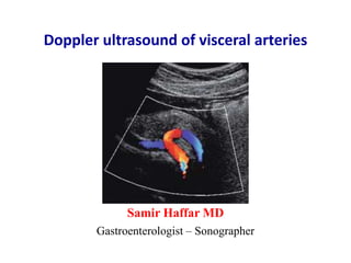
  • 1. Doppler ultrasound of visceral arteries Samir Haffar MD Gastroenterologist – Sonographer
  • 2. Doppler ultrasound of visceral arteries ① Normal Doppler ultrasound of visceral arteries ② Doppler of proximal abdominal aorta in cardiac diseases ③ Arterial stenosis and occlusion ④ Arterial aneurysm ⑤ Arterial pseudo-aneurysm ⑥ Arteriovenous fistula ⑦ Arterial dissection ⑧ Abdominal vascular compression syndromes
  • 3. ① Normal Doppler ultrasound of visceral arteries
  • 4. Optimization of color and spectral Doppler Color box Small box improved frame rate with better color resolution Doppler gain Just below the noise level Color scale (PRF) Low PRF more sensitive to low velocity flow but may lead to aliasing Beam steering Adjust to obtain satisfactory vessel angle Sample volume Usually two thirds of vessel lumen Wall filter Higher filter cuts out noise & slower velocity flow Keep filter at 50–100 Hz Focal zone Color flow optimized at level of focal zone Bhatt S et al. Ultrasound Clin 2007;2:437–453.
  • 5. Arteries scanned • Abdominal aorta: upper & lower • Celiac axis • Splenic artery • Hepatic artery • Superior mesenteric artery (SMA) • Inferior mesenteric artery (IMA) • Renal arteries Myers KA & Clough A. Making sense of vascular ultrasound. Arnold, London, 2004.
  • 6. Aorta, celiac axis, SMA & IMA SMA: superior mesenteric artery – IMA: inferior mesenteric artery https://radiologykey.com/ultrasound-assessment-of-the-splanchnic-mesenteric-arteries/
  • 7. All arise from anterior abdominal aorta Celiac axis is first major branch of abdominal aorta SMA origin is 1 – 2 cm distal to celiac axis IMA origin is 3 – 5 cm proximal to aortic bifurcation Myers KA & Clough A. Making sense of vascular ultrasound. Arnold, London, 2004. Aorta, celiac axis, SMA & IMA
  • 8. Transducer positions for scanning AA Thrush A, Hartshorne T. Peripheral vascular ultrasound: How, why and when. Elsevier Churchill Livingstone, London, 2nd edition, 2005. Sagittal or longitudinal Transverse Coronal
  • 9. Normal spectral Doppler of abdominal aorta Middle abdominal aorta Biphasic flow pattern Proximal abdominal aorta Triphasic flow pattern Antegrade systolic waveform Early diastolic reversal Low velocity antegrade flow Antegrade systolic velocity Early diastolic reversal No antegrade diastolic flow Schäberle W. Ultrasonography in vascular diagnosis. Springer-Verlag, Berlin, 2nd edition, 2011.
  • 10. Reverberation artefact Longitudinal gray scale US of proximal aorta Echogenic focus mimicking intraluminal thrombus (arrow) Secondary to reverberation artifact Bhatt S et al. Ultrasound Clin 2007;2:437–453. Longitudinal gray scale image of upper abdominal aorta
  • 11. Duplicated aorta or duplication artifact? Meuwly JY et al. Ultraschall Med 2011;32:233–236. Duplication image artifact frequent in lower abdomen: False cases of twin pregnancies Double intra-uterine devices Gray-scale US Duplicated aorta Color Doppler US 2 aortic lumen filled with color Tiny sliding probe to right Only one lumen filled with color
  • 12. Normal aortic bifurcation Transverse gray scale image Transverse color Doppler image Bhatt S et al. Ultrasound Clin 2007;2:437–453.
  • 13. Normal aortic bifurcation Longitudinal gray scale image Bhatt S et al. Ultrasound Clin 2007;2:437–453. Longitudinal color Doppler image Normal aortic bifurcationNormal aortic bifurcation
  • 14. Normal Doppler of celiac axis Forward flow in systole & diastole Low-resistance flow (RI: 0.74) Celiac axis divides into CHA & HA Ao: aorta; IVC: inferior vena cava; SV: splenic vein Transverse US of celiac axis Spectral Doppler of celiac axis RI: resistive index https://radiologykey.com/anatomy-and-normal-doppler-signatures-of-abdominal-vessels/
  • 15. CA: celiac axis – CHA: common hepatic artery – LGA: left gastric artery White RD et al. RadioGraphics 2015;35:879–898. Common celiac axis branching patterns Trifurcating CA Separate origin of LGA from aorta Bifurcating CA into CHA & LGA Quadrifurcating of celiac axis
  • 16. Normal hepatic artery waveform Forward flow in systole & diastole Low-resistance spectral pattern (RI: 0.67) RI: resistive index Swart J et al. Ultrasound Clin 2007;2:355–375.
  • 17. Common variations of hepatic artery “replaced” right hepatic artery originating from SMA Common hepatic artery originating from CA Quadrifurcating CA Separate left & right hepatic artery White RD et al. RadioGraphics 2015;35:879–898.
  • 18. Right hepatic artery from SMA Battaglia S et al. J Ultrasound 2010;13:49–56. Common hepatic artery gives rise to GDA & LHA Right hepatic artery originates directely from SMA SMA Branch of SMA CHA SMA LHA RHA GDA
  • 19. Common hepatic artery from SMA Battaglia S et al. J Ultrasound 2010;13:49–56. Common hepatic artery originates from superior mesenteric artery SMACHA LHARHA SMACHA
  • 20. Left hepatic artery from left gastric artery Battaglia S et al. J Ultrasound 2010;13:49–56. LHA originates from LGA Runs alongside venous ligament LGA LHA CHA RHA LHA LGA
  • 21. Normal splenic artery Normal Doppler signal of splenic artery PSV: 110 cm/s – EDV: 45 cm/s – RI: 0.59 Low resistance flow PSV: peak systolic velocity – EDV: end diastolic velocity – RI: resistive index https://radiologykey.com/anatomy-and-normal-doppler-signatures-of-abdominal-vessels/
  • 22. Sagittal ultrasound of SMA Ao: abdominal aorta CA: celiac artery SMA: superior mesenteric artery Zachrisson H et al. J Med Diagn Meth 2013, 3:2.
  • 23. Doppler angle for superior mesenteric artery Doppler angle can be improved from 75° to 25° by moving transducer distally & tilting it cranially Schäberle W. Ultrasonography in vascular diagnosis. Springer-Verlag, Berlin Heidelberg, 2nd edition, 2011.
  • 24. Transverse US of superior mesenteric artery Transverse US of pancreatic body and its dorsal landmark SMA surrounded by collar of fat (yellow arrow) A: Aorta; P: pancreas; splenic vein: white arrow Diagnostic ultrasound, Edited by CM Rumack, 5th edition, 2018, Elsevier
  • 25. Anatomic relationships of SMA Anterior to SMA: SV & pancreas Posterior to SMA: aorta Left to SMA: left renal vein Right to SMA: Right renal vein & IVC Surrounded by layer of echogenic fat SMA: superior mesenteric artery Revzin MV et al. Ultrasound Clin 2007;2:477–492.
  • 26. Normal spectral Doppler of SMA SMA: superior mesenteric artery Myers KA & Clough A. Making sense of vascular ultrasound. Arnold, London, 2004. Sharp systolic upstroke Variable diastolic component Low flow, reversed flow, or both Fasting state High-resistance flow Postprandial state Low-resistance flow Increased velocity High diastolic velocity Absence of reverse flow
  • 27. Common origin of CA and SMA Common origin of CA and SMA (arrow) Common celiac and SMA origin Color Doppler of abdominal aorta Sagittal MRA of abdominal aorta CA: celiac axis – MRA: magnetic resonance angiography – SMA: superior mesenteric artery Revzin MV et al. Ultrasound Clin 2007;2:477–492.
  • 28. Inferior mesenteric artery Schäberle W. Ultrasonography in vascular diagnosis. Springer-Verlag, Berlin, 2nd edition, 2011. Arises 3 – 5 cm above aortic bifurcation From left anterolateral aspect of the aorta Transverse scan with slight pressure & moving upward from bifurcation Scanned over length of 2 – 5 cm from its origin
  • 29. Color Doppler ultrasound of normal IMA Longitudinal view Normal inferior mesenteric artery (arrow) arising from anterior left lateral aspect of aorta AO: aorta – IMA: inferior mesenteric artery Revzin MV et al. Ultrasound Clin 2007;2:477–492.
  • 30. Inferior mesenteric artery and vein lying adjacent to aorta (arrows) IMA: inferior mesenteric artery Color Doppler ultrasound of normal IMA Transverse view
  • 31. Normal spectral Doppler of normal IMA PSV: 150 cm/sec Early diastolic reversed flow Low forward diastolic flow High resistance flow IMA: inferior mesenteric artery
  • 32. Normal spectral Doppler of IMA Mirk P et al. Abdom Imaging 1998;23:364–369. High systolic peak Early diastolic reflux Low velocity diastolic flow High pulsatility index: 5.46 Transverse & left sagittal US of infrarenal aorta IMA originates from left anterior aspect of aorta & descends along its left side with straight course in its proximal portion Doppler waveforms of IMA
  • 33. Normal spectral Doppler of IMA • Diameter: 2.8 ± 0.55 mm (1.3 – 4.3 mm) • Pic systolic velocity: 1.41 m/s ± 0.48 • Minimal diastolic velocity: 0.10 m/s ± 0.16 • Pulsatility index: 3.49 ± 0.49 (1.60 – 6.23) • Time-average velocity (TAV): 0.43 m/s ± 0.19 • Flow volume: 0.13 ± 0.06 L/min (0.05 – 0.29) Study of 116 patients without splanchnic vessel pathology Mirk P et al. Abdom Imaging 1998;23:364–369.
  • 34. Aorta and renal arteries https://radiologykey.com/ultrasound-assessment-of-the-splanchnic-mesenteric-arteries/
  • 35. Anatomy of aorta and renal vessels Lt kidney Rt kidney IVC Aorta SMA CA LRA LRV RRA RRV CA: celiac axis – IVC: inferior vena cava – LRA: left renal artery – LRV: left renal vein RRA: right renal artery – RRV: right renal vein – SMA: superior mesenteric artery
  • 36. Number of main renal arteries Özkan et al. Diagn Interv Radiol 2006;12:183–186. Angiographic evaluation of 855 consecutive patients No of vessels 1 2 3 4 Right 83% 15% 1% – Left 86% 12% 1% 0.2%
  • 37. Anatomy of main renal arteries Japan Society of Ultrasonics in Medicine. J Med Ultrasonics 2016;43:145–162.
  • 38. Branched structure of renal artery Japan Society of Ultrasonics in Medicine. J Med Ultrasonics 2016;43:145–162.
  • 39. Main renal arteries Schäberle W. Ultrasonography in vascular diagnosis. Springer-Verlag, Berlin, 2nd edition, 2011. Transverse scan with probe angulations
  • 40. Norma right renal artery Moukaddam H et al. Ultrasound Clin 2007;2:455–475. Transverse gray scale image Right main renal artery Transverse color Doppler image Right main renal artery
  • 41. Normal right renal artery Coronal image of inferior vena cava Moukaddam H et al. Ultrasound Clin 2007;2:455–475. RRA is the only vessel to course under the IVC Often slightly indents the IVC
  • 42. Longitudinal scan in left lateral decubitus Two right renal arteries (15%) Moukaddam H et al. Ultrasound Clin 2007;2:455–475.
  • 43. Pre-caval right renal artery RRA: right renal artery – IVC: inferior vena cava Hélénon O et al. EMC-Radiologie 2005;2:367–412. aorta IVC RRA
  • 44. Norma left renal artery Moukaddam H et al. Ultrasound Clin 2007;2:455–475. Proximal main left renal artery Proximal main left renal artery Color Doppler image LRA Gray scale image LRA
  • 45. Angle of insonation Difficult to estimate in tortuous or curved renal artery Correct angleIncorrect angle Schäberle W. Ultrasonography in vascular diagnosis. Springer-Verlag, Berlin Heidelberg, 2nd edition, 2011.
  • 46. Pulsed Doppler of main left renal artery Hélénon O et al. EMC-Radiologie 2005;2:367–412. Early systolic notch Low resistance flow (PSV 90 cm/sec)
  • 47. Early systolic notch Moukaddam H et al. Ultrasound Clin 2007;2:455–475. Some normal waveforms have early systolic notch 1. Measuring to point of PSV results in prolonged acceleration time 2. Excellent negative predictive value of stenosis > 60% Doppler of right main renal artery
  • 48. Limits in visualization of main renal arteries • Obesity • Overlying bowel gas • Dyspnea • Shadowing from arterial calcifications • Cardiac arrhythmias • Poor angle of Doppler insonation • Accessory renal arteries (small size) RA: renal arteries Moukaddam H et al. Ultrasound Clin 2007;2:455–475. Expert sonographers detect 80-90% of main RA CEUS improves success rate to 95%
  • 49. Adjustment of Doppler control Low flow settings for intra-renal arteries • Lowest pulse repetition frequency without aliasing • Small color box • Greatest gain without background noise • Lowest wall filter • High color priority
  • 50. Normal segmental & interlobar renal arteries Normal segmental renal arteries (long arrows) Color Doppler image of kidney Moukaddam H et al. Ultrasound Clin 2007;2:455–475. Normal interlobar renal arteries (short arrows)
  • 51. Study of interlobular arteries Perfusion study / Low PRF Hélénon O et al. EMC-Radiologie 2005;2:367–412. Cortical perfusion
  • 52. Normal pulsed Doppler waveform Renal segmental artery Sharp systolic upstroke Continuous forward diastolic flow Low resistance flow (low RI)
  • 53. Measurement of resistive index Resistive index: S – ED / S Normal 50 – 70% Abnormal > 80%
  • 54. Measurement of acceleration time (AT) • Length of time in second from onset of systole to peak systole • Normal value: < 0.07 sec (70 msec) Acceleration time (AT) or rise time (RT)
  • 55. Normal renal aortic ratio (RAR) Left renal artery Upper abdominal aorta Japan Society of Ultrasonics in Medicine. J Med Ultrasonics 2016;43:145–162. PSV: 80 cm/sec PSV: 120 cm/sec RAR: PSV of renal artery to aorta (80/120 = 0.67) Normal value ˂ 3
  • 56. Normal spectral Doppler of renal arteries • PSV of renal artery at origin: < 180 cm/s • RAR (PSV ratio of renal artery/aorta): < 3 • Resistive index (RI): < 0.70 • ∆ RI (right – left): < 0.15 • Acceleration time (AT): < 0.07 sec • Acceleration index (AI): > 3.5 m/s2 PSV: peak systolic velocity – RI: resistive index
  • 57. ② Doppler ultrasound of proximal abdominal aorta in cardiac diseases Window to left heart
  • 58. Abnormal spectral Doppler of proximal abdominal aorta TVI: Time Velocity Integral Fadel BM et al. Echocardiography 2014;00:1–5. Severe aortic regurgitation Holodiastolic flow reversal (arrows) Diastolic TVI (17 cm) close to systolic TVI (20 cm) Indicating a large regurgitant fraction
  • 59. Fadel BM et al. Echocardiography 2014;00:1–5. Coarctation of the aorta Tardus Parvus wave Abnormal spectral Doppler of proximal abdominal aorta Prolongation of acceleration time (arrow) Prolongation of deceleration time (arrowhead) Prominent diastolic forward flow No flow reversal
  • 60. Fadel BM et al. Echocardiography 2014;00:1–5. Aorto-iliac disease Mid systolic notching Notching of antegrade systolic waveform (arrow) Abnormal spectral Doppler of proximal abdominal aorta
  • 61. Fadel BM et al. Echocardiography 2014;00:1–5. Left ventricular assist device Monophasic forward flow throughout the cardiac cycle Lack of flow reversals Abnormal spectral Doppler of proximal abdominal aorta
  • 62. Fadel BM et al. Echocardiography 2014;00:1–5. Pulsus alternans Alternating high peak velocities (arrow) and low peak velocities (arrowhead) Abnormal spectral Doppler of proximal abdominal aorta
  • 63. Fadel BM et al. Echocardiography 2014;00:1–5. Pulsus paradoxus Significant respiratory variability in flow Inspiration decreases systolic velocity & duration (arrows) Abnormal spectral Doppler of proximal abdominal aorta
  • 64. 2D-imaging of abdominal aorta coupled with pulse Doppler imaging of proximal abdominal aorta adds substantially to value of TTE study TTE: trans-thoracic echocardiography
  • 65. ③ Arterial stenosis and occlusion
  • 66. Signs of stenosis on pulsed Doppler Sites of pulsed Doppler Features of pulsed Doppler Proximal to stenosis: Mid systolic notching At stenosis: Increased PSV/EDV compared to pre-stenotic segment Laminar flow Beyond stenosis: Post-stenotic turbulence Spectral broadening Loss of well demarcated spectral edge Distal to stenosis: Tardus-parvus wave
  • 67. Atherosclerotic abdominal aorta Revzin MV et al. Ultrasound Clin 2007;2:477–492. Luminal irregularity Sagittal US of abdominal aorta Marked calcification (arrow) of abdominal aorta (AO) Color Doppler of abd aorta
  • 68. Atherosclerotic stenosis of abdominal aorta Stenosis in middle aortic segment with significant turbulence Peak systolic velocity: 369 cm/s https://radiologykey.com/the-aorta-and-inferior-vena-cava/
  • 69. Abdominal aortic occlusion Kaschwich M et al. J Cardiovasc Surg 2017;58(2):313 20. Transverse US of lower abd aorta Longitudinal US of lower abd aorta
  • 70. Mid-aortic syndrome Hypoplasia of aorta, atrophy of aorta, atypical coarctation • Rare disease w narrowing in distal thoracic &/or abdominal aorta • Usually found in children and young adults • Causes: Takayasu arthritis, congenital developmental anomaly, neurofibromatosis, Williams syndrome • Renal artery involved in 90% of cases: arterial hypertension • Clinic: AHT, lower extremity claudication, chronic abdominal pain • Physical exam: abdominal bruit, reduced or absent femoral pulses, differential blood pressure between upper & lower extremities Han L et al. J Clin Ultrasound 2019;47:22–26.
  • 71. Refractory hypertension for 18 years in a 36-year-old man Mid-aortic syndrome PSV: peak systolic velocity – RI: resistivity index Han L et al. J Clin Ultrasound 2019;47:22–26. Color Doppler US of abdominal aorta Spectral Doppler of abdominal aorta Turbulent flow in narrowed suprarenal aorta (arrow) Spectral Doppler of right kidney Turbulent flow with high velocity PSV > 4.0 m/s Tardus-parvus waveform Low RI: 0.32
  • 72. Different types of celiac trunk stenosis * Medial Arcuate Ligament Syndrome (celiac artery compression syndrome) Schäberle W. Ultrasonography in vascular diagnosis. Springer-Verlag, Berlin, 2nd edition, 2011. Fibromuscular dysplasia rare Atherosclerosis most frequent MALS syndrome*
  • 73. Grading stenosis of celiac axis • Stenosis 50 – 69%: 43 patients PSV: > 240 cm/s (sens 87%, spec 83%, OA 86%) EDV: > 40 cm/s (sens 84%, spec 48%, OA 73%) PSV ratio1: 1.5 • Stenosis 70 – 99% : 62 patients PSV: > 320 cm/s (sens 80%, spec 89%, OA 85%) EDV: > 100 cm/s (sens 58%, spec 91%, OA 77%) PSV ratio1: 2.75 (1) PSV ratio of celiac axis to aorta AbuRahma AF et al. J Vasc Surg 2012;55:428-36. 105 patients with celiac artery stenosis on angiography
  • 74. Stenosis of celiac axis Aliasing and tissue bruit around a stenotic coeliac axis origin Longitudinal color Doppler UD of upper abdominal aorta
  • 75. Atherosclerotic stenosis of celiac artery Spectral tracing PSV: peak systolic velocity – EDV: end diastolic velocity Myers KA & Clough A. Making sense of vascular ultrasound. Arnold, London, 2004. PSV: 276 cm/sec – EDV: 134 cm/sec Stenosis ≥ 50%
  • 76. Celiac axis stenosis PSV: 318 cm/s indicating ≥ 70% stenosis Color Doppler of celiac axis Pulsed Doppler at stenotic site Color aliasing at CA origin (stenosis) Post-stenotic dilatation CA: celiac axis – PSV: peak systolic velocity
  • 77. Grading of SMA stenosis • Stenosis 50 – 69%: PSV > 280 cm/s EDV > 45 cm/s PSV ratio of SMA/aorta > 3.6 • Stenosis 70 – 99%: PSV > 395 cm/s EDV > 74 cm/s PSV ratio of SMA/aorta > 3.6 PSV: peak systolic velocity – EDV: end diastolic velocity – SMA: superior mesenteric artery Bird S et al. J Clin Ultrasound. 2019;47(5):267–271. 65 patients with SMA stenosis on CT angiography
  • 78. Severe stenosis of superior mesenteric artery PSV 390 cm/s indicating ≥ 70% stenosis Color and spectral Doppler of SMA PSV: peak systolic velocity https://thoracickey.com/clinical-evaluation-and-treatment-of-mesenteric-vascular-disease/
  • 79. Severe stenosis of superior mesenteric artery Color & spectral Doppler at stenotic site Color bruit outside vessel High PSV > 500 cm/s Stenosis ≥ 70% Post-stenotic color & spectral Doppler Post-stenotic turbulence: Typical “spike hairdo” waveform Mirror-image artifact “flow below baseline” at top of tracing Diagnostic ultrasound, Edited by Rumack CM, 5th edition, 2018, Elsevier
  • 80. Pulsed Doppler at origin of SMA Severe stenosis of superior mesenteric artery PSV: 422 cm/s Stenosis ≥ 70% Low-velocity rounded waveforms: tardus-parvus in post-stenotic zone Pulsed Doppler of mid-distal SMA Revzin MV et al. Ultrasound Clin 2007;2:477–492.
  • 81. Occlusion of superior mesenteric artery Revzin MV et al. Ultrasound Clin 2007;2:477–492. Thump artifacts Focal retrograde flow in collateral vessel (straight arrow) Occlusion of proximal SMA (straight arrow) Reconstitution by collaterals from CA (wavy arrow) US Doppler of SMA origin US Doppler of occluded segment
  • 82. Grading of IMA stenosis based on angiography IMA: inferior mesenteric artery – PSV: peak systolic velocity – EDV: end diastolic velocity AbuRahma AF et al. Vascular 2012; 20(3):145–149. Stenosis PSV cm/s EDV cm/s IMA/aortic systolic ratio Normal2 1051 < 50% stenosis3 ≥ 210 no flow or ≥ 20 ≥ 2.5 50 – 69% stenosis4 ≥ 250 ≥ 90 ≥ 4.5 ≥ 70% stenosis5 ≥ 270 ≥ 100 – (1): mean – (2): 45 patients – (3): 12 patients – (4): 8 patients – (5): 15 patients
  • 83. Severe stenosis of inferior mesenteric artery Color aliasing at origin of IMA (arrow) Pulsed Doppler of stenotic region: 332 PSV cm/s Consistent with hemodynamically severe stenosis (≥ 70%) IMA: inferior mesenteric artery Pellerito JS et al. J Ultrasound Med 2009;28:641–650.
  • 84. Mesenteric ischemia • Acute mesenteric ischemia: high morbidity and mortality Causes: SMA embolism, SMA thrombosis, non-occlusive ischemia Abdominal pain out of proportion to physical examination Differentiate from other common causes of acute abdominal pain Laboratory findings of little value: metabolic acidosis, high lactate,… • Chronic mesenteric ischemia: less common Severe stenosis or occlusion of 2 of 3 mesenteric vessels Complete occlusion of single artery may cause intestinal angina Clinical triad: postprandial abd pain, weight loss, food avoidance ACR: American College of radiology Expert Panels on Vascular Imaging and Gastrointestinal Imaging. J Am Coll Radiol 2018;15:S332-S340.
  • 85. Acute mesenteric ischemia Schäberle W. Ultrasonography in vascular diagnosis. Springer-Verlag, Berlin, 2nd edition, 2011. Extent of intestinal necrosis varies with level of occlusion Main trunk Vasa recta Non occlusive intestinal ischemia Ileo-colic artery/ Right colic artery 2nd & 3rd order branches
  • 86. Imaging of suspected acute mesenteric ischemia ACR Appropriateness Criteria ACR: American College of radiology – CTA: computed tomography angiography Expert Panels on Vascular Imaging and Gastrointestinal Imaging. J Am Coll Radiol 2018;15:S332-S340. Procedures Appropriateness category CTA abdomen & pelvis with IV contrast Usually appropriate CT abdomen & pelvis with IV contrast May be appropriate Arteriography abdomen May be appropriate (disagreement) MRA abdomen & pelvis w/o & with IV contrast May be appropriate (disagreement) Radiography abdomen May be appropriate US duplex Doppler abdomen May be appropriate CTA of abdomen and pelvis is the most appropriate choice
  • 87. Doppler US in acute mesenteric ischemia ACR appropriateness criteria • Detecting proximal celiac axis & SMA stenosis with high Se & Sp • Detecting proximal mesenteric arterial thrombosis • Detecting superior mesenteric or portal venous thrombosis • Challenges: bowel gas, obesity, vascular calcifications • Limited role: distal arterial embol, nonocclusive mesenteric ischemia • Length of examination and pain associated with applied pressure to abdomen during imaging may be limiting factors ACR: American College of radiology Expert Panels on Vascular Imaging and Gastrointestinal Imaging. J Am Coll Radiol 2018;15:S332-S340.
  • 88. Acute superior mesenteric artery occlusion Longitudinal US of SMA Transverse US of SMA https://radiopaedia.org/cases/acute-superior-mesenteric-artery-occlusion-on-ultrasound
  • 89. Superior mesenteric artery thrombosis Thrombus demonstrated within an expanded SMA (arrows) SMA: superior mesenteric artery https://radiopaedia.org/cases/superior-mesenteric-artery-thrombosis-1 Longitudinal US of SMA Transverse color Doppler of SMA
  • 91. Imaging of suspected chronic mesenteric ischemia ACR Appropriateness Criteria Procedures Appropriateness category CTA abdomen & pelvis with IV contrast Usually appropriate MRA abdomen & pelvis w/o & with IV contrast Usually appropriate Arteriography abdomen May be appropriate (disagreement) CT abdomen and pelvis with IV contrast May be appropriate MRA abdomen and pelvis without IV contrast May be appropriate US duplex Doppler abdomen May be appropriate ACR: American College of radiology – CTA: computed tomography angiography Expert Panels on Vascular Imaging and Gastrointestinal Imaging. J Am Coll Radiol 2018;15:S332-S340. CTA of abdomen and pelvis is the most appropriate choice
  • 92. ACR: American College of radiology Expert Panels on Vascular Imaging and Gastrointestinal Imaging. J Am Coll Radiol 2018;15:S332-S340. Doppler US in chronic mesenteric ischemia ACR appropriateness criteria • Useful initial screening tool for chronic mesenteric ischemia • Best performed in fasting state & early in the day to avoid bowel gas • Peak systolic velocity used for diagnosing stenosis SMA: 295 cm/s and 400 cm/s for 50% and 70% respectively Celiac artery: 240 cm/s
  • 93. Doppler of renal artery stenosis Gray scale imaging first • Kidneys Maximum renal length Echogenicity of renal cortex Thickness of renal cortex Masses – hydronephrosis – renal calculi • Aorta Plaque – thrombus – dissection – aneurysm • Adrenal glands
  • 94. Measurement of parenchymal & cortical thickness Cortical thickness: normal 8 – 10 mm Parenchymal thickness: normal 14 – 18 mm Tuma J et al. European course book: Genitourinary ultrasound. European Foundation of Societies of Ultrasound in Medicine & Biology. parenchyma cortex
  • 95. Sites for pulsed Doppler of renal arteries Aorta Ostium of main renal arteries Trunk of main renal arteries Hilum of both kidney Upper pole of both kidney Middle pole of both kidney Lower pole of both kidney
  • 96. • Aorta: PSV • Ostium of renal arteries: PSV • Renal aorta ratio: PSV ratio of renal artery to aorta • Trunk of renal arteries: PSV • Segmental or interlobar arteries: RI and AT Upper, middle and lower poles of both kidney Doppler of renal artery stenosis Ultrasound protocol AT: acceleration time –PSV: peak systolic velocity – RI: resistive index Japan Society of Ultrasonics in Medicine. J Med Ultrasonics 2016;43:145–162.
  • 97. Two main causes of renal artery stenosis Atherosclerosis (> 90%) FMD (< 10%) Age ˃ 50 years young Gender more common in males more common in females Location proximal 1 cm of main RA branching points middle of renal artery others (carotids) Post-stenotic dilatation rare frequent FMD: fibromuscular dysplasia
  • 98. Doppler features of renal artery stenosis Main renal artery Intra-renal arteries Focal color aliasing AT > 0.07 sec2 Color bruit Flat blood flow waveforms Turbulence beyond stenosis Δ RI (right – left) > 15% RAR > 3.5 PSV > 180 cm/sec1 EDV > 90 cm/sec 1 PSV >180 cm/s: ≥ 50% diameter reduction PSV > 300 cm/s: ≥ 60% diameter reduction 2 AT > 0.07 sec: > 85% diameter reduction AT: acceleration time – EDV: end diastolic velocity PSV: peak systolic velocity – RAR: renal aortic ratio – RI: resistive index Japan Society of Ultrasonics in Medicine. J Med Ultrasonics 2016;43:145–162.
  • 99. Renal artery stenosis Renal aortic ratio (RAR) Left renal artery Upper abdominal aorta PSV: peak systolic velocity Japan Society of Ultrasonics in Medicine. J Med Ultrasonics 2016;43:145–162. PSV: 410 cm/s PSV: 50 cm/s RAR: 410/50 = 8.20 (normal ˂ 3) Renal artery stenosis ≥ 60%
  • 100. PSV: 293 cm/sec – RI : 0.91 Controversial indication of PTA2 Aliasing in left renal artery Retro-aortic course of LRV LRV: left renal vein – PSV: peak systolic velocity – RI resistive index – PTA: angioplasty 1 Schäberle W. Ultrasonography in vascular diagnosis. Springer-Verlag, Berlin, 2nd edition, 2011. 2 Jaeger KA et al. Ultraschall in Med 2007;28:28–31. Renal artery stenosis
  • 101. Waveform of intra-renal blood flow AT: acceleration time Japan Society of Ultrasonics in Medicine. J Med Ultrasonics 2016;43:145–162. Normal flow patternIntra-renal artery Renal artery stenosis
  • 102. Acceleration time (AT) Normal ˂ 70 msec Japan Society of Ultrasonics in Medicine. J Med Ultrasonics 2016;43:145–162. AT: 40 msec AT: 65 msec AT: 140 msec Renal artery stenosis Normal AT Normal AT Prolonged AT
  • 103. Tardus Parvus wave • Mimics Aortic/mitral valve disease Aortic coarctation William syndrome Left ventricle dysfunction CV medications: after-load reducers • Exaggerating 25 mg captopril 1 hour before exam • Minimizing Age – HTN – DM (vessel compliance) CV: cerebrovascular – DM: diabetes mellitus – HTN: hypertension Moukaddam H et al. Ultrasound Clin 2007;2:455–475.
  • 104. Fibromuscular dysplasia Moniliform aspect of RRA Typical FMD in middle third of RRA Hélénon O et al. EMC-Radiologie 2005;2:367–412. PSV: 250 cm/sec No parallelism of RRA walls
  • 105. Fibromuscular dysplasia Japan Society of Ultrasonics in Medicine. J Med Ultrasonics 2016;43:145–162.
  • 106. Indications of PTRA in renal artery stenosis Still controversial • Hemodynamically significant renal artery stenosis and: Resistant hypertension despite use of ≥ 3 antihypertensive drugs Exacerbating hypertension Malignant hypertension Hypertension with idiopathic unilateral kidney atrophy Idiopathic pulmonary edema that suddenly develops Repeated heart failure Unstable angina Fibromuscular dysplasia • Patients with bilateral renal artery stenosis • Progressive chronic kidney disease in a single functioning kidney PTRA: percutaneous transluminal renal angioplasty Japan Society of Ultrasonics in Medicine. J Med Ultrasonics 2016;43:145–162.
  • 107. Renal artery thrombosis (complete) Irshad A et al. Semin Ultrasound CT MRI 2009;30:298–314. Transplanted kidney Absence of flow within kidney Power Doppler US Power Doppler US more medially Flow in iliac artery & proximal anastomotic artery
  • 108. Partial acute renal infarction Japan Society of Ultrasonics in Medicine. J Med Ultrasonics 2016;43:145–162. Gray scale image Power Doppler image
  • 110. Aneurysms, false aneursysm & arterial dissection Normal artery Arterial wall components True aneurysm Components of arterial wall "stretched" Hole in arterial wall with confined collection of blood False aneurysm Arterial dissection Hematoma between components of wall Zwiebel JW. Ultrasound assessment of aorta, iliac arteries and inferior vena cava. In: Introduction to vascular ultrasonography. Zwiebel JW ed, 5th edition, Elsevier Saunders, 2005.
  • 111. Selective screening for AAA • Selective screening 3 important risk factors: Males Age > 65 years History of smoking • Effectiveness of screening 4 RCTs including more than 125,000 men Reported results for up to 5 – 10 years of follow-up Reduction in mortality from 68% to 21% RCT: randomized controlled trial Lederle FA. Ann Intern Med 2003 ; 139 : 516 – 22.
  • 112. Abdominal aortic aneurysm (AAA) • Normal size of abdominal aorta 1.5 – 2.5 cm • Ectatic aorta 2.5 – 3 cm • Aortic aneurysm > 3 cm • Annual growth rate of aneurysms 0.33 cm/year measuring between 4 & 5.5 cm Bhatt S et al. Ultrasound Clin 2008;3:83–91.
  • 113. Abdominal aortic aneurysm (AAA) • Measurement • Location • Morphological evaluation • Possible evaluation of iliac arteries • Complications: rupture, thrombosis, dissection, embolization What to evaluate?
  • 114. Measurement of widest part Measurement technique of aneurysm Myers KA & Clough A. Making sense of vascular ultrasound. Arnold, London, 2004.
  • 115. Measuring diameter of AAA Incorrect measurement Correct measurement AAA: abdominal aortic aneurysm Schäberle W. Ultrasonography in vascular diagnosis. Springer-Verlag, Berlin, 2nd edition, 2011. Correct diameter measured by rotating transducer clockwise until round image of aorta comes into view
  • 116. Battaglia S et al. J Ultrasound 2010:13:107–117. Abdominal aortic aneurysm (swirling flow) Pseudo ‘‘Yin-Yang sign’’ Similarity in appearance to pseudo-aneurysm finding
  • 117. Classification of abdominal aortic aneurysms Classification Categories By location Suprarenal: Above origin of renal areteries (very rare) Juxtarenal: Where renal arteries originate Infrarenal: Below origin of RA (most common) Bhatt S et al. Ultrasound Clin 2007;2:437–453. By morphology Fusiform (most common) Hourglass Saccular By etiology Atherosclerotic (most common) Inflammatory (5% – 10%) Mycotic (1%): saccular, salmonella & SA, high mortality
  • 118. Classification of AAA by location Based on relation to renal arteries Suprarenal Suprarenal: Aneurysm involves origins of one or more visceral arteries Pararenal: RAs arise from aneurysm but aorta at level of SMA not aneurysmal Juxtarenal: Aneurysm originates just beyond origins of RAs Infrarenal: Aneurysm originates distal to renal arteries. Pararenal Juxtarenal Infrarenal AAA: abdominal aortic aneurysm
  • 119. Supra-renal abdominal aortic aneurysm Schuster H et al. Ultraschall Med 2009;30:528–543. Transverse US viewLongitudinal US view Inclusion of visceral & renal arteries Perfused lumen & narrow circular thrombus
  • 120. Infra-renal abdominal aortic aneurysm Distance between renal vein & upper limit of aneurysm LRV: left renal vein – SMA: superior mesenteric artery Thrush A, Hartshorne T. Peripheral vascular ultrasound: How, why and when. Elsevier Churchill Livingstone, London, 2nd edition, 2005. SMA LRV
  • 121. Classification of AAA by shape Fusiform Saccular Most frequent Double aneurysm Hourglass aorta AAA: abdominal aortic aneurysm
  • 122. Abdominal aortic aneurysm (fusiform) Transverse image Anteroposterior diameter from outer wall to outer wall Sagittal image Diameter measured in transverse image larger due to obliquity Myers KA & Clough A. Making sense of vascular ultrasound. Arnold, London, 2004.
  • 123. Abdominal aortic aneurysm (hourglass) Bhatt S et al. Ultrasound Clin 2007;2 437–453. Two discontinuous focal segments of aneurysmal dilatation Aortic diameter in between is normal in caliber
  • 124. Abdominal aortic aneurysm (saccular) Saccular or mycotic aneurysm Thrombus seen as low-level echoes within aneurysm Longitudinal image of abdominal aorta Abraham D et al. Emergency medicine sonography: Pocket guide. Jones & Bartlett Publishers, Boston, MA, USA, 1st edition, 2010.
  • 125. Complications of abdominal aortic aneurysm • Thrombosis • Dissection • Rupture • Embolization
  • 126. Complications of abdominal aortic aneurysm Partial thrombus Bhatt S et al. Ultrasound Clin 2007;2:437–453. Infrarenal AAA with thrombus occluding approximately two thirds of the lumen Longitudinal color Doppler US Transverse color Doppler US
  • 127. Abdominal aortic aneurysm (thrombus liquefaction) Area of thrombus liquefaction may be confused with dissection Large thrombus separate area of liquefaction from lumen Thrush A, Hartshorne T. Peripheral vascular ultrasound: How, why and when. Elsevier Churchill Livingstone, London, 2nd edition, 2005.
  • 128. Longitudinal color Doppler Transverse color Doppler Accorci F. Ultrasound in abdominal aorta and iliac artery atherosclerotic diseases. http://www.echodoppler-lessons.com/en/pathology-of-the-aorta-and-the-peripheral-arteries/ ultrasound-in-abdominal-aorta-and-iliac-arteries-atherosclerotic-disease Complications of abdominal aortic aneurysm Complete thrombus
  • 129. Bhatt S et al. Ultrasound Clin 2007;2:437–453. Longitudinal gray-scale US Echogenic band in aortic lumen Echogenic band in aortic lumen Transverse gray-scale US Transverse color Doppler US Anterior false lumen (blue) Posterior true lumen (red) Longitudinal color Doppler US Entry point (arrowhead) of intimal tear causing dissection Complications of AAA (dissection)
  • 130. AAA with peripheral thrombus Small hypoechoic area (wall rupture) Hypoechoic structure at upper end Presence of active bleeding No further imaging confirmation Taken directly to OR AAA: abdominal aortic aneurysm – OR: operating room Bhatt S et al. Ultrasound Clin 2007;2:437–453. Complications of abdominal aortic aneurysm Rupture (high mortality rate: 90%)
  • 131. Stent-graft expands to make firm circumferential contact with  ‘neck’ of relatively normal aorta between RA & upper end of AAA  each CIA below aneurysm Endovascular aortic aneurysm repair (EVAR) First performed by Parodi from Argentina in 1990 1 AAA: abdominal aortic aneurysm – CIA: common iliac artery –RA: renal arteries 1 Parodi JC et al. Ann Vasc Surg 1991;5:491–499. 2 Myers KA & Clough A. Making sense of vascular ultrasound. Arnold, London, 2004. Stent-graft
  • 132. Endoleak after EVAR Persistent flow in aneurysm lumen after procedure • Increase in aneurysmal diameter with risk of rupture • 20 – 40% at any time after graft placement1 • Lifelong surveillance 1st month, 6th month, yearly2 • Modalities CTA: gold standard CDUS/CEUS: acceptable alternative MRA – DSA CDUS: color Doppler ultrasound – CEUS: contrast-enhanced ultrasound CTA: computed tomography angiography – MRA: magnetic resonance angiography 1 Demirpolat G et al. J Clin Ultrasound 2011;39:263–269. 2 Stavropoulos SW et al. Radiology 2007;243:641. Determination of endoleak & aneurysmal size
  • 133. Endoleak following EVAR Type I Type II Type III Type IX Failure of proximal or distal attachment sites Flow through aortic or iliac branches common Perforation & tear in graft material rare Porosity of graft material resolved in 1 month EVAR: endo-vascular abdominal aneurysm repair White GH et al. J Endovasc Surg 1996 ;3:124–5. Carrafiello G et al. Cardiovasc Intervent Radiol 2006;29:969–974.
  • 134. Types of endoleak Type I: Distal attachment site Type II: Patent lumbar artery Thrush A et al. Peripheral vascular ultrasound. Elsevier, London, 2nd edition, 2005. Hartshorne T. Ultrasound 2006;14:34–42. Type II: Inferior mesenteric artery Type I: Proximal attachment site
  • 135. Endoleak type II over mesenteric artery Zimmermann H et al. Clin Hemorheology Microcirculation 2014;58:247–260. Aortic aneurysm with graft No endoleak seen in B-mode Type II endoleak (yellow arrow) over mesenteric artery Color Doppler ultrasoundGray scale ultrasound
  • 136. Zimmermann H et al. Clin Hemorheology Microcirculation 2014;58:247–260. Gray scale ultrasound Color Doppler ultrasound Aortic aneurysm with graft No endoleak seen in B-mode Endoleak type II over lumbar artery Type II endoleak (yellow arrow) over right lumbar artery
  • 137. Endovascular aortic aneurysm repair Mirror artifact Demirpolat G et al. J Clin Ultrasound 2011;39:263–269. Synchronous pulsatility with flow in patent graft Changing position while examining from different aspects Spectral analysis aids in reducing false positive Mirror image behind patent limbs of stent graft
  • 138. Iliac artery aneurysm • Most iliac artery aneurysms associated w distal aortic aneurysms Aortic aneurysm is the source of clinical concern • Isolated iliac artery aneurysm uncommon but may be deadly: Not palpated on physical examination even when large Rupture generates nonspecific abdominal or pelvic symptoms Nor recognized until patient becomes hypotensive or dies • Located in proximal or distal portion of common iliac arteries • Surgical repair or percutaneous stenting recommended if >3 cm Zwiebel JW. Ultrasound assessment of aorta, iliac arteries and inferior vena cava. In: Introduction to vascular ultrasonography. Zwiebel JW ed, 5th edition, Elsevier Saunders, 2005.
  • 139. Aortic and iliac artery aneurysms Aortic aneurysm & common iliac artery aneurysm Separated by relatively normal segment of proximal CIA (arrow) Longitudinal view of lower abdominal aorta
  • 140. Isolated common iliac artery aneurysm Transverse ultrasound of abdominal aortic bifurcation Left iliac artery aneurysm Pseudo Yin Yang flow pattern Battaglia S et al. J Ultrasound 2010;13:107–117.
  • 141. Isolated internal iliac artery aneurysm Zwiebel JW. Ultrasound assessment of aorta, iliac arteries and inferior vena cava. In: Introduction to vascular ultrasonography. Zwiebel JW ed, 5th edition, Elsevier Saunders, 2005. Flow in the mass (arrows) and apparent communication with IIA Gray scale ultrasound Color Doppler ultrasound Hypoechoic mass in pelvis near EIA (arrows) Initially thought to be nodal mass
  • 142. • Definition: Arterial dilatation > 1.5 times the size of original vessel with loss of parallelism of vascular walls • Size of visceral arteries: Celiac trunk: 0.79 ± 0.06 cm Common hepatic artery: 0.50 ± 0.04 cm Proper hepatic artery: 0.45 ± 0.03 cm Splenic artery: 0.46 ± 0.03 cm Visceral artery aneurysms Ibrahim F et al. Curr Treat Options Cardio Med 2018;20:97–111.
  • 143. Sites of visceral artery aneurysms Rare – discovered incidentally – high mortality in rupture Jesinger RA et al. Radiographics 2013;33:E71–96. Piasek E et al. J Ultrason 2018;18:148–151. Splenic artery: Hepatic artery: Superior mesenteric artery: Celiac artery: Inferior mesenteric artery: Gastric, intestinal, pancreatic, gastroduodenal: Renal artery 60% 20% 5% 4% 1% rare 1 – 10 % Splenic artery & hepatic artery most commonly involved
  • 144. Celiac artery aneurysm Piasek E et al. J Ultrason 2018;18:148–151. Transverse gray scale US of upper abdomen Aneurysm of celiac axis
  • 145. Revzin MV et al. Ultrasound Clin 2007;2:477–492. Celiac artery aneurysm Lumen of celiac axis dilated (arrow) Disturbed flow on color Doppler (pseudo Yin Yang flow)
  • 146. Celiac axis aneurysm with dissection 52-year-old male - CT for workup of testicular seminoma Celiac axis aneurysm 13 mm Aneurysmal dissection w true & false lumen PSV: peak systolic velocity – EDV: end diastolic velocity Errigo A et al. J Vascular Ultrasound 2018;42(2):82–84. Doppler of false lumen PSV: 402 cm/s EDV: 142 cm/s
  • 147. Extra-hepatic artery aneurysm Mansour MA et al. Vascular Diagnosis. Elsevier-Saunders, Philadelphia, 1st edition, 2005 Extrahepatic sacular hepatic artery aneurysm Pseudo Yin-Yang flow pattern
  • 148. Ignee A et al. Z Gastroenterol 2002;40:21–32. Gray-scale US Color & duplex US Pseudo Yin-Yang flow pattern Arterial flow on spectral Doppler Ovoid mass in porta hepatis Extra-hepatic artery aneurysm
  • 149. Intra-hepatic artery aneurysm O’Driscoll D et al. Br J Radiology 1999;72:1018–1025. 26-year-old man – abdominal pain after abdominal blunt trauma Hypoechoic lesion in right lobe Arterial flow on spectral Doppler Contrast-enhanced CTPulsed Doppler US Large aneurysm in right hepatic lobe
  • 150. • 3rd common intra-abdominal aneurysm after aorta & iliac arteries • Abnormal dilatation of splenic artery > 1 cm in diameter • Risk factors: pregnancy, portal hypertension, trauma, arterial degeneration (medial fibrodysplasia), atherosclerosis • Majority asymptomatic (80%) & discovered incidentally • Symptomatic in 20%: epigatric or LUQ pain • Spontaneous rupture: frequency 3% – mortality 25% • Risk of rupture: pregnancy, PHT, > 2 cm, liver transplantation Splenic artery aneurysm LUQ: left upper quadrant – PHT: portal hypertension Al-Habbal Y et al. Surgeon 2010;8:223–231.
  • 151. Splenic artery aneurysm Piasek E et al. J Ultrason 2018;18:148–151. Gray-scale US of splenic hilum Color Doppler US of splenic hilum Splenic artery aneurysmCystic mass in splenic hilum
  • 152. Gray scale ultrasound Wakui N et al. J Med Ultrasonics 2011:38:167–171. Splenic artery aneurysm Color Doppler ultrasound 2.2-cm cystic mass at splenic hilum (arrow) Color Doppler: pseudo Yin-Yang pattern Pulsatile waveform in cystic lesion
  • 153. Atherosclerotic splenic artery aneurysm Badea R et al. J Med Ultrasonics 2013;40:487–490. Turbulent color signal suggesting splanchnic aneurysm 4-cm hypoechoic mass on splenic artery trajectory with wall calcification Presence of splenic artery aneurysm CT angiographyGray scale US Color Doppler US
  • 154. Multiple splenic artery aneurysms Bagga B, Das CJ. BMJ Case Rep 2019;12:e228705. Transverse abdominal US Color spectral Doppler US Dilated splenic artery (asterisk) Multiple aneurysms (arrow) likely arising from it Arterial waveform in aneurysms confirming them to be arising from splenic artery
  • 155. Thrombosed splenic artey aneurysm mimicking pancreatic adenocarcinoma Round hypoechoic solid mass appears as aneurysmatic thrombosed dilatation arising from splenic artery Casadei R et al. JOP (Online) 2007;8(2):235-239. Transverse color Doppler US Non-contrast-enhanced CT scan Hypodense round solid mass of pancreatic body w calcified periphera spot simulating pancreatic neoplasm
  • 156. Splenic vein aneurysm • Splenic vein aneurysms are rare • Etiology: portal hypertension, congenital weakness in vein wall • Clinic: asymptomatic – usually incidental findings • Complication: rupture, thrombosis, compression adjacent structures • Appropriate management: unknown Noninvasive follow-up, plication, and aneurysm excision Torres G et al. J Vasc Surg 1999;29:719–21.
  • 157. Splenic vein aneurysm Cystic round mass in pancreatic tail Enhancing mass in left mid-abdomen Torres G et al. J Vasc Surg 1999;29:719–21. Transverse US image Abdominal CT (venous phase)
  • 158. Inferior mesenteric artery aneurysm Momin AA et al. J Clin Ultrasound 2008;36:42–44. Transverse color US of middle abdominal aorta Longitudinal US of middle abdominal aorta IMA aneurysm parallel to lower part of abdominal aorta AO: aorta VC: inferior vena cava Aneurysm: IMA aneurysm
  • 159. Renal artery aneurysm Japan Society of Ultrasonics in Medicine. J Med Ultrasonics 2016;43:145–162. Ultrasound of kidney Cystic mass at hilum Color Doppler US Renal artery aneurysm Pulsed Doppler US Low resistance arterial flow
  • 160. Indications to treat visceral aneurysms • Clinical symptoms: pain, embolism, rupture • > 2 cm in size or twice the size of the artery by consensus • Rapid increase in diameter of aneurysm • Specific location: duodenopancreatic arcades • Pregnant women or women of childbearing age • Non-atherosclerotic etiology (ie: connective tissue disease) • Patient requiring a liver transplant • Portal hypertension for splenic artery aneurysm Chiaradia M et al. Diag Inter Imaging 2015:96:796–806.
  • 161. Endovascular or open surgery approaches for visceral artery aneurysms • Endovascular approach: Used more often by surgeons Shorter hospital stay Lower rates of cardio-vascular complications Higher rates of re-intervention Low access site complication (<5%) Post-embolization syndrome: from 9% (renal) to 38% (splenic) Coil migration: from 8% (splenic) to 29% (renal) • Difference in mortality of 2 approaches not statistically significant Barrionuevo P et al. J Vasc Surg. 2019 May 21. doi: 10.1016/j.jvs.2019.02.024. Systematic review 80 observational studies – 2845 visceral aneurysms
  • 163. Pulsed Doppler of pseudo-aneurysm Middleton WD et al. Ultrasound Quarterly 2005;21:3–17. To-and-fro flow Typical triphasic flow
  • 164. “to-and-fro” flow of pseudo-aneurysm Middleton WD et al. Ultrasound Quarterly 2005;21:3–17. During systole “to” Flow enters PA via the neck Pseudo-aneurysm lumen enlarges During diastole “fro” Flow exits PA via the neck Pseudo-aneurysm lumen contracts
  • 165. Variations in ‘‘to-and-fro’’ flow in pseudoaneurysm Middleton WD et al. Ultrasound Quarterly 2005;21:3–17. Limited systolic flow More pronounced diastolic flow Diastolic flow decreases progressively Diastolic flow increases progressively Diastolic flow relatively limited Two distinct phases of diastolic flow Variations in duration & velocities of systolic & diastolic flow due to arrhythmia
  • 166. Pseudoaneurysm of abdominal aorta Ertürk H et al. J Clin Ultrasound 1999;27:202–205. “to-and-fro” flow pattern in neck of aortic pseudoaneurysm Turbulent flow in pseudoaneurysm Anterior displacement of normal-sized aorta (arrows) Transverse color Doppler US Longitudinal color duplex US
  • 167. Pseudoaneurysm of hepatic artery PSC: primary sclerosis cholangitis Parlak S et al. J Belg Society Radiology 2015;99(2):61–4. 50-year-old male, liver transplant for PSC, abnormal liver tests Pseudoaneurysm originating from hepatic artery (arrow) Classic Yin and Yang flow pattern in pseudoaneurysm Peripheral partial thrombus in pseudoaneurysm (arrowhead)
  • 168. Pseudoaneurysm of SMA Communication between SMA (arrow) & pseudoaneurysm Classic Yin and Yang flow pattern in pseudoaneurysm AO: aorta – SMA: superior mesenteric artery – SV: splenic vein 66-year-old male, chronic alcoholic pancreatitis, upper abd pain Săftoiu A et al. J Pancreas (Online) 2005; 6(1):29–35.
  • 169. Pseudoaneurysm of the kidney Secondary to renal biopsy https://iame.com/online/duplex_and_color_doppler_of_the_kidney/content.php Color Doppler US of kidney Puled Doppler at the neck “to-and-fro” flow patternPseudoaneurysm Yin and yang low pattern
  • 171. External iliac arteriovenous fistula Normal left external iliac artery Right external iliac artery Low resistance arterial flow Right external iliac vein Arterialized venous flow Normal left external iliac vein
  • 172. Kidney arterio-venous fistula First described in 1962 1 • Cause Iatrogenic (percutaneous procedure) –Trauma • Clinic Asymptomatic (80%) Gross hematuria – High output cardiac failure Thrombo-embolic episodes – RF – HTN • Evolution Most regress spontaneously in 6 months Some progress to life-threatening complication • Rx Asymptomatic: follow-up by Doppler Symptomatic: embolization Routine post-biopsy Doppler US & 6 months later 1 Fernstrom I et al. J Urol 1962;88:709. 2 J Clin Ultrasound 2008;36:377–380.
  • 173. Kidney arterio-venous fistula Feeding artery PRF:pulse repetition frequency Hélénon O et al. EMC-Radiologie 2005;2:367–412. Perivascular artifact in inferior pole “confetti phenomenon” Color Doppler US (high PRF) Low resistance arterial flow Arterialized venous flow Feeding artery & draining vein
  • 174. Arterio-portal fistula syndrome APFS • Fistula involving one or several arteries & the portal vein or one of its tributaries • Hepatic artery: 65% of cases Splenic artery: 10% of cases SMA or IMA: 25% of cases • Up to 1996, 75 cases of splenic arterio-venous fistula reported in the medical litterature Z Gastroenterol 1996;34:234–249.
  • 175. • Asymptomatic • Heart failure: Rare Protection of heart by liver • Intestinal ischemia: Steal phenomenon 20 % Abdominal pain, diarrhea, bleeding • Portal hypertension: Splenomegaly – varices – ascites 20 – 40% Arterio-portal fistula syndrome Clinical presentation
  • 176. Arterio-portal fistula syndrome Dilated splenic artery (14 mm) Low RI in splenic artery: 0.42 Arterialized flow in splenic vein Dilated SV behind pancreas (20 mm)
  • 178. Intimal dissection of abdominal aorta Schäberle W. Ultrasonography in vascular diagnosis. Springer-Verlag, Berlin Heidelberg, 2nd edition, 2011. Change in color coding due to position of re-entry site Color Doppler US Longitudinal & transverse scan Gray-scale US Longitudinal & transverse scan Intimal flap seen if sound beam strikes at perpendicular angle Search for involvement of visceral & iliac arteries
  • 179. Spontaneous isolated visceral arterial dissection • Dissection of CA, SMA, IMA or renal artery w/o aortic dissection • Artery: SMA – CA Less frequent – Both arteries: very rare • Dominant baseline profile: male smokers with hypertension • Causes: arterial wall pathology, connective tissue diseases, atherosclerosis • Clinic: severe sudden abdominal pain, vomiting • Diagnosis: CT angiography or digital subtraction angiography • Misdiagnosis: common & may lead to intestinal ischemic necrosis CA: celiac axis – CT: computed tomography – SMA: superior mesenteric artery IMA: inferior mesenteric artery Kim YW. Vascular Specialist International 2016;32(2):37–43.
  • 180. Aortic & superior mesenteric artery dissection Intimal flap in abdominal aorta & superior mesenteric artery Huang CY et al. J Med Ultrasound 2019;27:47–9. Longitudinal ultrasound of upper abdominal aorta
  • 181. Extension of aortic dissection to renal artery Aortic dissection with true lumen (red) and false lumen (blue) Right renal artery dissection with 2 lumens (double arrows) Japan Society of Ultrasonics in Medicine. J Med Ultrasonics 2016;43:145–162
  • 182. Types of SMA dissection SMA: superior mesenteric artery Yun WS et al. Eur J Vasc Endovasc Surg 2009;37: 572–577. Type I: Patent true & false lumens, visible entry and re-entry Type II-a: Patent true & false lumens, visible entry, no visible re-entry Type II-b: Patent true lumen, thrombosed false lumen, no visible re-entry Type III: Occluded true & false lumens, no visible entry & re-entry
  • 183. Type I spontaneous isolated SMA dissection Bao S et al. Exper Ther Med 2019;17:3489–4494. Long axis Color Doppler of SMA 48 year-old male with sudden abdominal pain for 4 h Short axis Color Doppler of SMA Perforation between true and false lumen Entry site & thrombosis in false lumen Perforation between true and false lumen Less blood flow & thrombosis in false lumen
  • 184. Bao S et al. Exper Ther Med 2019;17:3489–4494. 57 year-old female with sudden abdominal pain for 8 h Transverse US of SMALongitudinal US of SMA Ventral false lumen Dorsal true lumen Only entry site is visible Pepsi color flow in false lumen Spontaneous isolated SMA dissection (type II-a)
  • 185. Color Doppler USLongitudinal US of SMA Intimal flap in SMA (arrow) Thrombosis of false lumen (type II-b) Spontaneous isolated SMA dissection (type II-b) Huang CY et al. J Med Ultrasound 2019;27:47–9. 46-year-old male with epigastric pain for 3 days Transverse US of SMA Increased diameter of SMA: 16.8 mm
  • 186. Bao S et al. Exper Ther Med 2019;17:3489–4494. 57 year-old male with sudden abdominal pain for 5 h Spontaneous isolated SMA dissection (type II-b) Thrombotic false lumen Narrowed true lumen PSV in true lumen: 404 cm/sec Indicating severe stenosis
  • 187. Differential diagnosis of spontaneous isolated visceral arteries dissection • Atherosclerosis: More common in elderly Plaque at origin of artery w narrowed lumen & increased velocity • Embolism: Cardiac causes: atrial fibrillation or myocardial infarction Intra-luminal hypo-echoic solid tissue filling without blood flow • Abdominal aortic dissection involving visceral artery: Dissection between beginning & distal end of artery Normal abdominal aorta Bao S et al. Exper Ther Med 2019;17:3489–4494.
  • 188. ⑧ Abdominal vascular compression syndromes
  • 189. Syndromes Also known as Celiac artery compression syndrome Median arcuate ligament syndrome Dunbar syndrome Superior mesenteric artery syndrome Wilkie syndrome Cast syndrome Aorto-mesenteric syndrome Nutcracker renal syndrome Left renal vein compression syndrome May-Thurner syndrome Iliac vein compression syndrome Cockett syndrome Abdominal vascular compression syndromes
  • 191. Median arcuate ligament in relation to celiac artery Kim EN et al. JAMA Surgery 2016;151(5):471–7. Low-riding median arcuate ligament compared with normal anatomy
  • 192. • Patients undergo extensive evaluation for other diagnoses: Abdominal ultrasonography Abdominal computed tomography with 3D reconstruction Magnetic resonance angiography Upper gastrointestinal endoscopy Hepatobiliary iminodiacetic acid scanning • Duplex US used as preliminary assessment: Celiac axis tracks cephalad during expiration, leading to external compression and elevated velocities with post-stenotic dilatation Kim EN et al. JAMA Surgery 2016;151(5):471–7. Celiac artery compression syndrome Commonly considered a diagnosis of exclusion
  • 193. • Spectral Doppler of compressed segment of celiac axis: During quiet respiration, inspiration & expiration In supine position & upright positions • Spectral Doppler of abd aorta at diaphragmatic level • Deflection angle of celiac axis in supine during expiration Celiac artery compression syndrome Ultrasound protocol US: ultrasound – PSV: peak systolic velocity – EDV: end diastolic velocity Gruber H et al. Medical Ultrasonography 2012;14 (1):5– 9.
  • 194. Celiac artery compression syndrome Ultrasound features • Pulsed Doppler: PSV of celiac axis in supine during expiration ˃ 200 cm/s 1 PSV ratio (celiac axis in expiration/upper abdominal aorta) ˃ 3:1 1 Decreased PSV of celiac axis in supine during inspiration Normal PSV of celiac axis in upright position • Deflection angle: Celiac axis deflection angle in supine during expiration ˃ 50° 2 1 Ozel A et al. Medical Ultrasonography 2012;14 (2):154–157. 2 Gruber H et al. Medical Ultrasonography 2012;14 (1):5– 9.
  • 195. PSV: 155 cm/s EDV: 45 cm/s PSV: 128 cm/s EDV: 35 cm/s PSV: 170 cm/s EDV: 56 cm/s Quiet respiration Inspiration Expiration Normal Doppler ultrasound of celiac artery PSV:peak systolic velocity – EDV: end diastoliv velocity Wang XM et al. Ultrasound Med Biol 2018;44(1):243-250. PSV < 200 cm/s at rest, during inspiration and expiration
  • 196. PSV: 345 cm/s EDV: 134 cm/s PSV: 147 cm/s EDV: 55 cm/s PSV: 470 cm/s EDV: 160 cm/s Quiet respiration Inspiration Expiration Wang XM et al. Ultrasound Med Biol 2018;44(1):243-250. Celiac artery compression syndrome Triplex Doppler ultrasound Elevated PSV during quiet respiration, decreases in inspiration and increases in expiration
  • 197. Celiac artery compression syndrome Triplex Doppler of hepatic artery Elwertowski M et al. J Ultrasonography 2015;15: 85–95. Low-resistance flow in hepatic artery Normalization of flow during inspiration
  • 198. Celiac artery compression syndrome Triplex Doppler in splenic artery Elwertowski M et al. J Ultrasonography 2015;15: 85–95. Low-resistance flow in splenic artery Normalization of flow during inspiration
  • 199. Gruber H et al. Medical Ultrasonography 2012;14 (1):5– 9. Celiac artery compression syndrome Deflection angle of celiac axis Deflection angle of about 80° Normal ˂ 50° Maximum expirationMaximum inspiration
  • 200. Celiac artery compression syndrome CT angiography Normal celiac axis Wang XM et al. Ultrasound Med Biol 2018;44 (1):243–250. Inspiration Severe stenosis of celiac artery Expiration
  • 202. • Loss of mesenteric fat pad between aorta and SMA: Narrow angle between 2 vessels & compression of 3rd duodenum • Causes: severe weight loss (trauma, burns, anorexia nervosa) • Rare causes: scoliosis surgery, congenital short ligament of Treitz • Clinic: epigastric pain, abdominal distension, nausea, vomiting • Diagnosis: abdominal ultrasound or abdominal CT Reduced aorta-mesenteric angle (normal 45° – 60°) Reduced aorta-mesenteric distance: normal (10 – 20 mm) • Treatment: electrolytes correction, NG tube, nutritional support, naso-jejunal tube beyond 3rd duodenum, surgery if no improvement Superior mesenteric artery syndrome Kothari TH et al. Can J Gastroenterol 2011;25(11): 599–600.
  • 203. Superior mesenteric artery syndrome Duodenum surrounded by adipose tissue Loss of fatty cushion results in narrowing of aorto-mesenteric angle Angle < 22° is diagnostic of SMA syndrome
  • 204. Superior mesenteric artery syndrome Mathenge N et al. Clinical Anatomy 2014;27:1244–1252. Duodenal compression between aorta & middle colic artery Duodenal compression between aorta & superior mesenteric artery
  • 205. Superior mesenteric artery syndrome Kothari TH et al. Can J Gastroenterol 2011;25(11): 599–600. Aorta-SMA angle: 18° Longitudinal US of upper abdominal aorta Aorta-SMA distance: 6 mm Measured at level of duodenum
  • 206. CECT: contrast-enhanced computed tomography Fong JKK et al. AJR 2014;203:29–36. Dilated stomach & duodenum Narrowed as it travels under SMA Aorto-mesenteric distance 5 mm 17-year-old girl with of abdominal distention and vomiting Superior mesenteric artery syndrome CECT Aorto-mesenteric angle of 15° CECT Barium meal Vertical compression of mucosal folds & proximal dilatation of duodenum & stomach from vascular compression
  • 208. Schematic representations of main types of LRV Anterior LRV Posterior LRV Circumaortic LRV Most frequent Less frequent 0.8 – 7.1 %1 Rare LRV: left renal vein 1 Skeik N et al. Vasc Endovascular Surg. 2011;45:749-755. 2 Orczyk K et al. BioMed Research International 2017, article ID 1746570, 7 pages
  • 209. Types of LRV compression syndrome • Anterior LRV compression syndrome: most frequent Pre-aortic LRV compressed between SMA & abdominal aorta • Posterior LRV compression syndrome: rare Retro-aortic LRV compressed between aorta & lumbar vertebral body Rare: 11 reported patients with posterior NCS by 20111 • Circumaortic LRV compression syndrome: very rare Compression of both anterior and posterior LRV 1 Skeik N et al. Vasc Endovascular Surg 2011;45:749-755. 2 Lemasle P et al. Phlebolymphology 2017;24(2):79 – 87
  • 210. • Nutcracker phenomenon: compression of LRV between aorta & SMA Nutcracker syndrome: clinical manifestations of this phenomenon • Higher in females, 30-40 years, low BMI, increased hight • Manifestations: hematuria, proteinuria, left-sided flank pain, variocele • Diagnosis: Doppler US – CTA – MRA – phlebography • Conservative treatment: ACE inhibitors (Alacepril) – aspirin • • Endo-vascular treatment: stenting • Surgery: spleno-renal venous bypass - left renal vein transposition Nutcracker renal syndrome ( NCRS) CTA: computed tomography angiography – MRA: magnetic resonance angiography He Y et al. Urology 2014;83:12–17.
  • 211. Recommended US protocol in suspected NCS • Angle between aorta & SMA origin in longitudinal plane • AP diameter of LRV lumen at renal hilum and compressed portion • Flow velocity of LRV at renal hilum and compressed portion • Assess surrounding structures for causes of LRV compression • Assess & measure both kidneys looking for any pathology • Assess vasculature within inguinal region looking for incompetence • Scrotal varicocele in male & ovarian vein incompetence in female AP: antero-posterior – LRV: left renal vein – SMA: superior mesenteric artery Englund KM et al. AJUM 2018;21(2):75–78.
  • 212. US features of nutcracker renal syndrome • Reduced angle between aorta and SMA < 35° • Diameter ratio of LRV (hilum/aorto-mesenteric) > 5.0 • Velocity ratio of LRV (aorto-mesenteric/hilum) > 5.0 • Left-sided varicocele with vein diameter > 3 mm LRV: left renal vein – NCRS: nutcracker renal syndrome He Y et al. Urology 2014;83:12–17. Englund KM et al. AJUM 2018;21(2):75–78. NCRS may exist in non-distended or distended LRVs Normal flow can exist in a distended LRV
  • 213. Diagnostic accuracy of Doppler ultrasound in nutcracker syndrome Diameter ratio of LRV (hilum/aorto-mesenteric) > 5.0 + PSV ratio of LRV (aorto-mesenteric/hilum) > 5.0 Sensibility 80% Specificity 94% Diagnostic accuracy 83% LRV: left renal vein – PSV: peak systolic velocity Kim SH et al. Radiology 1996;198:93–97.
  • 214. Nutcracker renal syndrome Reduced aortic/SMA angle Englund KM et al. AJUM 2018;21(2):75–78. Angle between aorta and SMA: 18.8° Proposed aorta-SMA angle for diagnosis: usually < 35°
  • 215. LRV: left renal vein – SMA: superior mesenteric artery Englund KM et al. AJUM 2018;21(2):75–78. Nutcracker renal syndrome Compression of LRV between aorta & SMA LRV stenosed at aorto-mesenteric portion & dilated at hilar portion Proposed diameter ratio of LRV for diagnosis 4:1 Normal diameter of LRV: 4 – 5 mm
  • 216. Tight stenosis of LRV in aorto-mesenteric space Nutcracker renal syndrome Compression of LRV between aorta & SMA LRV: left renal vein – SMA: superior mesenteric artery Lemasle P et al. Phlebolymphology 2017;24(2):79 – 87
  • 217. Renal Doppler ultrasound of left renal vein Peak velocity in aorto-mesenteric stenosed portion: 194.8 cm/s Peak velocity in hilar portion: 21.3 cm/s Velocity ratio: 9.1 Nutcracker syndrome Velocity ratio of left renal vein Shin JI et al. Eur J Pediatr 2007;166:399–404. Entrapped portion of LRV Hilar portion of LRV
  • 218. Englund KM et al. AJUM 2018;21(2):75–78. Nutcracker renal syndrome Varicocele Antero-posterior diameter of vein: 3.3 mm
  • 219. Pelvic congestion syndrome Up to 30% of women w pelvic pain without clear cause • Mostly premenopausal multiparous women • Chronic pelvic pain lasting > 6 months: Not related to menstrual cycle Pain worsens when sitting or standing Pain relieved with lying down In some patients: pain with urination (dysuria) or during sexual activity (dyspareunia) https://vein.stonybrookmedicine.edu/disease/pelvic-congestion-syndrome
  • 220. Ultrasound features of pelvic congestion syndrome • Large vein crossing the uterine body • Presence of tortuous and dilated pelvic venous plexuses • Increased diameter & flow reversal of varicoceles in Valsava • Dilated arcuate veins communicating with pelvic varicose veins • Dilatation of left ovarian vein with reversed caudal flow Higher sensitivity when performed in upright position Poor correlation with venography for pelvic varices
  • 221. US of pelvic congestion syndrome Systematic review • Vein > 5 mm crossing the uterine body: Specificity: 91% (95% CI: 77–98%) • Pelvic varicoceles: Sensitivity: 100% (95% CI: 89–100%) Specificity: 83–100% (95% CI: 66–93%) Steenbeek MP et al. Acta Obstet Gynecol Scand 2018;97:776–786. Trans-vaginal ultrasound Trans-abdominal ultrasound • Reversed caudal flow in ovarian vein: Sensitivity: 100% (95% CI: 84–100%)
  • 222. Ultrasound in pelvic congestion syndrome Large vein crossing uterine body Large vein crossing uterine body from left to right with dilated pelvic varicocele Transvaginal ultrasound Park SJ et al. AJR 2004;182:683–688.
  • 223. Ultrasound in pelvic congestion syndrome Tortuous & dilated pelvic venous plexuses 48-year-old woman with chronic pelvic pain Multiple tubular structures Flow in tubular structures compatible with multiple tortuous dilated pelvic veins Transvaginal ultrasound image Color Doppler of same image Johnson NR. Pelvic congestion syndrome. UpToDate May 08, 2017.
  • 224. Ovarian vein anatomy Right ovarian vein: Drains directly into IVC Courses inferiorly in relation to pelvic venous plexus Left ovarian vein: Drains into left renal vein Courses inferiorly in relation to pelvic venous plexus Internal iliac veins: Communicate with pelvic venous plexus Knuttinen MG et al. AJR 2015; 204:448–458
  • 225. Transverse scan of normal ovarian vein Lemasle P et al. Phlebolymphology 2017;24(2):79–87 Ovarian vein located on medial side of psoas muscle Psoas muscle is the best anatomical landmark Ovarian vein located on anterior side of psoas muscle Lower part of ovarian vein Upper part of ovarian vein Psoas Psoas Iliac vessels Iliac vessels Ovarian vein Ovarian vein
  • 226. Longitudinal scan along left ovarian vein Labropoulos NL et al. Phlebology. 2017;32(9):608–619.
  • 227. Normal left ovarian vein Labropoulos NL et al. Phlebology. 2017;32(9):608–619. Normal left ovarian vein Duameter of 5.4 mm Longitudinal view of left ovarian vein Normal antegrade flow
  • 228. Reflux in left ovarian vein Labropoulos NL et al. Phlebology. 2017;32(9):608–619. Normal flow after compression distal to imaging site Retrograde flow after release of compression Retrograde flow in left ovarian vein Color duplex US of left ovarian vein
  • 230. Anatomy of May-Thurner syndrome LCIV: left common iliac vein – RCIA: right common iliac artery https://www.ardms.org/may-thurner-syndrome-what-sonographers-should-know/ Normal anatomy of iliac veins May-Thurner syndrome Compression of LCIV by RCIA
  • 231. • Stage 1: asymptomatic Iliac vein compression without structural vein changes • Stage 2: can also be asymptomatic Venous spur formations which restrict blood flow and increase risk for edema and deep vein thrombosis. • Stage 3: symptoms begin to emerge Obstruction causing deep vein thrombosis, edema, & formation of varicose veins Stages of May-Thurner syndrome https://www.ardms.org/may-thurner-syndrome-what-sonographers-should-know/
  • 232. May-Thurner syndrome Compression of left CIV by right CIA Stage 1 Stage 2 Stage 3 Diffuse atrophy of left CIV Cordlike obliteration of left CIV CIA: common iliac artery – CIV: common iliac vein Narese D et al. Transl Med UniSa 2015;12(5):19–28.
  • 233. • Second or third decade of life – More common in women • Acute presentation: left CIV thrombosis, iliac vein rupture, PE (rare) • Chronic presentation: chronic venous insufficiency, chronic leg pain, skin pigmentation, phlegmasia cerulea dolens, skin ulcers • Noninvasive diagnostic tests: color Doppler US – CT – MRI • Invasive diagnostic test: contrast venography – intravascular US • Medical therapy: anticoagulation with compression bandages • Endovascular therapy: percutaneous transluminal angioplasty + stent • Surgical therapy: failure of endovascular therapy Narese D et al. Transl Med UniSa 2015;12(5):19-28. May-Thurner syndrome
  • 234. 40-year-old woman with left lower extremity pain and swelling Acute May-Thurner syndrome Mathur M et al. Circulation 2014;129:824–825.
  • 235. Phlegmasia cerulea dolens (PCD) Surgical emergency Thrombosis involves deep, superficial, & collateral veins Thrombosis extends into capillaries in 40 – 60 % of patients Irreversible ischemia, necrosis & gangrene https://www.grepmed.com/images/489/phlegmasia-clinical-cerulea-dolens-liftl-dvt-im
  • 236. • Features of color Doppler US: - Small LRV in transverse view at level of crossing by right ICA - Asymmetry in comparison to contralateral vessel - Elevated velocity& continuous flow in stenotic segment - Diminished responses during valsalva & compression maneuvers • Study of iliac veins challenging in some patients: obesity – gas • Higher diagnostic accuracy of CT/MRI for venous compression Doppler US in May-Thurner syndrome Murphy EH et al. J Vasc Surg 2009;49:697–703. If diagnosis suspected by US: cross-sectional imaging is needed
  • 237. May-Thurner syndrome Compression of LCIV at level of RCIA (blue) Increased velocity near site of compression (51 cm/s) Continuous flow in normal respiration Murphy EH et al. J Vasc Surg 2009;49:697–703.
  • 238. Compression of left common iliac vein (LCIV) by right common iliac artery (RCIA) May-Thurner syndrome stage 1 Color Doppler ultrasound Power Doppler ultrasound https://www.ardms.org/may-thurner-syndrome-what-sonographers-should-know/ RCIA LCIV LCIV
  • 239. Huynh N et al. J Vasc Surg Cases 2016;2:46–9. Venous color Doppler US Same flow direction in left common femoral artery and vein Complete reversal of flow toward lower extremity in the vein Severe left lower extremity edema Two areas of skin ulceration on left anterior calf Cyanosis in left toes May-Thurner syndrome stage 3 Lower limbs
  • 240. Treatment of May-Thurner syndrome Self-expanding stent in left common iliac vein Narese D et al. Transl Med UniSa 2015;12(5):19-28. Palma operation with autologous saphenous vein graft Endo-vascular treatment Surgical treatment

Editor's Notes

  1. Accessory renal arteries from the aorta to the upper or lower poles of the kidney in 15 -24 %. In a study performed by Bude and colleagues, a hemodynamically significant stenosis isolated to an accessory renal artery was found in only 1.5% of patients undergoing angiography for evaluation of RVH. This study concluded that failure to evaluate accessory renal arteries should not negatively affect the usefulness of a noninvasive study for detecting RVH.
  2. Even expert sonographers detect only 80–90 per cent of renal arteries. Ultrasound contrast agents improve the technical success rate to 95 per cent.
  3. PSV is recommended, may be combined with RAR (and ΔRI) to improve specificity.
  4. End organ damage may have already occurred in patients who have a small kidney with a thin, echogenic renal cortex or an RI greater than 0.8 in the intraparenchymal renal arteries, and that improvement of blood pressure or renal function is less likely following intervention in such patients.
  5. abdominal aorta should be examined at least once in men of 65 – 75 years with history of smoking or family history of AA
  6. Green line Incorrect (not perpendicular to the main axis of the vessel) Red line Correct Black line Correct plane but not in the widest part of the aneurysm
  7. Distance between the renal arteries and upper limit of the aneurysm Distance between the renal arteries and upper limit of the aneurysm can be measured. In practice, this can be an extremely difficult or virtually impossible assessment to make. First, the presence of the aneurysm may obscure views of the upper abdominal aorta. Second, the renal arteries cannot usually be imaged with the probe in the longitudinal direction required to make this measurement. However, the position of the renal arteries can be estimated by identifying the SMA in the longitudinal plane, as the renal arteries should lie approximately 1.5 cm below the SMA origin. Accessory renal arteries may arise well below this point The left renal vein can act as another useful landmark, if it is found to be at the level of the renal arteries in a transverse image. Turning the transducer into a longitudinal plane, it is possible to identify the renal vein as it crosses over the top of the aorta. Other imaging techniques, such as CT, MRI or arteriography, are generally used to identify the position of the renal arteries in large aneurysms, especially with the increasing use of endovascular devices to repair aneurysms.
  8. Dumb-bell appearance: كرتان حديديتان يربط بينهما قضيب تمرن بها العضلات
  9. In September 1990 an Argentine surgeon, Dr Juan Parodi, performed the first endovascular aneurysm repair. Reference: Parodi JC, Palmaz JC, Barone HD: Transfemoral intraluminal graft implantation for abdominal aortic aneurysms. Ann Vasc Surg 1991;5:491–499.
  10. Computed tomographic angiography (CTA) is the gold standard for postoperative follow-ups. MRA, color Doppler ultrasonography(CDUS), CEUS, and digital subtraction angiography are alternative methods. The sensitivity of CDUS has been reported to be 25% to 100% compared with CTA as the gold standard. In a meta-analysis of 21 studies by Mirza et al, sensitivity of duplex ultrasound for endoleak detection was 77% and specificity was 94%. Ashoke et al reported similar results in their systematic review.
  11. Sonographic examination may require one to one and a half hours to perform. Reverse Trendelenberg position (feet approximately 15 to 20 degrees below the level of the heart). This allows visceral contents to descend into the abdomen, creating larger acoustic windows.
  12. Stent material can cause artifacts.
  13. To-and-fro pattern: Antegrade flow into the aneurysm during systole Retrograde flow out of the aneurysm during diastole. Note that the diastolic flow reversal persists throughout the entire diastolic portion of the cardiac cycle. Typical triphasic pattern: Antegrade flow in systole Short retrograde component in early diastole third phase of limited antegrade flow during mid diastole.
  14. “confetti phenomenon”: قصاصات الوق الملون تنثر على الناس في الكرنفالات والأعراس
  15. PCD occurs when thrombosis involves the deep, superficial, and collateral veins of the lower extremity, resulting in outflow obstruction, arterial insufficiency, massive extravascular fluid sequestration, and edema. Thrombosis extends into the capillaries in 40% to 60% of patients who have PCD, leading to irreversible ischemia, necrosis, and gangrene. PCD is a surgical emergency, and early diagnosis by ultrasound may expedite appropriate management.