Low Rate Call Girls Pune Esha 9907093804 Short 1500 Night 6000 Best call girl...
081 carotid artery plaque
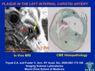
1. PLAQUE IN THE LEFT INTERNAL CAROTID ARTERY
T2W
CME HistopathologyIn Vivo MRI
450µm in-plane resolution
3 mm slice thickness
Fayad Z.A. and Fuster V. Ann. NY Acad. Sci. 2000;902:173-186
Imaging Science Laboratories
Mount Sinai School of Medicine
2. TEE
T2W
4.5 mm plaque
AHA TYPE Va (fibroatheroma) AORTIC PLAQUE
Fayad ZA et al.
Circ 2000;101;2503-2509
Imaging Science Laboratories
Mount Sinai School of Medicine
Plaque
Fibrous
Cap
3. Fayad ZA et al. Circ. 2000;102;506-510
Imaging Science Laboratories
Mount Sinai School of Medicine
X-ray Angiogram
high grade stenosis
LAD
LAD Wall
MR CORONARY WALL IMAGING
Editor's Notes
In vivo transverse T2-weighted fast SE MR imaging of a left internal carotid artery. Plaque characterization was based on information obtained from T1-, intermediate-, and T2-weighted MR images. Left: T2-weighted MR image (repetition time, two R-R intervals; echo time, 55 msec; 3-mm section thickness; 450-µm in-plane resolution) shows low-signal-intensity lipid core (lc), high-signal-intensity fibrous cap (fc), and very high–signal-intensity thrombus (t). l = arterial lumen. Right: Corresponding histopathologic section. (Mason-eosin stain; original magnification, X10).
Fayad ZA, Fuster V. Characterization of atherosclerotic plaques by magnetic resonance imaging. Ann N Y Acad Sci. 2000;902:173-86.
• In vivo magnetic resonance images of a 4.5 mm thick plaque in the descending thoracic aorta: A) T1-weighted; B) Proton density-weighted; C) T2-weighted; with the corresponding transesophageal echocardiography (TEE) image (panel D). The MR images show an example of an AHA type Va plaque with a dark area in the center (arrow) identified on the T2-weighted image as a lipid rich core (panel C). The lipid rich core is separated from the lumen by a fibrous cap. Plaque characterization was based on the information obtained from T1-, proton-density-, and T2- weighted MR images. Image resolution is 0.8 mm.