

Recommended
More Related Content
What's hot
What's hot (20)
Similar to Cleft lip and palate
Similar to Cleft lip and palate (20)
More from shekhar star
Recently uploaded
Recently uploaded (20)
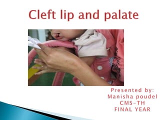
Cleft lip and palate
- 2. Introduction Incidence Embryological background Etiology Predisposing factor Associated syndromes Diagnostic consideration in cleft lip and palate Classification of cleft lip and palate Problems associated with cleft lip and palate Management of cleft lip and palate
- 3. The dictionary meaning of cleft is a crack, fissure,split or gap. Cleft lip and palate is a congenital birth defect which is characterized by complete or partial cleft of lip and/or palate Not life threatening unless associated with other syndrome Severity may vary from trace of notching of upper lip to complete non fusion of lip, primary palate and secondary palate
- 4. The approximate incidence is 1 in 700 live births, among them 25% are bilateral and 85% are associated with cleft palate. Isolated cleft palate occurs in 1 in 2000 live births . Negroids having least incidence (0.4/1000) and mongoloid and afghans(4.9/1000) having the highest incidence. Cleft lip is more common among males and cleft palate is more commoly among females. Unilateral clefts accounting for 80% of incidence and bilateral for remaining 20%. Among unilateral clefts, clefts involving left side are seen in 70% of cases.
- 5. 51 clefts was identified among 30,592 live births during year 2005 to 2010. Birth prevalance of cleft of lip +cleft palate was 1.64/1000 live birth per year. 19 clefts of lip alone(birth prevalance 0.61/1000 per year).
- 6. 21 cleft lip and palate (0.67/1000 per year) 11 cleft palate only(0.35/1000 per year) CLP and CL are common in male whereas CP is common in female. -Source:Article on prevalencene of CL And CP in tertiary hospital in Eastern Nepal by Singh VP,Sagatani R,Sagtani A
- 7. Face is formed by fusion of number of embryonic processes. Around the 4th week of intrauterine life, five brachial arches develop at the site of future neck.
- 8. The first arch, mandibular arch plays a role in development of nasomaxillary complex.
- 9. Mandibular arch gives rise to maxillary process from the dorsal end.
- 10. With the formation of nasal pits, frontonasal process gets divided into a medial nasal process and two lateral nasal processes. 5-6th week
- 11. Maxillary process fuse with medial and lateral nasal processes to form upper lip and primary palate respectively
- 12. Development of palate Development begins in 6th week Develops from- 1.Primary palate(from medial nasal process) 2.Secondary palate(from maxillary process)
- 13. Palate is formed by contibution of 1. Maxillary process 2.Palatal shelves given off by maxillary process 3.Fronto-nasal process Fronto-nasal process give rise to premaxillary region while palatal shelves form rest of palate.
- 14. Fusion of palatal shelves begin at 8th week which continues till 12/17th week. Initially the palatal shelves are covered by an epithelial lining. As they join epithelial cells degenerate. The connective tissue of the palatal shelves intermingle with each other resulting in their fusion.
- 15. Lower Lip is formed by the fusion of mandibular process of two sides. Upper Lip is derived from medial nasal and maxillary processes. Failure of merging between the medial nasal and maxillary processes at 5 weeks’gestation,on one or both sides,results in cleft lip.
- 17. If cleft of lip extends deep into primary palate, it results in cleft of alveolus too. Occasionally, cleft of lip and alveolus may have bands of soft tissue bridging across the two sides called simonnarts band.
- 18. Cleft of palate occurs in number of ways: Defective growth of palatal shelves Delayed or total failure of shelves to elevate and attain a horizontal position Lack of contact between shelves Post fusion rupture of shelves Failure of mesenchyme consolidation.
- 19. Isolated cleft of palate: It is a separate entity and appears to be under strong genetic influence. More common in females . Severity from mild notching of tip of uvula to cleft extending the soft palate alone or into secondary hard palate upto incisive foramen. Usually associated with pierre robin syndrome.
- 20. Submucous clefts: Present on palate as bony defects but covered with oral mucosa The submucosal clefts most often affect the posterior part of the palate at the posterior nasal spine.
- 21. And may be accidentally discovered on routine occlusal X-ray or could be located on careful clinical palpation of the palate. Occurs only in hard palate and continue to open cleft of soft palate or it may occur as a submucous cleft of the soft palate with or without notching into the hard palate.
- 22. Many workers are of view that clefts occur due to a number of causes and no single etiology can be pinpointed. o Heredity: Drilien reported that 1 in 3 children with clefts had some relatives with similar congenital defects. Family history is available in about 12- 20% of cases. o Tranforming growth factor alpha(TGFA) o Tranforming growth factor beta 3(TGFB3) o AP2 and MSX1 are the genes that have been identified as major role in the development of cleft lip and cleft palate.
- 23. o Environment: can be divided into 4 categories i) Womb environment ii) External environment iii) Nutrition- micronutrient and folic acid deficiencies iv) Drugs Certain drugs acts as teratogen that cause birth defects like - Anti-abortificant drugs -Anti emetics -Antiepilectic drugs( phenytoin, valproic acid) -Thalidomide - Dioxin, retinoic acid, maternal alcohol use, and maternal cigarette smoking
- 24. Some of the known teratogens are: Rubella virus Cortisone Mercaptopurine Methotrexate valium Infections like rubella, syphilis and toxoplasmosis Maternal alcohol use was found to cause interruption in the migration and differentiation of neural crest cells Embryos exposed to maternal smoking have increased risk of having clefts
- 25. o Multifactorial etiology: Recent studies show that etiology of cleft lip and palate cannot be attributed solely to either genetic or environmental factors. IT seems to involve more than one factor. They argue that unless a person is genetically susceptible, the environment factors may not by themselves cause clefts.
- 26. o Increased maternal age: women who conceive late have increased risk of having offsprings with clefts o Racial: Mongoloids have greatest incidence of clefts o Blood supply: Any factor that reduces blood supply to nasomaxillary areas during embryological development predisposes to clefts
- 27. Cleft lip may be associated with the following syndromes: • Down’s syndrome • Wardenburg’s syndrome (abnormities of pigmentation of hair,iris and skin,deafness) • Vander Woude’s syndrome(lips pits) • Orofacial digital syndrome • Treacher collins syndrome • Pierre Robin syndrome • Klippel-Feil syndrome
- 28. According to certain studies, up to 30% of cleft lips and palates and 50% of pure palatal clefts may be related to a syndrome. (Marazita 2002,Calzolari at al.2007)
- 29. Ultrasonography and 3D ultrasonography enables utero diagnosis of clefts especially in 3rd trimester.
- 30. 1.Time for parent education on the management of the baby to be born. 2.Allows psychological preparation of the parents and allow them to have realistic expectations. 3.It gives opportunity to investigate the presence of other chromosomal abnormalities. 4.Gives parent the choice of continuing the pregnancy.
- 31. 5.Helps in getting prepared for the neonatal feeding and care. 6.Opportunity for fetal surgery.
- 32. Hereditary risk of children of parents with cleft or slibling of children with cleft is approx. 4%. Risk is significantly smaller(0.6%) for second degree relatives.
- 33. If more than one family member has a cleft,this increases the risk of inheritance. (Harper 2004,Sivertsen et al. 2008)
- 34. Cleft of lip is denoted as CL. Cleft of lip along with alveolus is denoted as CLA. complete cleft extending from a unilateral cleft lip and primary palate passing through the mid-palatal suture to the soft tissue uvula is denoted as UCLP( unilateral cleft lip and palate).
- 35. Bilateral cleft of lip and premaxilla which extends down to the bifid uvula is denoted as bilateral complete cleft of the lip and palate (BCLP). Isolated cleft of palate can be abbreviated as CPO.
- 36. Classification system can broadly classified into: 1. Typical and Atypical orofacial clefts 2.Syndromic and Non-Syndromic Clefts Atypical clefts Lateral transverse Tessier type of facial cleft Median clefts
- 37. Non-Syndromic clefts individuals have no other physical or developmental anomalies though they may show subnormal growth or other parameters. It has been suggested that about 70% of cases of CL/P and 50% of CPO are non- syndromic.
- 38. Syndromic cleft : May appear as a part of congenital anomalies (MCA). In a multiple congenital anomaly syndrome, cleft patients can be further grouped into: 1. Monogenic syndrome, 2. Chromosomal aberrations, 3. Part of an association, or part of a complex of multiple congenital anomalies of unknown aetiology. 4. Teratogenic syndromes.
- 39. 1. Davis and Ritchie classification Morphological classification based on the location of cleft relative to alveolar process Group I- prealveolar clefts involving only lips and are subclassified as - Unilateral - bilateral - Median Group II- post alveolar clefts that comprises hard and soft palate clefts upto the alveolar ridge Group III- alveolar clefts. Complete clefts involving the palate, alveolar ridge and lips.can be subdivided into-
- 40. can be subdivided into Unilateral Bilateral Median 2) Veau’s classification Group 1 – cleft involving soft palate only Group 2 - cleft of hard and soft palate extending upto incisive foramen Group 3 – complete unilateral clefts involving soft palate, hard palate, lips and alveolar ridge Group 4 - complete bilateral clefts affecting the soft palate, hard palate, lips and alveolar ridge
- 41. Fogh Anderson (1942) Group 1: cleft of lip. Can be subdivided into single – unilateral or median clefts double - bilateral clefts Group 2 - cleft of lip and palate Single – unilateral cleft double - bilateral cleft Group 3 – clefts of palate extending upto incisive foramen
- 42. Schuchart and pfelter’s symbolic classification -It makes use of chart made up of a vertical block of three pairs of rectangles with an inverted triangle at bottom - Inverted triangle represents soft palate while the rectangles represent the lip, alveolus and hard palate as we go down. - Areas affected by clefts are shaded on chart.
- 43. Kernahan’s stripped ‘Y’ classification - Symbolic classification - Uses a stripped ‘Y’ having numbered blocks - Each block represents a specific area on the oral cavity Block 1 and 4 – lip Block 2 and 5 – alveolus Block 3 and 6 - hard palate anterior to incisive foramen Block 7 and 8 – hard palate posterior to incisive foramen Block 9 – soft palate
- 44. Lahshal classification simple classification presented by okriens in 1987. L – lip A – alveolus H - hard palate S - soft palate H - hard palate A – alveolus L - lip
- 45. Goslon yardstick Three of the authors have mentioned clinical features that they considered to be most important in categorizing the severity of malocclusion. They are a) Anteroposterior arch relationship b) Vertical labial segment relationship c) Transverse relationship a)Anteroposterior arch relationship - Class III incisor relationship is the least satisfactory and severe form whereas class II, div I is favorable for orthodontic correction b) Vertical labial segment relationship
- 46. A deep overbite is preferred over an openbite. c) Transverse Relationship: - Canine crossbites are considered worse than molar crossbite Goslon index Group 1 – (Excellent) no orthodontic treatment required Group 2 – (Good) minimum orthodontic treatment required Group 3 – ( Fair) Complex orthodontic treatment required Grooup 4 – ( Poor) Complex orthodontic treatment required along with surgery
- 47. 1.Prenatal Growth Various forces which influence the facial growth in utero are: a)Over maxillary segment on non-cleft side: Pull of lip and cheek muscles Tongue pressure Relatively unstrained nasal septum growth b)Over maxillary segment on cleft side: Instrinsic Deficiency Pressure from alar base due to stretching of the nostrils.
- 48. Due to above mentioned forces,deficiency produced in cleft lip and palate babies are: a)Incomplete unilateral cleft lip and palate: -Severe deviation of midline away from cleft. -Smaller maxillary segment shows retro- positioning or growth inhibition and collapse. -Nose is deviated towards normal side. b)In bilateral cleft lip and palate cases -Premaxilla tilts forward and/or shifts to one side due to tongue pressure.
- 49. 2.Facial growth in unrepaired cleft lip and palate Cases -A functionally normal facial skeleton develops in unrepaired cases expect for presence of local bony defect due to intrinsic deficiency in immediate area of cleft. -Due to absence of normal lip pressure abnormal development of dento-alveolar process can occur in vicinity of cleft lip.
- 50. 3.Facial Growth following Surgical Repair a)Effect of lip repair: -Tight upper lip following repair significantly inhibits the facial growth in antero posteior direction. -Increased tightness in lower part of repaired lip near free border leads to retroclination of dentoalveolar structures.
- 51. b)Effect of palate repair: -Palate repair may inhibit the growth of maxilla and due to scar contracture,reduction in maxillary arch may occur.
- 52. 1)Dental problems 2) Aesthetic problems 3) Hearing and speech problems 4) Psychological problems
- 53. 1) Dental problems Congenitally missing teeth( mostly upper lateral incisors) Presence of supernumerary, neonatal and natal teeth Ectopically erupted tooth Enamel hypoplasia Microdontia, macrodontia Fused teeth Gemination, dilaceration
- 54. Tendency towards class III skeletal pattern Posterior and anterior cross bite Deep bite Spacing/ crowding Protruding premaxilla 2) Esthetic problems Facial disfigurement Orofacial structures can be malformed and congenitally missing Deformities of nose can also occur
- 55. 3) Hearing and speech problems -Children with cleft often have associated speech and language disorder. - Assessment of speech therapist is required as early as 9 months of age - Patient with delayed development of speech may have receptive language problems that arise because of the collection of fluid in middle ear - Hearing loss may also occur due to ossicular malformation
- 56. - Speech problems are more severe if surgery is delayed - Usually an operated cleft palate patient present short palate, decreased mobility of soft palate due to scarring and presence of oronasal fistula, all which contribute to velopharyngeal insufficiency 4)Psychological problems Patients are under a lot of psychological stress.
- 57. Multidisciplinary cleft palate team
- 58. Birth to 1 month Initial assessment Presurgical orthopaedics 3 months Primary lip repair 9 to 18 months Palate repair 2 years Speech assessment 3-5 years Lip revision surgery 8-9 years Initial interventional orthodontics in preparaton for alveolar bone grafting Continuing speech therapy
- 59. 10 years Alveolar bone grafts 12-14 years Definitive orthodontics 16 years Nasal revisional surgery 17-20 years Orthognathic surgery Advanced conservative treatment
- 60. Parents are usually not prepared to face this problem. Sometimes, they have a feeling of guilt that they had done something wrong during pregnancy. They should be informed that there is nothing known which they could have done earlier to prevent its occurrence. The management regarding surgery,dentistry and speech therapy should be explained properly along with possible outcomes.
- 61. The cleft surgeon/plastic surgeon undertakes primary and secondary repair. The speech therapist monitors speech from 9 months onwards and institutes measures for normal development of the speech. The audiologist quantifies and locates the cause of hearing problems. The orthodontist monitors dental development, occlusion, skeletal problems and institute interceptive therapy, dentofacial orthopaedics and prepares for secondary alveolar bone graft.
- 62. Oral surgeon: Secondary alveolar bone graft and orthognathic surgery if required. Plastic surgeon: Correction of nose deformity, secondary deformities of lip and scar revision. Specialist cleft nurse who can monitor the neonates during early days after birth and give feeding advice. Clinical geneticist to resolve genetic basis of the cleft, and advice on recurrence risk.
- 63. Paediatric dentist/dentist to maintain and monitor dental health and oral hygiene. ENT surgeon/audiologist to take care of recurrent ear infections, insert grommets and hearing tests With the increasing advances in treatment of CLP, interdisciplinary approach is considered effective than multidiscilpinary team.
- 64. Immediately on birth Feeding and psychological problems are the biggest issues. A cleft lip or palate makes feeding of baby more difficult. The major problems with feeding a baby with cleft are problems with sucking and with formula coming through the nose. The following specialists are required to evaluate the child: Neonatologist, paediatrician Feeding specialist nurse Geneticist to assess for syndromic associations Clinical psychologist.
- 65. Newborn children with clefts present the risk of aspiration and airway obstruction which may lead to acute asphyxia in children with small mandibles like in Pierre Robinson syndrome. Such cases may require tracheostomy at birth.
- 66. Feeding dificulties are common, so specialized feeding bottles such as haberman feeder and Mead Johnson bottle are helpful. Haberman feeder Mead Johnson bottle
- 67. Recurrent chest and throat infections: care by the neonatologist/ and paediatrician. First few months Primary surgery of lip and anterior palate: done by a cleft surgeon who could be an oral surgeon/plastic surgeon/ paediatric surgeon.
- 68. Management of cleft lip and palate can be divided into following stages: Stage I- treatment done from birth to 18 month of age Stage II- from 18 th month to 5th year of life( primary dentition stage) Stage III- treatment carried out during mixed dentition stage from 6th to 11th year of life Stage IV - treatment done during permanent dentition stage ( 12-18 years)
- 69. m- months, y- years
- 70. Stage I treatment: Includes: i. Fabrication of a passive obturator ii. Presurgical orthopedics iii. Surgical management of cleft lip iv. Surgical management of cleft palate Passive maxillary obturator: Is an intraoral prosthetic device that fills the palatal clefts and provides false roofing against which child can suckle Reduces the feeding difficulties like insufficient suction, choking, excessive air intake
- 71. Obturator is fabricated using cold cure acrylic after selective blocking of all the undesirable undercuts Clasp aid in retention, in case insufficient retention, wings made of thick wire can be imbedded in acrylic and made to follow cheek contour extraorally.
- 72. Presurgical orthopaedics 1. It facilitate the creation of good functioning palate. 2. Normalize tongue position. 3. Help in speech development. 4. Improve symmetry of nose and cleft of maxilla. 5. Psychologically boost patient and parents as the patient get continued supervision.
- 73. Surgical lip closure: Primary closure of lip is undertaken at age of 3 months or 10 weeks when child is fit to undergo general anaesthesia. Millard has suggested rule of 10 . Approximately 10 weeks of age 10 pounds (4.54 kg) Bood haemoglobin not less than 10 gram% Two techniques have been popular: a. Tenninson’s triangular flap procedure b. Millard’s rotation flap Other technique is: -Veau repair
- 74. Where a=rotational flap b=advancement flap c=columella flap a and c are planned on medial side of Cleft.After full thickness Of lip is cut along the marking which is filled by b planned on lateral side In this method minimal Tissue is discarded and the Result can be modified during surgery.
- 75. Veau repair
- 76. Surgical palate closure: Should be attempted between 12-24 months of age. Facilitates normal speech, hearing and swallowing.
- 77. The tension of lip closure centralises premaxilla and then the other side of lip is closed at 4 months of age. Vomerine flaps from right and left sides are used to close the anterior palate, which is done at 8-12 months using von langenback technique. Lip revision and columella lengthening are done at age of three years.
- 78. The Oslo protocol evolved at the Oslo Cleft Centre, which is one of the two centralized care centres in Norway. Their protocol does not follow preoperative orthopaedics. Millard’s procedure is carried out for lip repair at the age of 3 months.
- 79. In cases with an associated cleft of the alveolus and palate, a cranial based single layer vomer flap is sutured under the alveolus palate periostium at the time of lip closure. In light of the present knowledge, ongoing research and different long-term and inter- centre studies, the Oslo protocol has been observed to generate good treatment outcome.
- 80. Remaining hard and soft palate closure is done at the age of 18 months by von Langenbeck pattern palatoplasty. Alveolar bone grafting is done at the age of 8-10 years.
- 81. Stage two treatment: Comprises treatment carried out during primary dentition period. Procedures carried out during this phase: Adjustment of intraoral obturator to accommodate the erupting deciduous teeth. To maintain a check on eruption pattern and timing. Oral hygiene instructions. Restoration of decayed teeth.
- 82. Orthodontic treatment is not normally recommended for primary dentition as it may damage permanent dentition follicles. However , in patients with: Moderately underdeveloped maxilla and no class III hereditary defect reverse headgear treatment should be advocated at age of 4 – 7 years.
- 83. Parents should understand the value of tooth brushing . Parents may be nervous to brush in region of cleft especially following primary lip and palate surgery. They should be shown in detail about how to brush. A low fluoride children toothpaste containing no more than 600 ppm fluoride is recommended for children under 6 years. Twice brushing daily is recommended. In addition, twice yearly professional application of topical fluoride varnish is useful.
- 84. Stage three treatment Carried out during mixed dentition phase. As in the early years, the main emphasis throughout the mixed dentition stage should be on prevention of dental disease. In this phase, secondary alveolar bone grafting is common.
- 86. A child with cleft palate may need surgery after intial cleft palate repair to replace missing bone in front of mouth and roof to the mouth. Successful grafting provides osseous envionment to permit spontaneous eruption of canine in grafted area and so should be undertaken after eruption of permanent incisors but before eruption of permanent canine. Alveolar bone grafting provides bony bridge to cleft in alveolar area.
- 87. Benefits of SABG: 1.Provides bony support for alar base to minimize nasal deformity. 2.Elimination of oronasal and nasolabial fistulae,hence avoiding nasal reflux of fluid and air.
- 88. 3.Stabilization of maxillary segments,so as to facilitate future secondary corrective osteotomy if required. 4.Facilitation of teeth eruption into cleft site and achieve orthodontic movement adjacent to cleft site.
- 89. Timing of SABG: It is done at the age when growth inhibition effects of surgery are minimized and it can help maxillary canine and lateral incisor to erupt through the cancellous bone. It is done in mixed dentition stage after eruption of permanent incisors but before eruption of permanent canine.
- 90. Assessment for the need for bone graft Required careful clinical and radiological assessment Teeth in the vicinity of cleft area need to be assessed All retained deciduous teeth, supernumerary teeth and rudimentary teeth are usually extracted before bone graft
- 91. Pre bone graft orthodontics Maxillary arch expansion is performed preparatory to secondary bone grafting for which quad helix is appliance of choice. Nowadays repetitive weekly protocol of alternate rapid maxillary expansion and constriction are performed.
- 92. Clinical result of maxillary protraction using 2- hinged expander, repetitive weekly Protocol of Alt-RAM and intraoral protraction springs
- 93. Surgical technique Two surgeons work simultaneously, one on donor site and other on host site. It involves incision around margin of cleft alveolus Full thickness mucoperiosteal flap is raised to allow space for bone graft. Gingival mucoperiosteal is the most recommended one.
- 94. Iliac bone is harvested, packed in cleft alveolus space. the flap is then sutured to ensure complete seal. Post bone graft follow up requires retention of the expansion either by full bonded appliance or by reinserting a passive expansion appliance. some include surgical exposure of canine and orthodontic traction.
- 95. Orthodontic procedures carried out during mixed dentition phase are: 1. Correction of anterior crossbite using removable or fixed appliance can be used like Z springs 2. Buccal segment crossbites can be treated using quad helix and expansion screws which are pre bone graft orthodontics
- 96. Consist of treatment during permanent dentition. Presence of permanent dentition usually signs for the definitive orthodontic treatment. All local irregularities like crowding, spacing, crossbites and overjet overbite problems are corrected. Patients with hypoplastic maxilla may be given facemask to advance maxilla.
- 97. Regular oral hygiene monitoring and instruction is necessary. Dietary counselling. Patients are to be made aware of excessive sugar intake. Following orthodontic treatment procedures, the patients should on retention phase to maintain orthodontic correction
- 98. Prosthesis can be given in case of missing teeth
- 99. fig: Intraoral distraction device and segmental osteotomy for interdental distraction osteogenesis
- 101. Cleft lip surgery Unilateral - Dehisence - Infection -Thin white roll -tension Bilateral -Dehisence -Thin white roll
- 102. Cleft palate repair -Fistula Velopharyngeal incompetence -Continued VPI -Stenotic side ports Alveolar bone grafting -Infected donor site #Hematoma -Failed grafts #Dehisence #Palatal prosthesis
- 103. Midfacial advancement Le fort osteotomies -Malocclusion -Infection -Necrosis Rhinoplasty -Alar stenosis
- 104. In cleft palate patients due to abnormal function of eustachian tube there is an increased risk of otitis media. The parents are counselled for possible hearing loss.
- 105. ENT specialist, Audiologist and speech specialist work together to note the middle ear problems and progress in speech. Speech therapy is started from 6 months of age and if needed continued till adulthood.
- 106. Roughly 1 in 4 patients with CLP develop defects in growth of upper mandible and midface. Resulting severe malocclusion can have a major detrimental impact on mandible function and facial appearance,which can be pschyological difficult for teenagers. A growth defect in maxilla cannot be corrected through orthodontics alone, but orthognathic surgery is required to correct alignment of maxilla.
- 107. An maxilla growth defect is most often treated by Le fort I osteotomy. Distraction of maxilla,whereby maxilla is gradually pulled to desired position,is also possibility. Severe growth defect may require both procedures: distraction during the growth stage and osteotomy towards the end of growth.
- 108. Mandible osteotomy is sometimes required to correct the facial structures. The nasal deformity typical of CP may be more pronounced as a result of LE Fort 1 osteotomy. A thorogh rhinoplasty operation is thus performed at this stage.
- 109. Nasal surgery(rhinoplasty) and lip surgery(revision cheiloplasty) may be necessary to improve the appearance and function of nose and lip which have been distorted with growth after initial surgery. The nose may appear flattened or there nay be asymmetry of the nose.
- 110. There may be nasal obstruction due to a small nostril or deviated septum. Surgery to revise the appearance of lip and nose may take place before the child starts school or during teenage years,depending on recommendation of plastic surgeon.
- 111. Children with repaired cleft palate may have a resulting condition referred to as “VPI” (Velopharyngeal Incompletence). This means that too much air escapes through the nose during speech,resulting in nasal speech. This occurs because the repaired soft palate is too short or does not move adequately.
- 112. This condition is diagnosed primarily by the trained ear of speech pathologist. However,special diagnostic procedures such as nasoendoscopy and videofluoroscopy of speech may be required to directly visualize the soft palate during speech. This helps in directing the type of intervention ,which is the most appropriate. .
- 113. With the goal of successful communication for the child with cleft lip and palate, the speech pathologist regularly monitors the development of using and understanding language and the development of speech abilities including pronunciation of words,the sound of voice and amount of nasality during speech Operation to improve the fuction of soft palate are pharyngeal flap or pharyngoplasty procedures. In this operations,some of the tissue from palate and back of throat are repositioned to help close off the escape of air through the nose.
- 114. The key to successful rehabilitation of cleft lip and palate include flexibility and a interdisciplinary approach. Patient should be treated with sympathy and concern. Parents should not panic with the condition rather should provide special attention to such child .
- 115. - Orthodontics the art and science fifth edition S.I Bhalajhi -Textbook of pedodontics Shova Tandon - Orthodontics –diagnosis and management of malocclusion and dentofacial deformities Om Prakash Kharbanda
Editor's Notes
- The maxillary process grows ventro-medio-cranial to the main part of the mandibular arch which is now called the “MANDIBULAR PROCESS”. Thus at this stage the primitive mouth or stomodeum is overlapped from above by the frontal process,below by the mandibular process & on either side by the maxillary process.
- The stomodeum is thus overlapped superiorly by the fronto-nasal process. The mandibular arches of both The sides form the lateral walls of the stomodeum. The mandibular arch gives off a bud from its dorsal end called the “MAXILLARY PROCESS”.
- The ectoderm overlying the fronto-nasal process shows bilateral localized thickenings above the stomodeum. These are called the “NASAL PLACODES”.These placodes soon sink and form the nasal pits. The formation of these nasal pits divides the fronto-nasal process into two parts: a)The medial nasal process & b)The lateral nasal process
- C