1. Cerebral Cortex Advance Access published August 1, 2011
Cerebral Cortex
doi:10.1093/cercor/bhr199
WIP Is a Negative Regulator of Neuronal Maturation and Synaptic Activity
A. Franco1, S. Knafo2,3, I. Banon-Rodriguez1, P. Merino-Serrais2, I. Fernaud-Espinosa2, M. Nieto1, J.J. Garrido2,3,4, J.A. Esteban3,
F. Wandosell3,4 and I.M. Anton1,4
1
Centro Nacional de Biotecnologı´ a (CNB-CSIC), 28049 Madrid, Spain, 2Instituto Cajal (CSIC), 28002 Madrid, Spain, 3Centro de
Biologı´ a Molecular ‘‘Severo Ochoa’’ (CSIC-UAM), Universidad Autonoma de Madrid, 28049 Madrid, Spain and 4CIBERNED, Centro
´
Investigacion Biomedica en Red de Enfermedades Neurodegenerativas, 28031 Spain
´ ´
A. Franco and S. Knafo contributed equally to this work
Address correspondence to Dr Ines M. Anton, Cellular and Molecular Department, Centro Nacional de Biotecnologı´ a (CNB-CSIC), Darwin 3, 28049
´ ´
Madrid, Spain. Email: ianton@cnb.csic.es.
Wiskott--Aldrich syndrome protein (WASP) --interacting protein factors (NPF), such as neural Wiskott-Aldrich syndrome protein
(WIP) is an actin-binding protein involved in the regulation of actin (N-WASP). The Arp2/3 complex nucleates actin, inducing
polymerization in cells, such as fibroblasts and lymphocytes. branching and elongation, and with N-WASP, it mediates
Despite its recognized function in non-neuronal cells, the role of neurite elongation (Suetsugu, Hattori, et al. 2002; Pinyol et al.
WIP in the central nervous system has not been examined 2007) and neurite branching (Kakimoto et al. 2004). N-WASP
Downloaded from cercor.oxfordjournals.org at Universidad Autonoma de Madrid. Facultad de Medicina on August 2, 2011
previously. We used WIP-deficient mice to examine WIP function interacts with WASP-interacting protein (WIP), a broadly
both in vivo and in vitro. We report here that WIP2/2 hippocampal expressed proline-rich protein that regulates N-WASP function
neurons exhibit enlargement of somas as well as overgrowth of as NPF and whose deficiency modifies actin polymerization
neuritic and dendritic branches that are more evident in early kinetics and the density of the subcortical actin network
developmental stages. Dendritic arborization and synaptogenesis, (Anton et al. 2007). Through WASP/N-WASP--dependent or
which includes generation of postsynaptic dendritic spines, are --independent mechanisms, WIP participates in a wide variety
actin-dependent processes that occur in parallel at later stages. of cellular functions, including signaling, endocytosis, and actin
WIP deficiency also increases the amplitude and frequency of cytoskeleton remodeling (Anton et al. 2007). WIP deficiency in
miniature excitatory postsynaptic currents, suggesting that WIP2/2 mice alters the immune response, reducing T and mast cell
neurons have more mature synapses than wild-type neurons. These activity and increasing B cell function (Anton et al. 2002;
findings reveal WIP as a previously unreported regulator of Kettner et al. 2004). Moreover, WIP null mice have a pro-
neuronal maturation and synaptic activity. gressive immunological disorder of autoimmune nature, with
evident ulcerative colitis, interstitial pneumonitis, glomerular
Keywords: dendritic spine, electrophysiology, neuritogenesis, N-WASP, nephropathy with IgA deposits, autoantibodies, and joint
synapse inflammation that lead, all together, to premature death
(Curcio et al. 2007). Although molecular details of WIP-
WASP/N-WASP inter-action have been studied extensively
(Volkman et al. 2002; Ho et al. 2004; Dong et al. 2007; Peterson
Introduction et al. 2007), few data are available on its functional impact and
Neuronal cytoarchitecture is first established through neurito- even fewer regarding the central nervous system, where the
genesis, a process in which neurons extend their neurites to role of WIP has not been previously addressed.
form a functional network during neuronal development (de Using the WIP knockout mouse as a tool, here, we describe
Curtis 2007). Neuron morphology greatly determines the final that loss of this protein impacts neurite and dendrite dynamics
complexity of the nervous system and is essential for the signal and morphology, both in early and in late developmental stages,
flow that underlies information integration and processing. It is in vitro and in vivo. Gross examination of WIP–/– brain revealed
therefore important that neuritogenesis occurs at the right changes in forebrain and hippocampal size. Extensive analysis
place and time for correct establishment of synaptic contacts of WIP–/– hippocampal neuron development showed premature
with proper targets (de Curtis 2007). Several environmental neuritogenesis. Finally, electrophysiological and immunocyto-
cues converge on common coordinated intracellular pathways to chemical analyses demonstrated modified synaptic activity of
modulate neuritogenesis. Such intracellular events involve sig- WIP–/– mature neurons. These studies show that WIP is an
naling transduction, exocytic and endocytic mechanisms related essential negative regulator in the control of the cytoskeletal
to membrane trafficking and cytoskeletal rearrangements. events that underlie neuronal and synaptic development.
Neurite initiation and outgrowth are based on the capacity
of the neuronal cytoskeleton, constituted mainly of actin Materials and Methods
microfilaments (MF) and tubulin microtubules (MT), to
assemble and disassemble in response to extracellular signals Mice
(Luo 2002; Conde and Caceres 2009). The polarized growth of Wild-type (WT) and WIP KO SV129/BL6 mice (Anton et al. 2002) were
neurites requires the initial depolymerization of actin MF housed in specific pathogen-free conditions at the animal facility of the
(Bradke and Dotti 1999), stabilization of MT (Ferreira and Centro de Biologı´ a Molecular ‘‘Severo Ochoa,’’ Madrid, Spain. The
mouse colony was maintained by continuous mating of heterozygous
Caceres 1989), and accumulation of a number of specific females with heterozygous males for more than 20 generations. To
proteins (Wiggin et al. 2005). Actin polymerization is con- obtain control or WIP–/– embryos/litters, we mate control male and
trolled by the actin-related protein (Arp2/3) complex and by female or WIP–/– male and female mice. Handling of mice and all
the action of actin-binding proteins and nucleation-promoting manipulations were carried out in accordance with national and
Ó The Author 2011. Published by Oxford University Press. All rights reserved.
For permissions, please e-mail: journals.permissions@oup.com
2. European Community guidelines and were reviewed and approved by a 403 oil-immersion lens (NA, 1.3). Metamorph 6.2r6 (Universal
the institutional committee for animal welfare. All quantification was Imaging) software was used to process the time-lapse captured images.
conducted in a genotype-blind manner.
Morphometry
Image stacks (physical size 76.9 3 76.9 lm) were imported to the
Brain Lysates and Western Blot
confocal module of Neurolucida 7.1 (MicroBrightfield, Inc., Williston,
Control or WIP–/– brains were homogenized in lysis buffer (20 mM 4-(2-
VT), and neuronal dendritic trees were traced by drawing the dendrites
hydroxyethyl)-1-piperazineethanesulfonic acid, pH 7.4, 100 mM NaCl, 5
and the bifurcation points. Sholl analysis was performed for each traced
mM ethylenediaminetetraacetic acid, 1% Triton X-100, 100 mM NaF, 1
neuron by automatically calculating the number of dendritic intersec-
mM Na3VO4, and the Complete Protease Inhibitor Cocktail, Roche
tions and the dendritic length at 10-lm interval starting from the soma.
Diagnostics), and soluble extracts were resolved by sodium dodecyl
Total dendritic length and total number of intersections and branches for
sulfate--polyacrylamide gel electrophoresis after determination of
each neuron were also calculated as an index of dendritic complexity.
protein concentration by Bradford analysis (BioRad). Proteins were
Soma area was determined by drawing soma contours while tracing cells.
then transferred to nitrocellulose filters, which were blocked and
incubated with a mouse monoclonal antibody (mAb) specific to WIP
(1/1000, 3D10; a generous gift of Prof. R. Geha, Children’s Hospital, Miniature Excitatory Postsynaptic Currents
Boston, MA). After exposure to a specific secondary antibody, antibody Miniature excitatory postsynaptic currents (mEPSC) were recorded
binding was visualized by enhanced chemiluminiscence substrate from dissociated hippocampal neurons bathed in artificial cerebro-
(Amersham Biosciences). spinal fluid (containing 119 mM NaCl, 2.5 mM KCl, 1 mM NaH2PO4, 11
mM glucose, 26 mM NaHCO3, 2.5 mM CaCl2, and 1.3 mM MgCl2) in the
presence of 1 lM tetrodotoxin and 100 lM picrotoxin (at 29 °C).
Primary Hippocampal Cultures Spontaneous activity was recorded for 3 min for each cell. mEPSC were
Downloaded from cercor.oxfordjournals.org at Universidad Autonoma de Madrid. Facultad de Medicina on August 2, 2011
identified using pClamp software and corrected by eye on the basis of
Neurons their kinetics.
Primary hippocampal cultures were prepared as described (Dotti et al.
1988; Kaech and Banker 2006). Briefly, hippocampi from E18 mouse
embryos (WT and WIP–/–) were washed and digested with 0.25% trypsin Morphology and Unbiased Stereology
(15 min, 37 °C). The tissue was then dissociated, resuspended in
minimun essential medium with 10% horse serum, and plated on poly- Perfusion
L-lysine--coated coverslips (1 mg/ml) at a density of 6 3 103 cells/cm2 Three-month-old male mice (WT, n = 6; WIP–/–, n = 6) were
for imaging at early times (up to 24 h after plating) and at a density of 4 3 anesthetized with pentobarbital (0.04 mg/kg) and perfused trans-
103 cells/cm2 for electrophysiological recordings. In some experiments, cardially with PBS (20 ml) followed by 100 ml 4% PFA (pH 7.4) in the
neurons were allowed to adhere to the substrate (1 h) and then same buffer. Brains were postfixed in the same fixative (24 h).
incubated with 2 or 5 lM wiskostatin (BIOMOL International) in
dimethyl sulfoxide (DMSO). In all cases, after 3 h, plating medium was Volumetric Analyses
replaced with neurobasal medium supplemented with B27 (Gibco). For Brains were sectioned coronally at a thickness of 50 lm to facilitate
long-term culture, at this time, neuron-including coverslips were measurements. Strict morphological criteria were used in all mice to
transferred into dishes containing an astrocyte monolayer, with determine the boundaries of these brain regions. Briefly, hippocampal
neurons oriented facing the glia but without contacting them. outlines encompassed the CA1--3 fields of Ammon’s horn and the
subiculum but not the presubiculum or fimbria hippocampus. The
DNA Constructs and Transient Transfection dentate gyrus was measured separately. Starting with one of the
pLVWIP-GFP was obtained by digestion of pcDNA3WIP-GFP (kindly sections, randomly selected across brains, 1 of every 6 sections was
provided by Prof. N. Ramesh, Children’s Hospital, Boston, MA) and by analyzed through the extent of a hemisphere of the brain. One
cloning the insert into pLV; control pLVGFP was generated in a similar hemisphere of each brain was analyzed. Using this sampling strategy,
manner. Suspended WT (cortical and hippocampal) or WIP–/– (cortical) 7--10 histological sections per brain were analyzed. All volumetric
neurons were nucleofected using a 6 lg DNA/100 ll suspension quantifications were performed with an Olympus BX51 with a 1.253
(Amaxa pulser; Lonza, Germany). Cells were maintained in suspension objective, a motorized XYZ axis computer-controlled stage (Prior
(4 h) to permit exogenous gene expression and then plated for 24 h Scientific, Houston, TX), a digital video camera (JVC), and Stereo-
before fixation for immunofluorescence analysis. Investigator, a stereology software package (version 8.03, MicroBright-
Field). When calculating hippocampus volume, the boundaries were
Immunofluorescence defined and the volumes determined with Stereo-Investigator software.
At 3 h, 1 and 22 DIV (days in vitro) postplating, cells were fixed in 4%
paraformaldehyde (PFA) in phosphate-buffered saline (PBS) (pH 7.4; 20 Intracellular Injection of Lucifer Yellow
min, room temperature). Cells were then permeablilized with 0.1% Coronal sections (150 lm) were cut on a vibratome. Sections were
Triton X-100 and labeled with phalloidin-tetramethylrhodamine iso- prelabeled with 4,6-diamidino-2-phenylindole, and a continuous cur-
thiocyanate (Sigma) and antibodies to tyrosinated a-tubulin (1/400; rent used to inject individual cells with lucifer yellow (8% in 0.1 M Tris
T9028, Sigma), MAP2 (MT-associated protein, 1/400; 514, Sanchez buffer, pH 7.4). Dye was injected into neurons in the dentate gyrus until
Martin et al. 1998), PSD-95 (5 lg/ml; 75-028, NeuroMab), or GFP (1/ the individual dendrites of each cell could be traced to an abrupt end at
100, 11814460; Roche); secondary fluo-rescent antibody or biotinylated the distal tips and the dendritic spines were readily visible, indicating
antibody and labeled streptavidin were then added. Cells were that the dendrites were completely filled. The sections were then
excluded from analysis if they showed obvious features of toxicity, processed, first with rabbit anti-lucifer yellow (Cajal Institute,
such as neurite fragmentation/blebbing or vacuoles in the cell body. 1/400 000 in stock solution of 2% Bovine serum albumin, 1% Triton
X-100, 5% sucrose in PBS) and then with Alexa 488-conjugated
Imaging secondary antibody (Molecular Probes, 1/1000, 4 h). Sections were
Confocal images were acquired digitally on a confocal LSM510 Meta mounted on a glass slide in fresh ProLong Gold antifade reagent
microscope (Zeiss) coupled to an inverted Axiovert 200 microscope (Invitrogen, Eugene, OR; 24 h, room temperature in the dark) and
(Zeiss). Image stacks (logical size 1024 3 1024 pixels) consisted of 10 sealed with nail polish.
image planes acquired through a 403 (numerical aperture (NA), 1.3) or
a 633 oil-immersion lens (NA 1.4). Confocal Microscopy
Time-lapse images of phase-contrast fields were captured on an Imaging was performed on a Leica laser scanning multispectral
inverted Axiovert 200 microscope (Zeiss) equipped with a mono- confocal microscope (TCS SP5) using an argon laser. Image stacks
chrome CCD camera and ultrafast filter change. Image stacks (logical (physical size 76.9 3 76.9 lm, logical size 1024 3 1024 pixels) consisted
size 1024 3 1024 pixels) consisted of 6 image planes acquired through of 100--350 image planes acquired through a 633 glycerol-immersion
Page 2 of 12 WIP Regulates Neuronal Maturation d
Franco et al.
3. lens (NA, 1.3; working distance, 280 lm; refraction index, 1.45) with murine brain and corroborate previous mRNA studies (Ramesh
a calculated optimal zoom factor of 3.2 and a z-step of 0.14 lm (voxel et al. 1997; Tsuboi 2006) as well as the data included in the Allen
size, 75.1 3 75.1 3 136.4 nm). These settings and optics represent the atlas (http://www.brain-map.org/).
highest resolution currently possible with confocal microscopy. For
each neuron (5 neurons per mouse, 55 neurons total), 1--5 randomly
selected dendrites were scanned from soma to tip, and stacks were Brain Hypertrophy in WIP-Deficient Mice
processed with a 3D blind deconvolution algorithm (Autodeblur; To assess general effects of WIP deficiency on the mouse brain,
Autoquant, Media Cybernetics) for 10 iterations to reduce the out-of- we determined volumes of the hippocampus and of the rest of
focus light. the murine forebrain (3-month-old male mice). Contours of the
structures of interest were drawn on Nissl-stained serial sec-
Statistical Analysis tions with the aid of Stereo-Investigator software to yield the
Quantification was performed in a blind fashion by independent volume of each. There was a significant increase (30%) in
researchers. For statistical analyses, we used the two-tailed Student t- forebrain volume in WIP-deficient brains (n = 5) compared
test to compare means, the chi-square test to compare nominal with WT littermates (n = 6, Fig. 1B--D and Supplementary Fig.
variables, and the two-way analysis of variance to compare Sholl analysis
1). The hippocampus proper (without the dentate gyrus) and
or the Kolmogorov--Smirnov test to compare cumulative frequency
analysis. Results are shown as mean ± standard error of the mean. the dentate gyrus itself were also hypertrophied in WIP–/– brain
(Fig. 1E,F). WIP deficiency thus causes general macrocephaly
that includes hippocampal hypertrophy.
Results
Downloaded from cercor.oxfordjournals.org at Universidad Autonoma de Madrid. Facultad de Medicina on August 2, 2011
Enhanced Early Neuronal Development in WIP –/– Neurons
WIP Is Expressed in the WT Mouse Brain The enlarged brain observed in WIP–/– mice relative to their WT
The expression pattern of WIP messenger RNA (mRNA) suggests littermates raised the possibility that the greater brain volume
that WIP is ubiquitously expressed in all mouse tissues, including could be due to increased neuritic branching since dendrites
brain (Ramesh et al. 1997; Tsuboi 2006). To date, however, the and axon collaterals account for most of the brain volume
levels of endogenous WIP protein have only been analyzed in (Acebes and Ferrus 2000). To test this hypothesis at the cellular
´
fibroblasts, myoblasts, and hematopoietic-derived cells (reviewed level, we examined the effect of WIP deficiency on early
in Anton et al. 2007). To study WIP expression in brain, we probed neuronal development. Primary hippocampal neurons from
western blots of cell lysates from 3 regions of the adult mouse brain control and WIP–/– embryos were grown at very low density so
(cortex, hippocampus, and olfactory bulb), using the WIP-specific that their neurite arbors did not overlap to avoid confusion
mAb 3D10 (Koduru et al. 2007). This mAb recognized 2 protein about which neurites protruded from each cell body. Cells
bands in cell lysates from each of the regions examined (Fig. 1A), were labeled with anti-tyrosinated a-tubulin and fluorescent
which could represent 2 WIP isoforms and/or be the result of phalloidin (Fig. 2A,B). As a measure of neuronal development,
posttranslational modifications of WIP. Neither band was present we quantified the fraction of neurons at each developmental
in WIP-deficient tissue. These data demonstrate WIP expression in stage at 3 h postplating. At stage 1, the MT-containing soma is
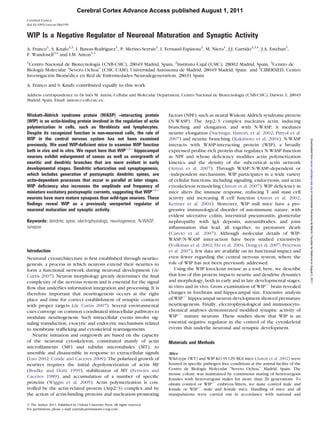
Figure 1. Brain hypertrophy in WIP-deficient mice. (A) Western blot indicates WIP expression in WT mouse brain (cortex, hippocampus, and olfactory bulb; 80 lg total protein/
lane) but not in WIPÀ/À mouse brain. Glyceraldehyde-3-phosphate dehydrogenase labeling confirmed equivalent protein loading in cortical samples. (B) Representative WT (left)
and WIPÀ/À brain (right) at 3 months of age. Note: the enlargement of the WIPÀ/À brain. Scale bar, 4 mm. (C) Representative Nissl staining showing hippocampus enlargement
in WIPÀ/À mice. Scale bar, 500 lm. (D--F) Average volumes of forebrain (D), hippocampus (E), and dentate gyrus (F) are increased in WIPÀ/À mice. P values were determined
with Student’s t-test; *P 0.05; **P 0.01.
Cerebral Cortex Page 3 of 12
4. Downloaded from cercor.oxfordjournals.org at Universidad Autonoma de Madrid. Facultad de Medicina on August 2, 2011
Figure 2. WIP deficiency accelerates development of dissociated hippocampal neurons. (A) Representative confocal images of WT- and WIPÀ/À-dissociated hippocampal
neurons fixed at 3 h postplating and stained for F-actin (phalloidin-tetramethylrhodamine isothiocyanate, red) and MT (Alexa488-anti-tyrosinated tubulin [TT], green). At this time
point, the WIPÀ/À neuron population showed more advanced development compared with WT neurons. Scale bar, 10 lm. (B) High magnification images of the inset in A. Scale
bar, 5 lm. (C) Neuron classification by developmental stage (3 h postplating) showed significantly lower frequency of WIPÀ/À at developmental stage 1 compared with WT
Page 4 of 12 WIP Regulates Neuronal Maturation d
Franco et al.
5. surrounded by flattened actin-rich lamellipodia, while in stage (pLVGFP; Fig. 3C). At 24 h postplating, while 31% of GFP-
2, the lamellipodia are transformed into equidistant neurites expressing neurons were in stage 1, the frequency of stage 1
(Dotti et al. 1988). Stage 1+ is defined as cells with incipient WIP-GFP--expressing neurons was 80% (Fig. 3D). The de-
tubulin-rich small protrusions. We found a clear difference in velopmental delay at 24 h induced by WIP overexpression was
the distribution of these populations (Fig. 2C); there was confirmed in cortical embryonic neurons (Supplementary Fig.
a significant decrease (35%) in the frequency of WIP–/– 2). This effect was sustained over time, since 48 h after plating,
neurons classified as developmental stage 1 compared with 22.6% of neurons with increased WIP levels remained at stage 1
WT neurons (n = 4 cultures and n = 150 neurons/genotype, compared with none of the GFP-expressing neurons.
Fig. 2C). Conversely, there was an increase (45%) in the We used rescue experiments to confirm the WIP contribution
percentage of WIP-deficient neurons at stage 2. As another to neuron development. WIP–/– neurons were nucleofected with
measure of neuronal development, we quantified the number a lentiviral-based vector coding for WIP-GFP or control GFP, and
of neurites arising from each neuron at 3 h postplating and 24 h after plating, the fraction of cells in stage 1 or 2 was
found a significant increase (36%) in the average number of quantified (Fig. 3E,F; WIP-GFP--expressing WIP–/– neurons). The
neurites arising from WIP–/– (n = 172) compared with WT population distribution of rescued neurons was indistinguishable
neurons (n = 151, Fig. 2D). This finding was confirmed in time- from that of WT neurons and differed significantly from that of
lapse images made with phase-contrast microscopy for 3 h GFP-expressing WIP–/– neurons. WIP-GFP expression in WIP –/–
after plating (Fig. 2E and Supplementary Videos 1 and 2). neurons also reversed the phenotype, as the mean number of
These data suggest that, at 3 h after plating, WIP–/– neurons are primary neurites (Fig. 3G and Supplementary Fig. 3) and neuritic
Downloaded from cercor.oxfordjournals.org at Universidad Autonoma de Madrid. Facultad de Medicina on August 2, 2011
in a more advanced developmental stage than WT neurons. bifurcations (Supplementary Fig. 3) were similar to those of GFP-
This implies enhanced early neuronal development in the positive WT neurons.
absence of WIP. These findings indicate that WIP acts as a negative regulator
of neuronal differentiation and that neuronal development can
be modulated bidirectionally by WIP levels.
N-WASP Activity and Fine-Tuned WIP Levels Regulate Neuronal
Development
To test whether this accelerated neuronal development Enhanced Dendritic Maturation in WIP –/– Neurons
continues at later developmental times, we quantified the To test whether the WIP regulatory role persists in differen-
fraction of neurons that remains at developmental stage 1 at 24 h tiated neurons, we also imaged cultured hippocampal neurons
postplating. Cultured hippocampal neurons were fixed and at 22 DIV (Fig. 4A). For quantitative analysis of the branching
labeled with anti-tyrosinated a-tubulin antibody and fluores- pattern of the neuritic or dendritic tree at the distinct neu-
cent phalloidin and imaged by confocal microscopy. As ronal developmental stages, we traced neurons (1 and 22 DIV)
observed at 3 h postplating, we found a decrease of 48% in with Neurolucida software. Sholl analysis was used to measure
the frequency of WIP–/– neurons at developmental stage 1 the number of neurites/dendrites crossing circles at various
(Fig. 3A left and Fig. 3B), implying that WIP–/– neurons still radial distances from the soma (Sholl 1953). Both neuronal
show accelerated development at this time. types showed addition of dendritic branches over time (Fig.
To determine whether this phenotype depends on the 4B,D), although the number of crossings was significantly
activity of N-WASP, an actin NPF with WIP-binding capacity, higher in WIP–/– neurons compared with WT neurons at both
we incubated cultures (23 h) with wiskostatin, a selective time points. This finding implies that WIP–/– neurons show
reversible N-WASP inhibitor (Peterson et al. 2004). Wiskosta- higher ramification of their neuritic (1 DIV) and dendritic
tin is a cell-permeable N-alkylated carbazole derivative that arbor (22 DIV, Fig. 4D). Nevertheless, the difference in the
selectively blocks actin filament assembly by binding to N- number of crossings between phenotypes, which was 82% at
WASP, stabilizing the autoinhibited conformation, and pre- 1 DIV, decreased to 45% at 22 DIV (Supplementary Fig. 4A).
venting activation of the Arp2/3 complex (Wegner et al. The increased ramification was reflected in greater total
2008). Wiskostatin produced a dose-dependent increase in neuritic or dendritic length of WIP–/– neurons at both time points
the fraction of stage 1 neurons, indicating impairment in (Fig. 4C,E), a difference that was also reduced in mature neurons
neuronal development after blockade of N-WASP-dependent (from 109% to 40%; Supplementary Fig. 4B). This difference was
filament assembly (Fig. 3A,B) (da Silva and Dotti 2002; even smaller in neurons of adult mice (see below).
Dent et al. 2007). Inhibition of N-WASP activity with 2 lM Soma size determines the number of dendrites and associated
wiskostatin induced a phenotype in WIP–/– neurons resem- branches in neurons (Kernell and Zwaagstra 1989; Kollins and
bling that of untreated control neurons, and with 5 lM Davenport 2005). To analyze whether WIP deficiency alters
wiskostatin, WIP–/– neurons no longer showed a lower soma area, we quantified this parameter in WT and WIP–/–
frequency of stage 1 neurons (Fig. 3A,B). This finding suggests neurons. Similar to our observations for the dendritic arbor,
that N-WASP activity is essential in mediating the accelerated soma area was significantly larger in WIP–/– neurons (Fig. 4F,G),
development of WIP–/– neurons. but this difference attenuated from 40% to 27% with time
To further evaluate the role of WIP in neurite sprouting, WT (Supplementary Fig. 4C). The effect of WIP deficiency on
neurons were nucleofected with lentiviral constructs for over- promoting neuritic or dendritic arborization and on soma size is
expression of WIP-GFP (pLVWIP-GFP) or GFP as control thus greater in early developmental stages.
neurons, implying an average more advanced developmental stage for WIPÀ/À neurons. Chi-square test, *P 5 0.0194. (D) The average number of primary neurites arising from
WIPÀ/À somas was significantly larger compared with WT neurons (3 h postplating), again indicating the more advanced developmental stage of WIPÀ/À neurons at this time.
Data derived from quantification of 150 neurons in each experimental group (from 4 separate experiments). Student’s t-test; ***P 0.001. (E) Selected images from a phase-
contrast time-lapse series of control (WT; Supplementary Video1) and WIP-deficient (WIPÀ/À; Supplementary Video2) hippocampal embryonic neurons cultured on poly-L-lysine
over a 3-h period.
Cerebral Cortex Page 5 of 12
6. Downloaded from cercor.oxfordjournals.org at Universidad Autonoma de Madrid. Facultad de Medicina on August 2, 2011
Figure 3. Inhibition of N-WASP prevents WIPÀ/À phenotype, WIP overexpression delays neurite protrusion, and WIP reexpression reverts the phenotype. (A) Representative images
of dissociated WT and WIPÀ/À hippocampal neurons, alone or wiskostatin-treated, fixed at 24 h postplating and stained for F-actin (phalloidin-tetramethylrhodamine isothiocyanate,
red) and tyrosinated MT (tyrosinated tubulin [TT], green). As at 3 h postplating (Fig. 2), a typical 24-h WIPÀ/À neuron showed more advanced development than a WT neuron. Scale
bar, 10 lm. (B) Percentage of cells at developmental stage 1, alone or wiskostatin-treated. Cells were dissociated and cultured without additives (control), with DMSO, or with
wiskostatin (2 or 5 lM). Wiskostatin (2 lM) caused a significant increase in the frequency of WIPÀ/À neurons at stage 1, equivalent to the frequency for untreated WT neurons. A
higher wiskostatin concentration (5 lM) prevented differentiation of both WT and WIPÀ/À neurons. Student’s t-test; *P 0.05; **P 0.01. (C) Images of phalloidin-stained (red)
hippocampal neurons expressing GFP (left) or GFP-WIP (right) at 24 h postplating. (D) The percentage of cells at developmental stage 1 was greater in WIP-overexpressing cells,
implying that WIP overexpression delays neurite formation. Student’s t-test; *P 0.05. (E) Representative images of cortical control WT neurons nucleofected to express GFP and of
WIPÀ/À neurons nucleofected to express GFP or WIP-GFP. Cortical neurons were fixed at 24 h postplating and stained for F-actin (phalloidin-tetramethylrhodamine isothiocyanate, red)
and anti-GFP (green). A typical WIPÀ/À neuron--expressing WIP-GFP showed similar development to that of a WT neuron. Scale bar, 10 lm. (F) Percentage of cells at developmental
stage 1 after WIP reexpression. The fraction of cells at stage 1 was quantified at 24 h postplating. Distribution of rescued WIPÀ/À neurons was similar to that of WT neurons,
confirming the role of WIP in neuronal development. Student’s t-test; **P 0.01. (G) Mean number of primary neurites per GFP-expressing WT neuron is similar to that of WIP-GFP--
expressing WIPÀ/À neurons and differs significantly from that of WIPÀ/ÀGFP-expressing neurons. Student’s t-test; ***P 0.001; ns, not significant.
Enhanced Neuronal Ramification in Adult WIP –/– Mice (Fig. 5A) and studied the structure of granule cells in the
To test whether WIP deficiency produces a long-lasting effect dentate gyrus. Sholl analysis showed significantly more cross-
on neuronal maturation in vivo, we evaluated dendritic archi- ings in the distal part of the dendrite in WIP–/– mice (n = 59
tecture in adult (3-month-old) mice after injection of lucifer neurons from 6 mice) compared with WT mice (n = 39
yellow into hippocampal neurons of fixed brain sections neurons from 5 mice, Fig. 5B). This increased number of
Page 6 of 12 WIP Regulates Neuronal Maturation d
Franco et al.
7. Downloaded from cercor.oxfordjournals.org at Universidad Autonoma de Madrid. Facultad de Medicina on August 2, 2011
Figure 4. Increased ramification in WIPÀ/À neurons in vitro. (A) Representative images of dissociated WT and WIPÀ/À hippocampal neurons fixed at 1 and 22 DIV and stained
with anti-tyrosinated-a-tubulin (1 DIV) or anti-MAP2 (22 DIV) antibodies. Note the greater ramification of WIPÀ/À neurons at both 1 and 22 DIV. (B and C) Sholl analysis of traced
WT and WIPÀ/À neurons showed more ramification in WIPÀ/À neurons (B), reflected as greater total neuritic or dendritic length for these cells (C). Two-way analysis of variance;
**P 0.01; ***P 0.001. (D) Total number of crossings at 1 and 22 DIV. Note the increase with time in the total number of crossings for both genotypes. (E) Total neuritic
or dendritic length at 1 and 22 DIV. Note the increase over time in the total dendritic length for both genotypes. (F) Representative images of WT and WIPÀ/À somas
demonstrating increased soma surface in WIPÀ/À neurons. (G) WIPÀ/À neuronal somas are larger than WT neuronal somas at 1 and 22 DIV. (D, E, and G) Student’s t-test;
**P 0.01; ***P 0.001.
Cerebral Cortex Page 7 of 12
8. crossings reflects a larger number of ramifications at the outer neuronal development. We found that, in hippocampal primary
molecular layer of the dentate gyrus. As a result of the higher neurons, loss of WIP accelerates the onset of neuritogenesis and
dendritic ramification, the dendritic length in this layer is also increases neuritic and dendritic branching. Moreover, lack of
increased in WIP–/– mice (Fig. 5C ), leading to greater total WIP enhances PSD-95 accumulation in hippocampal dendritic
dendritic length (18%) in WIP–/– mice (Fig. 5D). Cumulative spines and increases neuronal synaptic activity. Conversely, WIP
frequency analysis indicated that WIP–/– mice have a sub- overexpression blocks the initiation of neuritogenesis, suggesting
population of highly ramified neurons not detected in WT that WIP is a bidirectional regulator of neuronal development.
mice (Fig. 5E ). WIP deficiency thus leads to enhanced WIP Modulates Neuritic Branching During Neuronal
dendritic ramification in the adult mouse. Nonetheless, the Development
magnitude of change in total dendritic length of the adult
Marked dendritic elaboration normally begins in neuronal
WIP–/– mouse is smaller than in early developmental stages.
cultures 2--3 days after axonal outgrowth (Nowakowski and Rakic
1979; Dotti et al. 1988). Our experiments showed that immature
Enhanced Synaptic Maturation in WIP –/– Neurons
WIP–/– neurons begin to emit neurites starting at the first day in
During later stages of neuronal development, dendritic culture, much earlier than control neurons. After cell cycle exit
arborization and synaptogenesis occur in parallel (Cantallops and before neuronal polarization, cortical postmitotic neurons
et al. 2000; Cline 2001). The actin cytoskeleton has a pivotal make a transition through a multipolar stage, when multiple
role in both processes. In the context of synaptogenesis, actin neurites emerge rapidly from the cell body (Barnes and Polleux
dynamics control the morphogenesis and function of the den- 2009). This morphological transition is also controlled by WIP
Downloaded from cercor.oxfordjournals.org at Universidad Autonoma de Madrid. Facultad de Medicina on August 2, 2011
dritic spines, small actin-rich protrusions from dendritic shafts during brain development, as GFP in utero electroporation shows
(Ethell and Pasquale 2005). As we found that WIP modulates a notable increase in the percentage of multipolar cells in WIP–/–
dendritic arborization, we examined the possibility that WIP
embryos, in parallel to a reduction in the fraction of round and
deficiency fosters synaptic maturation. We measured the effects
unipolar neurons (not shown). In addition, neurites in immature
of WIP deficiency on spontaneous miniature (action-potential WIP–/– neurons are more branched than neurites of WT neurons.
independent) postsynaptic currents (mEPSC) in dissociated
Dendritic branching is also increased in WIP–/– mature neurons,
hippocampal neurons (22 DIV) in the presence of 1 lM
although to a lesser extent than at early neuronal developmental
tetrodotoxin (Fig. 6A). Both the amplitude and the frequency stages (Fig. 4 and Supplementary Fig. 4). Sholl analysis demon-
of mEPSC were significantly increased in WIP–/– compared with
strated that the initial morphological features acquired by
WT neurons (n = 17 and 15 neurons, respectively; Fig. 6B,C ). immature WIP–/– neurons determine the pattern of dendritic
mEPSC amplitude is indicative of the postsynaptic strength of branching of the more differentiated cells. We therefore suggest
individual functional synapses, whereas mEPSC frequency that WIP acts as a negative modulator that controls the precise
depends on synapse number and presynaptic properties timing of the correct onset of dendritic development. Our results
(Turrigiano et al. 1998; Han and Stevens 2009). WIP deficiency point to the possibility that some pathologies detected in mature
functionally increases both the strength and possibly the neurons might have their origin, yet unknown, in altered
number of individual synapses, suggesting that at this time, processes that occur during early neuronal development.
WIP –/– neurons show either a more mature phenotype or more
abundant and/or enlarged dendritic spines. WIP versus Other Negative Regulators of Dendritic
To examine whether WIP deficiency resulted in an increased Branching
number of structural synapses, we stained dissociated hippo- Numerous molecules are involved in the positive control of
campal neurons at the same developmental stage (22 DIV) for the neuritic or dendritic outgrowth and branching, many of them
postsynaptic marker PSD-95, known to drive maturation of related to the control of cytoskeleton dynamics (McAllister 2000;
glutamatergic synapses (El-Husseini et al. 2000). WIP–/– neurons Urbanska et al. 2008). Only a minority of these molecules, such as
showed increased fluorescence intensity of individual PSD-95 PTEN (Kwon et al. 2001; Jaworski et al. 2005) and RhoA (Negishi
puncta, indicating greater PSD-95 accumulation in spines in the and Katoh 2002; da Silva et al. 2003), have been identified as
absence of WIP (WT, n = 117 spines from 6 neurons; WIP–/–, n = negative regulators of these processes. Similarly to our findings,
168 spines from 7 neurons, Fig. 6D,E ). Frequency analysis conditional adult Pten mutant mice show enlargement of cortex
showed that in WIP–/– neurons, staining of some PSD-95--positive and hippocampus associated with dendritic hypertrophy and
spines is particularly intense, whereas such a population was not increased soma size (Kwon et al. 2001, 2006). At difference from
found in WT neurons (Fig. 6F ). Positive spine density was the WIP–/– phenotype observed here, PTEN is not necessary for
nonetheless similar in WIP–/– and WT neurons (Fig. 6G ). As WIP–/– initiation of neuritogenesis (Lachyankar et al. 2000). The most
neurons are substantially more branched than WT neurons (and striking difference between WIP- and PTEN-mediated negative
their total dendritic length is therefore increased), the total regulation thus appears to depend on the temporal window of
number of PSD-95--positive spines is significantly increased in neuronal development in which both proteins operate. PTEN
WIP–/– neurons (Fig. 6H). These neuronal cultures were does not control early stages of neuritogenesis, and its influence
maintained over WT astrocytes, excluding possible astrocyte on neuronal structure increases progressively throughout the
involvement in the synaptic modulation derived from the lack of animal’s life. In contrast, WIP modulates neuronal development
WIP. These results indicate that WIP deficiency increases the and synaptogenesis early in postnatal life, and its effects are
number and size of structural and functional synapses. attenuated as neuronal maturation progresses.
Similarly to the absence of WIP, RhoA inhibition increases
both the number of primary neurites and total neuritic length
Discussion in immature dissociated hippocampal neurons, whereas ex-
Here, we present the first report of a role for WIP in the brain, pression of a constitutively active form of RhoA arrests cells in
showing a specific role for WIP as a negative regulator of murine stage 1 (da Silva et al. 2003). In mature neurons, transfection of
Page 8 of 12 WIP Regulates Neuronal Maturation d
Franco et al.
9. Downloaded from cercor.oxfordjournals.org at Universidad Autonoma de Madrid. Facultad de Medicina on August 2, 2011
Figure 5. Increased ramification in WIPÀ/À neurons in vivo. (A) Representative projection images of lucifer yellow--injected granule neurons of 3-month-old WT and WIPÀ/À
mice. Scale bar, 25 lm. (B and C) Sholl analysis of traced WT and WIPÀ/À granule neurons showed greater ramification in the distal part of WIPÀ/À neurons (B). This is reflected
as greater dendritic length for these neurons (two-way analysis of variance; *P 0.05) (C). (D) Total dendritic length is greater in neurons from WIPÀ/À mice. Student’s t-test;
*P 0.05. (E) Cumulative frequency curves of total dendritic length indicating a shift toward higher values in neurons from WIPÀ/À mice. Kolmogorov--Smirnov test; *P 0.05.
a dominant-negative RhoA construct does not affect dendritic Possible Mechanisms for WIP-Mediated Neurite Sprouting
morphology (Nakayama et al. 2000), whereas WIP–/– neurons and Branching
still show enhanced dendritic arborization. The effect of WIP Sprouting of primary neurites and interstitial branching from
on neuronal maturation thus appears to be longer lasting than neuritic or dendritic shafts follows the same sequence as
that of other negative modulators of neuritic and dendritic cortical cytoskeletal rearrangements (Wu et al. 1999; Dent et al.
outgrowth and branching. 2007). For the extension of a single filopodium, which is
Cerebral Cortex Page 9 of 12
10. Downloaded from cercor.oxfordjournals.org at Universidad Autonoma de Madrid. Facultad de Medicina on August 2, 2011
Figure 6. WIP deficiency increases the strength and the number of individual synapses. (A) Representative trace (calibration: 20 pA, 500 ms) and event (calibration: 10 pA, 2
ms) of mEPSC recorded from a WT and a WIPÀ/À neuron. (B and C) Cumulative frequency representation of the amplitude (B) and frequency (C) of mEPSC, showing significant
shifts to higher values in WIPÀ/À neurons. Kolmogorov--Smirnov test; ***P 0.001. (D) Representative projection confocal images of WT and WIPÀ/À dendrites, stained with
anti-MAP2 (blue) and -PSD-95 antibodies (green). Note that there are intense dendritic spines in WIPÀ/À but not in WT dendrites. (E) Average intensity of PSD-95--positive spines
in WT and WIPÀ/À neurons. Student’s t-test; ***P 0.001. (F) Cumulative frequency representation of the intensity of PSD-95--positive spines. Observe the subpopulation of
particularly intense PSD-95--positive spines in WIPÀ/À but not in WT neurons. Kolmogorov--Smirnov test; ***P 0.001. (G) Sholl analysis of PSD-95--positive spine density in WT
and WIPÀ/À neurons. (H) The number of PSD-95--positive spines was calculated as a function of the distance from soma (Sholl analysis) by multiplying spine density for each
distance by total dendritic length at the same distance. Two-way analysis of variance; ***P 0.001.
subsequently stabilized by MT invasion, F-actin and MT must Our data indicate that WIP modulates neuritogenesis
undergo local depolymerization triggered by extracellular signals through its previously described ability to maintain N-WASP
(Acebes and Ferrus 2000; da Silva and Dotti 2002; Luo 2002).
´ in its autoinhibited state (Martinez-Quiles et al. 2001). N-WASP
WIP inhibits F-actin depolymerization (Martinez-Quiles et al. promotes neurite outgrowth and regulates neurite branching
2001), which could explain the neuritogenesis arrest found in in hippocampal neurons (Suetsugu, Hattori, et al. 2002; Abe
WIP-overexpressing neurons. et al. 2003; Pinyol et al. 2007). At the molecular level, N-WASP
Page 10 of 12 WIP Regulates Neuronal Maturation d
Franco et al.
11. acts as a signal integration device that can precisely target actin Daniel Gallego for electrophysiological signal processing, Jose Ramon
´ ´
polymerization to membrane sites at which PI(4,5)P2 and Valverde for statistical assistance and Catherine Mark for editorial
activated Src kinases and Cdc42 are located (Prehoda et al. assistance. Conflict of Interest : None declared.
2000; Suetsugu, Miki, et al. 2002). Cdc42 stimulates the actin-
polymerizing activity of N-WASP, creating free barbed ends
References
from which actin polymerization can take place (Miki,
Suetsugu, et al. 1998), leading to filopodium formation (Miki, Abe T, Kato M, Miki H, Takenawa T, Endo T. 2003. Small GTPase Tc10
and its homologue RhoT induce N-WASP-mediated long process
Sasaki, et al. 1998). In the absence of WIP, N-WASP would be
formation and neurite outgrowth. J Cell Sci. 116:155--168.
more easily released from inhibition and be hyperactivated to Acebes A, Ferrus A. 2000. Cellular and molecular features of axon
´
initiate premature filopodium formation. This interpretation is collaterals and dendrites. Trends Neurosci. 23:557--565.
supported by pharmacological inhibition of N-WASP by wiskos- Anton IM, de la Fuente MA, Sims TN, Freeman S, Ramesh N, Hartwig JH,
tatin, which blocked the accelerated neuritogenesis seen in WIP- Dustin ML, Geha RS. 2002. WIP deficiency reveals a differential role
deficient neurons (Fig. 3). for WIP and the actin cytoskeleton in T and B cell activation.
Immunity. 16:193--204.
Anton IM, Jones GE, Wandosell F, Geha R, Ramesh N. 2007. WASP-
WIP Modulates Synaptic Activity
interacting protein (WIP): working in polymerisation and much
We report here that mEPSC amplitude and frequency are more. Trends Cell Biol. 17:555--562.
increased in WIP–/– neurons, suggesting both stronger AMPA Barnes AP, Polleux F. 2009. Establishment of axon-dendrite polarity in
(alpha-amino-3-hydroxy-5-methyl-4-isoxazole propionic acid) developing neurons. Annu Rev Neurosci. 32:347--381.
Downloaded from cercor.oxfordjournals.org at Universidad Autonoma de Madrid. Facultad de Medicina on August 2, 2011
receptor--mediated synaptic currents and more functional Bradke F, Dotti CG. 1999. The role of local actin instability in axon
synapses in these cells (Fig. 6). In addition, we describe an formation. Science. 283:1931--1934.
increase in PSD-95 accumulation at individual spines in WIP–/– Cantallops I, Haas K, Cline HT. 2000. Postsynaptic CPG15 promotes
synaptic maturation and presynaptic axon arbor elaboration in vivo.
neurons. These 2 events are probably linked since PSD-95 Nat Neurosci. 3:1004--1011.
drives the maturation of excitatory synapses with the in- Cline HT. 2001. Dendritic arbor development and synaptogenesis. Curr
corporation of AMPA receptors (El-Husseini et al. 2000). Opin Neurobiol. 11:118--126.
Indeed, the contribution of AMPA receptors to synaptic Conde C, Caceres A. 2009. Microtubule assembly, organization and
transmission increases gradually with neuronal maturation dynamics in axons and dendrites. Nat Rev Neurosci. 10:319--332.
(Mammen et al. 1997; Petralia et al. 1999). The increase in Curcio C, Pannellini T, Lanzardo S, Forni G, Musiani P, Anton IM. 2007.
PSD-95 accumulation at individual WIP–/– spines might suggest WIP null mice display a progressive immunological disorder that
resembles Wiskott-Aldrich Syndrome. J Pathol. 211:67--75.
that the PSD (and therefore the synapse) is enlarged in the da Silva JS, Dotti CG. 2002. Breaking the neuronal sphere: regulation of
absence of WIP. Given that the synaptic area correlates the actin cytoskeleton in neuritogenesis. Nat Rev Neurosci.
positively with the number of synaptic AMPA receptors (Nusser 3:694--704.
et al. 1998), our electrophysiological findings are strengthened da Silva JS, Medina M, Zuliani C, Di Nardo A, Witke W, Dotti CG. 2003.
by the morphological analysis of PSD-95 in spines. RhoA/ROCK regulation of neuritogenesis via profilin IIa-mediated
In conclusion, this study sheds light on the molecular events control of actin stability. J Cell Biol. 162:1267--1279.
that control early neuronal development by presenting WIP as de Curtis I. 2007. Intracellular mechanisms of neuritogenesis. New
York: Springer.
a previously undescribed regulator that prevents premature
Dent EW, Kwiatkowski AV, Mebane LM, Philippar U, Barzik M,
dendritic and synaptic maturation. Rubinson DA, Gupton S, Van Veen JE, Furman C, Zhang J, et al.
2007. Filopodia are required for cortical neurite initiation. Nat Cell
Biol. 9:1347--1359.
Supplementary Material
Dong X, Patino-Lopez G, Candotti F, Shaw S. 2007. Structure-function
Supplementary material can be found at: http://www.cercor. analysis of the WIP role in T cell receptor-stimulated NFAT
oxfordjournals.org/. activation: evidence that WIP-WASP dissociation is not required
and that the WIP NH2 terminus is inhibitory. J Biol Chem.
282:30303--30310.
Funding Dotti CG, Sullivan CA, Banker GA. 1988. The establishment of polarity
Grants from Consejo Superior de Investigaciones Cientı´ ficas- by hippocampal neurons in culture. J Neurosci. 8:1454--1468.
Comunidad de Madrid (CCG08-CSIC/SAL-3471), CSIC (PIE2- El-Husseini AE, Schnell E, Chetkovich DM, Nicoll RA, Bredt DS. 2000.
00720I002), and the Spanish Ministry of Education and Science PSD-95 involvement in maturation of excitatory synapses. Science.
290:1364--1368.
(BFU2007-64144 and BFU2010-21374) to I.M.A., from Centro de
Ethell IM, Pasquale EB. 2005. Molecular mechanisms of dendritic spine
Investigacion Biomedica en Red Enfermedades Neurodegener-
´ ´ development and remodeling. Prog Neurobiol. 75:161--205.
ativas (Instituto de Salud Carlos III), the Plan Nacional DGCYT Ferreira A, Caceres A. 1989. The expression of acetylated microtubules
(SAF2009-12249-C02-01) to F.W. and SAF2010-15676 to S.K., during axonal and dendritic growth in cerebellar macroneurons
and by an institutional grant from the Fundacion Ramon Areces.
´ ´ which develop in vitro. Brain Res Dev Brain Res. 49:205--213.
A.F. was a recipient of an FPU MEC fellowship (AP2005-3405), Han EB, Stevens CF. 2009. Development regulates a switch between
I.B. held a contract from the Comunidad Autonoma de Madrid
´ post- and presynaptic strengthening in response to activity
deprivation. Proc Natl Acad Sci U S A. 106:10817--10822.
and S.K., a Ramon y Cajal contract.
´
Ho HY, Rohatgi R, Lebensohn AM, Le M, Li J, Gygi SP, Kirschner MW.
2004. Toca-1 mediates Cdc42-dependent actin nucleation by
Notes activating the N-WASP-WIP complex. Cell. 118:203--216.
We thank Chiara Ragazzini and Sonia Perez for their excellent technical
´ Jaworski J, Spangler S, Seeburg DP, Hoogenraad CC, Sheng M. 2005. Control
assistance and Javier de Felipe for his contribution to the morphomet- of dendritic arborization by the phosphoinositide-3’-kinase-Akt-
ric analysis with NeuroLucida Software. We are grateful to Raif Geha mammalian target of rapamycin pathway. J Neurosci. 25:11300--11312.
and Narayanaswamy Ramesh for the 3D10 mAb and to Lola Ledesma Kaech S, Banker G. 2006. Culturing hippocampal neurons. Nat Protoc.
and Carlos Dotti for helpful advice on the manuscript. We acknowledge 1:2406--2415.
Cerebral Cortex Page 11 of 12
12. Kakimoto T, Katoh H, Negishi M. 2004. Identification of splicing Peterson FC, Deng Q, Zettl M, Prehoda KE, Lim WA, Way M,
variants of Rapostlin, a novel RND2 effector that interacts with Volkman BF. 2007. Multiple WASP-interacting protein recognition
neural Wiskott-Aldrich syndrome protein and induces neurite motifs are required for a functional interaction with N-WASP. J Biol
branching. J Biol Chem. 279:14104--14110. Chem. 282:8446--8453.
Kernell D, Zwaagstra B. 1989. Dendrites of cat’s spinal motoneurones: Peterson JR, Bickford LC, Morgan D, Kim AS, Ouerfelli O,
relationship between stem diameter and predicted input conduc- Kirschner MW, Rosen MK. 2004. Chemical inhibition of N-WASP
tance. J Physiol. 413:255--269. by stabilization of a native autoinhibited conformation. Nat Struct
Kettner A, Kumar L, Anton IM, Sasahara Y, de la Fuente M, Pivniouk VI, Mol Biol. 11:747--755.
Falet H, Hartwig JH, Geha RS. 2004. WIP regulates signaling via the Petralia RS, Esteban JA, Wang YX, Partridge JG, Zhao HM, Wenthold RJ,
high affinity receptor for immunoglobulin E in mast cells. J Exp Med. Malinow R. 1999. Selective acquisition of AMPA receptors over
199:357--368.
postnatal development suggests a molecular basis for silent
Koduru S, Massaad M, Wilbur C, Kumar L, Geha R, Ramesh N. 2007. A
synapses. Nat Neurosci. 2:31--36.
novel anti-WIP monoclonal antibody detects an isoform of WIP that
Pinyol R, Haeckel A, Ritter A, Qualmann B, Kessels MM. 2007.
lacks the WASP binding domain. Biochem Biophys Res Commun.
Regulation of N-WASP and the Arp2/3 complex by Abp1 controls
353:875--881.
Kollins KM, Davenport RW. 2005. Branching morphogenesis in neuronal morphology. PLoS One. 2:e400.
vertebrate neurons. Austin (TX): Landes Bioscience. Prehoda KE, Scott JA, Mullins RD, Lim WA. 2000. Integration of multiple
Kwon CH, Luikart BW, Powell CM, Zhou J, Matheny SA, Zhang W, Li Y, signals through cooperative regulation of the N-WASP-Arp2/3
Baker SJ, Parada LF. 2006. Pten regulates neuronal arborization and complex. Science. 290:801--806.
social interaction in mice. Neuron. 50:377--388. Ramesh N, Anton IM, Hartwig JH, Geha RS. 1997. WIP, a protein
Kwon CH, Zhu X, Zhang J, Knoop LL, Tharp R, Smeyne RJ, associated with wiskott-aldrich syndrome protein, induces actin
polymerization and redistribution in lymphoid cells. Proc Natl Acad
Downloaded from cercor.oxfordjournals.org at Universidad Autonoma de Madrid. Facultad de Medicina on August 2, 2011
Eberhart CG, Burger PC, Baker SJ. 2001. Pten regulates neuronal
soma size: a mouse model of Lhermitte-Duclos disease. Nat Genet. Sci U S A. 94:14671--14676.
29:404--411. Sanchez Martin C, Diaz-Nido J, Avila J. 1998. Regulation of a site-specific
Lachyankar MB, Sultana N, Schonhoff CM, Mitra P, Poluha W, phosphorylation of the microtubule-associated protein 2 during the
Lambert S, Quesenberry PJ, Litofsky NS, Recht LD, Nabi R, et al. development of cultured neurons. Neuroscience. 87:861--870.
2000. A role for nuclear PTEN in neuronal differentiation. Sholl DA. 1953. Dendritic organization in the neurons of the visual and
J Neurosci. 20:1404--1413. motor cortices of the cat. J Anat. 87:387--406.
Luo L. 2002. Actin cytoskeleton regulation in neuronal morphogenesis Suetsugu S, Hattori M, Miki H, Tezuka T, Yamamoto T, Mikoshiba K,
and structural plasticity. Annu Rev Cell Dev Biol. 18:601--635. Takenawa T. 2002. Sustained activation of N-WASP through
Mammen AL, Huganir RL, O’Brien RJ. 1997. Redistribution and sta- phosphorylation is essential for neurite extension. Dev Cell.
bilization of cell surface glutamate receptors during synapse 3:645--658.
formation. J Neurosci. 17:7351--7358. Suetsugu S, Miki H, Takenawa T. 2002. Spatial and temporal regulation
Martinez-Quiles N, Rohatgi R, Anton IM, Medina M, Saville SP, Miki H, of actin polymerization for cytoskeleton formation through Arp2/3
Yamaguchi H, Takenawa T, Hartwig JH, Geha RS, et al. 2001. WIP complex and WASP/WAVE proteins. Cell Motil Cytoskeleton.
regulates N-WASP-mediated actin polymerization and filopodium 51:113--122.
formation. Nat Cell Biol. 3:484--491. Tsuboi S. 2006. A complex of Wiskott-Aldrich syndrome protein with
McAllister AK. 2000. Cellular and molecular mechanisms of dendrite mammalian verprolins plays an important role in monocyte
growth. Cereb Cortex. 10:963--973.
chemotaxis. J Immunol. 176:6576--6585.
Miki H, Sasaki T, Takai Y, Takenawa T. 1998. Induction of filopodium
Turrigiano GG, Leslie KR, Desai NS, Rutherford LC, Nelson SB. 1998.
formation by a WASP-related actin-depolymerizing protein N-WASP.
Activity-dependent scaling of quantal amplitude in neocortical
Nature. 391:93--96.
neurons. Nature. 391:892--896.
Miki H, Suetsugu S, Takenawa T. 1998. WAVE, a novel WASP-family
Urbanska M, Blazejczyk M, Jaworski J. 2008. Molecular basis of dendritic
protein involved in actin reorganization induced by Rac. EMBO J.
17:6932--6941. arborization. Acta Neurobiol Exp (Wars). 68:264--288.
Nakayama AY, Harms MB, Luo L. 2000. Small GTPases Rac and Rho in Volkman BF, Prehoda KE, Scott JA, Peterson FC, Lim WA. 2002. Structure
the maintenance of dendritic spines and branches in hippocampal of the N-WASP EVH1 domain-WIP complex: insight into the
pyramidal neurons. J Neurosci. 20:5329--5338. molecular basis of Wiskott-Aldrich Syndrome. Cell. 111:565--576.
Negishi M, Katoh H. 2002. Rho family GTPases as key regulators for Wegner AM, Nebhan CA, Hu L, Majumdar D, Meier KM, Weaver AM,
neuronal network formation. J Biochem. 132:157--166. Webb DJ. 2008. N-wasp and the arp2/3 complex are critical
Nowakowski RS, Rakic P. 1979. The mode of migration of neurons to regulators of actin in the development of dendritic spines and
the hippocampus: a Golgi and electron microscopic analysis in synapses. J Biol Chem. 283:15912--15920.
foetal rhesus monkey. J Neurocytol. 8:697--718. Wiggin GR, Fawcett JP, Pawson T. 2005. Polarity proteins in axon
Nusser Z, Lujan R, Laube G, Roberts JD, Molnar E, Somogyi P. 1998. Cell specification and synaptogenesis. Dev Cell. 8:803--816.
type and pathway dependence of synaptic AMPA receptor number Wu GY, Zou DJ, Rajan I, Cline H. 1999. Dendritic dynamics in vivo
and variability in the hippocampus. Neuron. 21:545--559. change during neuronal maturation. J Neurosci. 19:4472--4483.
Page 12 of 12 WIP Regulates Neuronal Maturation d
Franco et al.