1. By
Dr. Shreeraj Shah
Associate Professor,
Dept. of Pharmaceutical Technology,
L.J. Institute of Pharmacy,
Ahmedabad
1
2. Content
Introduction
General characteristics of Niosomes
Advantage of Niosomes
Disadvantage of Niosomes
Structure of Niosomes
Contrast and similarity between Niosomes and Liposomes
Methods of Preparation
Factors affecting the physicochemical properties of Niosomes
Types of Niosomes
Characterization of Niosomes
Therapeutic Applications
Marketed products
Future Prospects
Conclusion
2
References
3. Introduction
• Development of new drug, improving safety and efficacy of existing drugs is
difficult, expensive and time consuming
• At present, no available drug delivery system behaves ideally achieving all
the lofty goals
• Encapsulation of the drug in vesicular structures is one of the promising
system
• such as liposomes, niosomes, transferosomes, ethosomes and pharmacosomes
etc
• It delivers drug directly to the site of action, leading to reduction of drug
toxicity with no adverse effects 3
4. •Vesicular drug delivery reduces the cost of therapy by
improving bioavailability of medication and also solves the
problems of drug insolubility, instability and rapid degradation
•Niosomes are a novel drug delivery system, in which the
medication is encapsulated in a vesicle, composed of a bilayer
of non-ionic surface active agents and hence the name
niosomes
•The niosomes are very small, and microscopic in size. Their
size lies in the nanometric scale
4
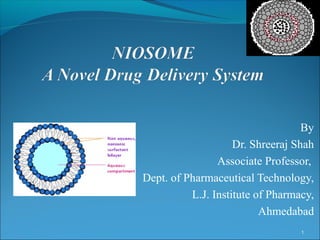
5. Definition
• Niosomes are synthetic microscopic vesicles
consisting of an aqueous core enclosed in a bi
layer consisting of cholesterol and one or more
nonionic surfactants
• Vesicles are prepared from self assembly of
hydrated non ionic surfactants molecules
5
6. General Characteristics of Niosome
•Biocompatible, biodegradable, non-toxic, non immunogenic and
non-carcinogenic
•The ability of nonionic surfactant to form bilayer vesicles is
dependant on the HLB value of the surfactant, the chemical structure
of the components and the critical packing parameter
•Niosomes can be characterized by their size distribution studies
• High resistance to hydrolytic degradation
•The properties of niosome depends both on composition of the
bilayer & on method of their production
6
7. Advantages of Niosomes
Targeted drug delivery can be achieved using niosomes the drug is delivered directly to
the body part where the therapeutic effect is required
Reduced dose is required to achieve the desired effect
Subsequent decrease in the side effects
The therapeutic efficacy of the drugs is improved by reducing the clearance rate, targeting
to the specific site and by protecting the encapsulated drug
Niosomes are amphiphillic i.e. both hydrophilic and lipophillic in nature and can
accommodate a large number of drugs with a wide range of solubilities
Improve the oral bioavailability of poorly soluble drugs
Enhance the skin permeability of drugs when applied topically
provide advantage of usage through various routes viz. oral, parentral, topical, ocular etc.
The bilayers of the niosomes protect the enclosed Active pharmaceutical ingredient from
the various factors present both inside and outside the body
The surfactants used and also the prepared niosomes are biodegradable, biocompatible
and non-immunogenic
They are osmotically active and stable
7
8. Disadvantages of Niosome
•Aqueous suspension of niosome may exhibit fusion, aggregation
leaching or hydrolysis of entrapped drug, thus limiting the shelf life
of niosome dispersion.
• Time consuming
• Requires specialized equipment
• Inefficient drug loading
8
9. Structure of Niosomes
•Niosomes are microscopic lamellar structures
•Basic structural components are
Non ionic surfactant
Cholesterol
Charge inducing molecule
•A number of non-ionic surfactants used are:
polyglycerol alkyl ether, glucosyl dialkyl ethers, crown ethers, ester
linked surfactants, polyoxyethylene alkyl ether and a series of spans and
tweens
9
10. Non-ionic Surfactants
The non-ionic surfactants orient themselves in bilayer lattices
where the polar or hydrophobic heads align facing aqueous
bulk (media) while the hydrophobic head or hydrocarbon
segments align in such a way that the interaction with the
aqueous media would be minimized.
To attain thermodynamic stability, every bilayer folds over
itself as continuous membrane i.e. forms vesicles so that
hydrocarbon/water interface remains no more exposed.
10
11. Mainly following types of non-ionic surfactants are used for the formation of niosomes:
Alkyl Ethers: L’Oreal described some surfactants for the preparation of niosomes
containing drugs/chemicals as
1) Surfactant-I (molecular weight (MW 473)) is C16 monoalkyl glycerol ether with
average of three glycerol units.
2) Surfactant-II (MW 972) is diglycerol ether with average of the seven glycerol units.
3) Surfactant III (MW 393) is ester linked surfactant.
Other than alkyl glycerol, alkyl glycosides and alkyl ethers bearing polyhydroxyl head
groups are also used in formulation of niosomes.
Alkyl Esters: Sorbitan esters are most preferred surfactant used for the preparation of
niosomes amongst this category of surfactants. Vesicles prepared by the polyoxyethylene
sorbitan monolaurate are relatively soluble than other surfactant vesicles. For example
polyoxyethylene (polysorbate 60) has been utilized for encapsulation of diclofenac
sodium. A mixture of polyoxyethylene-10-stearyl ether : glyceryl laurate : cholesterol (27 :
15 : 57) has been used in transdermal delivery of cyclosporine-A.
Alkyl Amides: Alkyl amide (e.g. galactosides and glucosides) have been utilized to
produce niosomal vesicles.
Fatty Acid and Amino Acid Compounds: Long chain fatty acids and amino acid moieties
have also been used in some niosomes preparation. 11
13. Cholesterol
Steroids are important components of the cell membrane and their
presence in membrane affect the bilayer fluidity and permeability.
Cholesterol is a steroid derivative, which is mainly used for the
formulation of niosomes. Although it may not show any role in the
formation of bilayer, its importance in formation of niosomes and
manipulation of layer characteristics can not be discarded.
In general, incorporation of cholesterol affects properties of niosomes
like membrane permeability, rigidity, encapsulation efficiency, ease of
rehydration of freeze dried niosomes and their toxicity.
It prevents the vesicle aggregation by the inclusion of molecules that
stabilize the system against the formation of aggregates by repulsive
steric or electrostatic forces that leads to the transition from the gel to
the liquid phase in niosome systems. As a result of this, the niosome
becomes less leaky in nature. 13
14. Charge inducing molecule
Some charged molecules are added to niosomes to increase stability
of niosomes by electrostatic repulsion which prevents aggregation
and coalescence. The negatively charged molecules used are
diacetyl phosphate (DCP) and phosphotidic acid. Similarly,
stearylamine (STR) and stearyl pyridinium chloride are the well
known positively charged molecules used in niosomal
preparations.
Only 2.5-5 mol % concentration of charged molecules is tolerable
because high concentration can inhibit the niosome formation
14
16. Contrast of Niosomes Vs liposomes
Niosome Liposome
1. Less Expensive 1. More expensive
2. Chemically Stable 2. Chemically unstable
3. Niosomes are prepared 3. liposomes are prepared
from uncharged single- from double-chain
chain surfactant phospholipids
4. They do not require 4. They require special
special storage and storage, handling &
handling purity of natural
phospholipid is variable.
5. Non ionic drugs carriers 5. The ionic drugs carriers
are safer are relatively toxic &
unsuitable
16
17. Similarity of niosomes and liposomes
• However Niosomes are similar to liposomes in functionality.
• Niosomes also increase the bioavailability of the drug and
reduce the clearance like liposomes.
• Niosomes can also be used for targeted drug delivery, similar
to liposomes.
• As with liposomes, the properties of the niosomes depend
both- on the composition of the bilayer, and the method of
production used.
17
18. Method of Preparation
1. Ether Injection (LUV) Based on the vesicle size, niosomes
can be divided into three groups.
2. Hand Shaking Method (MLV) These are small unilamellar vesicles
(SUV, size=0.025-0.05 μm),
3. The “Bubble” Method multilamellar vesicles (MLV,
size=>0.05 μm), and large
4. Reverse Phase Evaporation (LUV) unilamellar vesicles (LUV,
size=>0.10 μm).
5. Sonication (SUV)
6. Multiple membrane extrusion method
7. Trans Membrane pH Gradient Drug Uptake Process (remote
Loading) (MLV)
8. Microfluidization method (SUV)
9. Formation of Niosomes From Proniosomes 18
19. General steps of Niosome preparation
• Hydration of mixture of surfactant/lipid at elevated
temperature
• Sizing of niosomes
• Removal of unentrapped material from vesicles
19
20. Common stages of all methods of preparation of Niosomes
Cholesterol + Non ionic surfactant
Dissolve in organic solvent
Solution in organic solvent
Drying
Thin film
Dispersion (Hydration)
Niosome suspension
20
21. 1. Ether injection method
• This method is based on slow injection of surfactant : cholesterol
solution in ether through 14 gauge needle into a preheated aqueous
phase maintained at 600C
• Vaporization of ether resulting into a formation of ether gradient at
ether-water interface which leads to formation of single layered
vesicles
• Depending upon the conditions used, the diameter of the vesicle
range from 50 to 1000 nm
21
23. 2. Hand shaking method
(Thin film hydration technique)
• Surfactant and cholesterol are dissolved in a volatile organic solvent
(diethyl ether, chloroform or methanol) in a round bottom flask
• The organic solvent is removed under vaccum at room temperature
using rotary evaporator leaving a thin layer of solid mixture
deposited on the wall of the flask
• The dried surfactant film can be rehydrated with aqueous phase at
temperature slightly above the phase transition temperature of the
surfactant used, with gentle agitation
• This process forms large multilamellar niosomes 23
25. 3. The “Bubble” Method
•It is novel technique for the one step preparation of liposomes and
niosomes without the use of organic solvents
•The bubbling unit consists of round-bottom flask with three necks
positioned in water bath to control the temperature
•Water-cooled reflux and thermometer is positioned in the first and
second neck and nitrogen supply through the third neck
• Cholesterol and surfactant are dispersed together in this buffer (pH 7.4)
at 70°C, the dispersion mixed for 15 seconds with high shear
homogenizer and immediately afterwards “bubbled at 70°C using
nitrogen gas
25
26. 4. Reverse Phase Evaporation Technique (REV)
•Cholesterol and surfactant (1:1) are dissolved in a mixture of ether
and chloroform
•An aqueous phase containing drug is added to this and the resulting
two phases are sonicated at 4-5°C. The clear gel formed is further
sonicated after the addition of a small amount of phosphate buffered
saline (PBS)
•The organic phase is removed at 40°C under low pressure. The
resulting viscous niosome suspension is diluted with PBS and heated
on a water bath at 60°C for 10 min to yield niosomes
26
28. 5. Sonication
•In this method, an aliquot of drug solution in buffer is added to the
surfactant/cholesterol mixture in a 10-ml glass vial.
•The mixture is probe sonicated at 60°C for 3 minutes using a sonicator.
•The resultant vesicles are of small unilamellar type niosomes.
6. Multiple membrane extrusion method
• In this method, a mixture of surfactant, cholesterol and diacetyl phosphate is prepared
and then solvent is evaporated using rotary vacuum evaporator to leave a thin film. The
film is then hydrated with aqueous drug solution and the suspension thus obtained is
extruded through the polycarbonate membrane (mean pore size 0.1 mm) and then
placed in series up to eight passages to obtain uniform size niosomes.
•Good method of controlling niosome size. 28
30. 7. Trans membrane pH gradient (inside acidic) Drug
Uptake Process (remote Loading)
•Surfactant and cholesterol are dissolved in chloroform
• The solvent is then evaporated under reduced pressure to get a thin film on the
wall of the round bottom flask
•The film is hydrated with 300 mM citric acid (pH 4.0) by vortex mixing.
•The multilamellar vesicles are frozen and thawed 3 times and later sonicated.
To this niosomal suspension, aqueous solution containing 10 mg/ml of drug
is added and vortexed
• The pH of the sample is then raised to 7.0-7.2 with 1M disodium phosphate
• This mixture is later heated at 60°C for 10 minutes to give niosomes
30
31. 8. Microfluidization Method
In this method two fluidized streams (one containing drug
and the other surfactant) interact at ultra high velocity, in
precisely defined micro channels within the interaction
chamber in such a way that the energy supplied to the
system remains in the area of niosomes formations. This is
called submerged jet principle. It results in better
uniformity, smaller size and reproducibility in the
formulation of niosomes
31
32. 9. Formation of Niosomes from Proniosomes
•Another method of producing niosomes is to coat a water-soluble
carrier such as sorbitol with surfactant
• The result of the coating process is a dry formulation. In which each
water-soluble particle is covered with a thin film of dry surfactant. This
preparation is termed “Proniosomes”
•The niosomes are formed by the addition of aqueous phase at T > T m
and brief agitation
T = Temperature.
Tm = mean phase transition temperature
32
33. Separation of unentrapped drugs
The removal of unentrapped solute from the vesicles can be
accomplished by various techniques, which include:
Gel Filtration
The unentrapped drug is removed by gel filtration of niosomal dispersion through a
Sephadex-G-50 column and elution with phosphate buffered saline or normal saline.
Dialysis
The aqueous niosomal dispersion is dialyzed in a dialysis tubing against phosphate
buffer or normal saline or glucose solution
Centrifugation
The niosomal suspension is centrifuged and the supernatant is separated. The pellet is
washed and then resuspended to obtain a niosomal suspension free from unentrapped
33
drug.
34. Factors affecting the physicochemical properties of
niosomes
Membrane Additives
Stability of niosomes can be increased by the number of
additives into niosomal formulation along with surfactant and
drugs. The membrane stability, morphology and permeability of
vesicles are affected by numbers of additives e.g. addition of
cholesterol in niosomal system increases the rigidity and
decreases the drugs permeability through the membrane
Temperature of Hydration
Shape and size of niosome is also influenced by the hydration
temperature. Assembly of the niosomes vesicles is affected by
the temperature change of niosomal system. Temperature change
can also induce the vesicle shape transformation 34
35. Properties of Drugs
The drug entrapment in niosomes is affected by molecular weight, chemical
structure, hydrophilicity, lipophilicity as well as the hydrophilic lipophilic
balance (HLB) value of the drug. Vesicle size may increase due to entrapment
of drug. Drug particle interact with the surfactant head groups, which may
increase charge on polymer and thus cause repulsion of the surfactant bilayer
which leads to increase in vesicle size.
Amount and Type of Surfactant
As the HLB value of surfactants like span 85 (HLB 1.8) to span 20 (HLB 8.6)
increased, the mean size of niosomes also increases proportionally. It is due to
the fact that surface free energy decreases with increase in hydrophilicity of
surfactant. Alkyl chain is present in well ordered structure in gel state, while
in the liquid state the structure of bilayer is more disordered.
Entrapment efficiency is also affected by phase transition temperature i.e.
span 60 having higher TC, provide better entrapment efficiency. Entrapment
efficiency of the niosomes is affected by the HLB value for e.g. niosomes
have high entrapment efficiency at HLB value 8.6 but HLB value 14 to 17 is
35
not suitable for niosomes formulation
36. Effect of the nature of drug on the formation of niosomes
36
37. Cholesterol Content and charge
Hydrodynamic diameter and entrapment efficiency were found to be increased due to
cholesterol content in the niosomal bilayer. Cholesterol can act by two ways. First, it can
increase the chain order of liquidated bilayer and second, by decreasing the chain order of
the gel state bilayer.
Release rate of drug decreases and rigidity of bilayer increases due to high concentration of
cholesterol
The presence of charge tends to increase the interlamellar distance between successive
bilayers in multilamellar vesicle structure and leads to greater overall entrapped volume
Structure of Surfactants
The geometry of vesicles to be formed from surfactant is affected by its structure, which is
related to critical packing parameter (CPP)
where V= hydrophobic group volume
Ic = the critical hydrophobic group length
a0 = the area of hydrophilic head group
spherical micelles formed if CPP<0.5, inverted micelles is formed if CPP>1. 37
38. Method of Preparation
The average size of acyclovir niosomes prepared by hand-shaking process
was larger (2.7m m) as compared to the average size of niosomes 1.5m m
prepared by ether injection method which may be attributed to the passage of
cholesterol and span-80 solution through an orifice into the drug solution.
Reverse phase evaporation can be used to produce smaller size vesicles.
Vesicles with smaller size and greater stability can be produced by
microfluidization method. Niosomes obtained by transmembrane pH gradient
(inside acidic) drug uptake process showed greater entrapment efficiency and
better retention of drug
Resistance to Osmotic Stress
Diameter of niosomal vesicles was found to be decreased when niosomal
suspension is kept in contact with hypertonic salt solution. There is slow
release with slight swelling of vesicles, which is due to inhibition eluting
fluids from vesicles, followed by faster release, which may be due to decrease
in mechanical strength under osmotic stress
38
39. Types of Niosomes
Proniosomes
Proniosomes are the niosomal formulation containing carrier and
surfactant, which requires to be hydrated before being used. The
hydration results in the formation of aqueous niosome dispersion.
Proniosomes decreases the aggregation, leaking and fusion problem
associated with niosomal formulation
Aspasomes
Combination of acorbyl palmitate, cholesterol and highly charged
lipid diacetyl phosphate leads to the formation of vesicles called
aspasomes. Aspasomes are first hydrated with water/aqueous
solution and then sonicated to obtain the niosomes. Aspasomes can
be used to increase the transdermal permeation of drugs. Aspasomes
have also been used to decrease disorder caused by reactive oxygen
species as it has inherent antioxidant property 39
40. Vesicles in Water and Oil System (v/w/o)
It has been reported that the emulsification of an aqueous
niosomes into an oil phase form vesicle in water in oil emulsion
(v/w/o). This can be prepared by addition of niosomes
suspension formulated from mixture of sorbitol monostearate,
cholesterol and solulan C24 (poly-24-oxyethylene cholesteryl
ether) to oil phase at 60°C. This results in the formation of
vesicle in water in oil (v/w/o) emulsion which by cooling to
room temperature forms vesicle in water in oil gel (v/w/o gel).
The v/w/o gel thus obtained can entrap proteins/proteinous drugs
and also protect it from enzymatic degradation after oral
administration and controlled release. The immunogenic
properties of v/w/o gel and w/o gel reported that both exhibit
immunoadjuvant tendency. In this system aqueous niosomes
(v/w) are emulsified into the oily phase
40
41. Niosomes in Carbopol Gel
Niosomes were prepared using drug, spans and cholesterol. The
niosomes thus obtained were then incorporated in carbopol-934
gel (1% w/w) base containing propylene glycol (10% w/w) and
glycerol (30% w/w). It was observed that the mean flux value
and diffusion co-efficient were 5 to 7 times lower for niosomal
gel as compared to plain drug gels. Moreover, carrageenan
induced paw edema inhibition (i.e. 66.68 ± 5.19%) was higher by
niosome formulation as compared to plain gel.
Niosomes of Hydroxyl Propyl Methyl Cellulose
In this type, a base containing 10% glycerin of hydroxy propyl
methyl cellulose was first prepared and then niosomes were
incorporated in it. The bioavailability and reduction of paw
edema induced by carrageenan was found to be higher by this
niosomal system than the plain formulation of flurbiprofen. 41
49. Therapeutic applications of Niosomes
The applications of niosomes can be mainly classified into
three categories
1) For Controlled Release of Drugs
2) To Improve the Stability and Physical
Properties of the Drugs
3) For Targeting and Retention of Drug in
Blood Circulation
49
50. 1)To Prolong the Release Rate of Drugs
1.1
For Controlled Release
The release rate of drugs like withaferin and gliclazide from the
niosomes was found slower as compared to other dosage forms
1.2 In Ophthalmic Drug Delivery
Experimental results of the water soluble antibiotic gentamicin
sulphate showed a substantial change in the release rate. Beside
this, the percent entrapment efficiency of gentamicin sulphate
was altered when administered as niosomes. Also, as compared
to normal drug solution, niosomes of drug show slow release
Niosomal formulation containing timolol maleate (0.25%)
prepared by chitosan coating exhibited more effect on intra
ocular tension with fewer side effects as compared to the
marketed formulation.
50
51. 2) To Improve the Stability and Physical
Properties of the Drugs
2.1 To Increase Oral Bioavailability
With the formulation of niosomes, the oral bioavailability of the acyclovir as
well as griseofulvin was increased as compared to the drug alone. Similarly,
the absorptivity of poorly absorbed peptide and ergot alkaloid can be
increased by the administration in the bile duct of rats when administered as
micellar solution together with the POE-24-cholesteryl ester
2.2 For Improvement of Stability of Peptide Drugs
8-arginin vasopressin, 9-glycinamide-ω
The in vitro release of insulin from niosomes formulated by span 40 and span
60 in simulated intestinal fluid was lower than the niosomes formulated by
span 20 and span 80.
Niosomes prepared by the span 60 has high resistance against proteolytic
enzyme and exhibit good stability in storage temperature and in presence of
sodium deoxycholate 51
52. 2.3 To Promote Transdermal Delivery of Drugs
Many drugs such as lidocaine, estradiol, cyclosporine etc. are used for topical
and transdermal drug delivery system by formulating them as niosomes
The niosomes of natural compound, ammonium glycyrrhizinate were
formulated for effective anti-inflammatory activity using new non-ionic
surfactant, bola surfactat-span 80-cholesterol (2 : 3 : 1 ratio).
Experimental study showed that the bola niosomes were able to promote the
intracellular uptake of ammonium glycerrhizinic acid
2.4 As a Tool for Improvement of Stability of Immunological Products
Important tool for immunological selectivity, low toxicity and more stability
of the incorporated active moiety
2.5 To Improve Anti-inflammatory Activity
Niosomal formulation of diclofenac sodium prepared with 70% cholesterol
showed greater anti-inflammatory effect as compared to the free drug
Similarly, nimesulide and flurbiprofen showed greater activity than the free
drug
52
53. 3.For Targeting and Retention of Drug in
Blood Circulation
3.1 For Increased Uptake by A431 Cells [a model cell line (epidermoid
carcinoma) used in biomedical research]
Chitosan based vesicles incorporating transferrin and glucose as ligand have been reported.
These vesicles bind CoA (co-A) to their surface. Chitosan containing vesicles are then taken
up by A431 cells and the uptake was found to be enhanced by transferrin.
3.2 For Liver Targeting
Methotrexate was reported to be selectively taken up by liver cells after administration as a
niosomal drug delivery system.
3.3 To Improve the Efficacy of Drugs in Cancer Therapy
Niosomes can also be used as a suitable delivery system for the administration of drugs like
5-FU.
Niosomes of doxorubicin prepared from C16 monoalkyl glycerol ether with or without
cholesterol, exhibited an increased level of doxorubicin in tumor cells, serum and lungs, but
not in liver and spleen
Niosomal preparation of methotrexate exhibited greater antitumor activity as compared to
plain drug solution
53
54. 3.4 In Treatment of Localized Psoriasis
In the treatment of localized psoriasis, niosomes of methotrexate taking
chitosan as polymer have shown promising results
3.5 In Leishmaniasis
The leishmaniasis parasite mainly infects liver and spleen cells. The commonly used
drugs, antimonials, may damage the body organ like heart, liver, kidney etc. The
efficiency of sodium stibogluconate has been found to be enhanced by incorporation in
niosomes. The additive effect was observed for two doses given on successive days.
Moreover, the distribution of antimony in mice showed the higher level of antimony in
liver after its intravenous (i.v.) administration via niosomes drug formulation.
3.6 In Diagnostic Imaging
It has been studied that niosomes can also act as a carrier radiopharmaceuticals and
showed site specificity for spleen and liver for their imaging studies using 99mTc labelled
DTPA (diethylene triamine pentaacetic acid) containing niosomes.
Conjugated niosomal formulations of gadobenate with (N-palmitoyl-glucosamine,
NPG), PEG 4400 and both PEG and NPG can be used to increased tumor targeting of a
paramagnetic agent
54
55. 3.7 Carrier for Haemoglobin
Niosomes play an important role as a carrier for haemoglobin. The
niosomal haemoglobin suspension was found to give superimposable
curve on free haemoglobin curve
Usefulness of Niosomes in Cosmetics
Niosomes of N-acetyl glucosamine are prepared due to its potential in the delivery of
hydrophilic and hydrophobic drugs in topical form and improved penetration into the
skin. Prepared formulations improved the extent of drug localized in the skin, as
needed in hyperpigmentation disorders.
Elastic niosomes showed increased permeation through the skin which will be
beneficial for topical anti-aging application.
suitable for skin moisturising and tanning products
Niosomes were prepared as possible approach to improve the low skin penetration and
bioavailability shown by conventional topical vehicle for minoxidil.
Niosomes with added solubilizers enhanced the permeation of ellagic acid (a potent
antioxidant phytochemical substance which has limited use due to poor
biopharmaceutical properties, low solubility and low permeability) into the skin with
55
increased efficacy of ellagic acid
59. Marketed products
Lancome has come out with a variety of anti-ageing
products which are based on niosome formulations.
L’Oreal is also conducting research on anti-ageing
cosmetic products. A picture of the Lancome anti-ageing
formulation is below
LANCOME FOUNDATION CREAM NIOSOME+
59
60. Future Prospects
• Niosomes represent a promising drug delivery
module.
• There is lot of scope to encapsulate toxic anti-cancer drugs, anti
infective drugs, anti AIDS drugs, anti-inflammatory drugs, anti
viral drugs, etc. in niosomes and to use them as promising drug
carriers to achieve better bioavailability and targeting properties
and for reducing the toxicity and side effects of the drugs.
• The ionic drugs carriers are relatively toxic and unsuitable
whereas niosomal carriers are safer.
60
61. • In addition, handling and storage of niosomes require no special
conditions.
• Vesicular drug carriers like niosomes can be transported by
microphages which are known to infiltrate tumour cells.
• It may be possible to take advantage of these activated macrophage
system in delivering the anti tumour agents within vesicles more
quantitatively to tumour sites.
61
62. Conclusion
In nutshell, as a drug delivery device, compared to liposomes,
niosomes are osmotically active and are quite stable chemically by
their own as well as improve the stability of the drug so entrapped
and delivered. They do not require special conditions for handling,
protection or storage and industrial manufacturing. Beside this,
they offer flexibility in structural characteristics (composition,
fluidity, size), and can be designed as desired.
Niosomes offer various advantages over other drug delivery
devices and have found applicability in pharmaceutical field.
It was thus concluded that niosomes are very effective drug
delivery tools for incorporation/targeting of various therapeutically
active moieties and the onus lies on future scientists to effectively
harness its potential in diverse application areas for the benefit of
mankind. 62
63. References
1. Sanjay K. Jain and N.K. Jain Controlled and novel drug delivery
system
2. Dr. Rakesh S. Patel niosomes as a unique drug delivery system,
www.pharmainfo.net
3. Mithal, B. M., A text book of pharmaceutical formulation, 6 th
Edn., vallabh prakashan, 6, 306-307
4. International journal of pharmaceutical Science and
Nanotechnology volume 1,issue 1, April-June 2008
5. Shailendra Kumar Singh et.al, Niosomes: A Controlled and
Novel Drug Delivery System, Biol. Pharm. Bull. 34(7) 945—953
(July, 2011) 63