Skin cancers
•Download as PPTX, PDF•
91 likes•59,416 views

Skin cancer is the most commonly diagnosed cancer. The three main types are basal cell carcinoma (BCC), squamous cell carcinoma (SCC), and malignant melanoma. BCC is the most common type and usually appears as a waxy nodule, while SCC can metastasize if not treated early. Malignant melanoma is the most lethal type and risk factors include ultraviolet light exposure and family history. Early detection of skin lesions is important, and treatment may involve surgical excision, Mohs surgery, radiation, or chemotherapy depending on the cancer type and stage. Education about skin cancer signs and protecting skin from the sun are also important.

Recommended
More Related Content
What's hot
What's hot (20)
Viewers also liked
Viewers also liked (20)
Similar to Skin cancers
Similar to Skin cancers (20)
More from Siva Nanda Reddy
More from Siva Nanda Reddy (20)
Recently uploaded
Recently uploaded (20)
Skin cancers
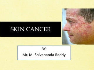
- 1. SKIN CANCER BY: Mr. M. Shivananda Reddy
- 2. • Skin cancer is the most commonly diagnosed cancer. • Skin cancers are either non-melanoma or melanoma. • A persistent skin lesion that does not heal is highly suspicious for malignancy and should be examined by a health care provider. • Early detection and treatment can often lead to a highly favourable prognosis. • The visibility of the skin lesions increases the likelihood of early detection and diagnosis
- 3. Risk Factors for Skin Cancer • Damage in the ozone layer • Fair-skinned and fair-haired people due to insufficient skin pigmentation • Prolonged exposure to sun light. • Exposure to chemical pollutants(arsenic, nitrates, coal, tar and pitch, oils and paraffins) • History of x-ray therapy for acne or benign lesions • Scars from severe burns • Chronic skin irritations • Immunosuppression • Genetic factors
- 4. Types of skin cancers: The most common types of skin cancer are: • Basal Cell Carcinoma (BCC) • Squamous cell (epidermoid) carcinoma (SCC). • Malignant melanoma
- 5. Basal Cell Carcinoma (BCC) • BCC is the most common type of skin cancer. • It generally appears on sun-exposed areas of the body and is more prevalent in regions where the population is subjected to intense and extensive exposure to the sun. • The incidence is proportional to the age of the patient and the total amount of sun exposure, and it is inversely proportional to the amount of melanin in the skin.
- 6. • Basal cell carcinoma (BCC) is a locally invasive malignancy arising from epidermal basal cells. It is the most common type.
- 7. Clinical manifestations: • BCC usually begins as a small, waxy nodule with rolled, translucent, pearly borders; telangiectatic vessels may be present. • As it grows, it undergoes central ulceration and sometimes crusting. • The tumors appear most frequently on the face. • BCC is characterized by invasion and erosion of adjoining tissues. • It rarely metastasizes, but recurrence is common.
- 8. BCC
- 9. Squamous Cell Carcinoma (SCC) • Squamous cell carcinoma (SCC) is a malignant neoplasm of keratinizing epidermal cells. • It frequently occurs on sun-exposed skin or at the base of skin lesion. • SCC is less common than BCC. • SCC can be highly aggressive, has the potential to metastasize, and may lead to death if not treated early and correctly.
- 10. Clinical manifestations: • The lesions may be primary, arising on the skin and mucous membranes, or they may develop from a precancerous condition, such as scarred or ulcerated lesions. • SCC appears as a rough, thickened, scaly tumor that may be asymptomatic or may involve bleeding. • The border of an SCC lesion may be wider, more infiltrated, and more inflammatory than that of a BCC lesion. • Exposed areas, especially of the upper extremities and of the face, lower lip, ears, nose, and forehead, are common sites
- 12. Management of BCC & SCC The treatment method depends on the tumor location; the cell type, location, and depth; the cosmetic desires of the patient; the history of previous treatment; whether the tumor is invasive, and whether metastatic nodes are present. The management of BCC and SCC includes • Surgical excision • Mohs’ micrographic surgery • Electrosurgery • Cryosurgery • Radiation therapy
- 13. Surgical excision • The primary goal is to remove the tumor entirely. • When the tumor is large, reconstructive surgery with use of a skin flap or skin grafting may be required. • A pressure dressing applied over the wound provides support.
- 14. Mohs’ Micrographic Surgery.: • The procedure removes the tumor layer by layer. • The first layer excised includes all evident tumor and a small margin of normal-appearing tissue. • The specimen is frozen and analyzed by section to determine if all the tumor has been removed. • If not, additional layers of tissue are shaved and examined until all tissue margins are tumor free. • In this manner, only the tumor and a safe, normal-tissue margin are removed.
- 15. Electro surgery • Electro surgery is the destruction or removal of tissue by electrical energy. • The current is converted to heat, which then passes to the tissue from an electrode.
- 16. Cryosurgery • Cryosurgery destroys the tumor by deep freezing the tissue. • A thermocouple needle apparatus is inserted into the skin, and liquid nitrogen is directed to the center of the tumor until the tumor base is −40°C to −60°C.
- 17. Radiation Therapy • Expose the tumor surface to ionizing and non-ionizing radiation to kill the cancerous cells
- 19. Malignant Melanoma • A malignant melanoma is a cancerous neoplasm in which abnormal melanocytes are present in the epidermis and the dermis (and sometimes the subcutaneous cells). • Most lethal of all the skin cancers • Most melanomas arise from cutaneous epidermal melanocytes
- 20. RISK FACTORS: • The cause of malignant melanoma is unknown • Ultraviolet rays are strongly suspected, based on indirect evidence such as the increased incidence of melanoma in countries near the equator • Family history
- 21. Clinical Manifestations • Superficial spreading melanoma occurs anywhere on the body. • It usually affects middle-aged people and occurs most frequently on the trunk and lower extremities. • The lesion tends to be circular, with irregular outer portions. • The margins of the lesion may be flat or elevated and palpable.
- 22. • The malignant melanoma may appear in a combination of colors, with hues of tan, brown, and black mixed with gray, blue- black, or white. • Sometimes a dullpink rose color can be seen in a small area within the lesion.
- 24. Management • Surgical excision is the treatment of choice for small, superficial lesions. • Deeper lesions require wide local excision, after which skin grafting may be needed. • Regional lymph node dissection is commonly performed to rule out metastasis.
- 25. • Immunotherapy: • Monoclonal antibodies directed at melanoma antigens. • Autologous immunization against specific tumor cells. • Chemotherapy may be used for metastatic melanoma
- 26. Nursing diagnoses • Acute pain related to surgical excision and grafting • Anxiety and depression related to possible life- threatening consequences of melanoma and disfigurement • Deficient knowledge about early signs of melanoma • Risk for infection related to break in the skin barrier
- 27. Other Malignancies of the Skin • Kaposi’s sarcoma • Basal and Squamous cell carcinomas in the immunocompromised population
- 28. KAPOSI’S SARCOMA • Kaposi’s sarcoma (KS) has received renewed attention since its association with HIV infection and AIDS. • Most patients have nodules or plaques on the lower extremities that rarely metastasize beyond the lower extremities. • This KS is chronic, relatively benign, and rarely fatal.
- 29. Basal and Squamous cell carcinomas in immunocompromised • The incidence of basal cell carcinoma and squamous cell carcinoma is increased in all immuno-compromised individuals, including those infected with HIV. • Clinically, the tumors have the same appearance as in non–HIV-infected people; however, in HIV patients, the tumors may grow more rapidly and recur more frequently.