

Recommended
More Related Content
What's hot
What's hot (20)
Viewers also liked
Viewers also liked (20)
Similar to Sarcoma of oral cavity
Similar to Sarcoma of oral cavity (20)
More from Sk Aziz Ikbal
More from Sk Aziz Ikbal (20)
Recently uploaded
Recently uploaded (20)
Sarcoma of oral cavity
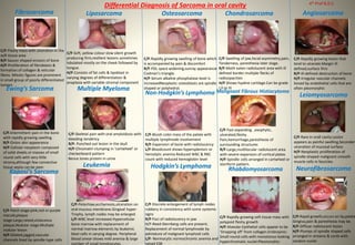
- 1. Fibrosarcoma C/F-Fleshy mass with ulceration in the soft tissue area R/F-Saucer shaped erosion of bone H/F-Proliferation of fibroblasts & formation of collagen & reticular fibres. Mitotic figures are prominent in small group of poorly differentiated tumors Liposarcoma C/F-Soft, yellow colour slow silent growth producing firm,resilient lesions sometimes lobulated mostly on the cheek followed by tongue. H/F-Consists of fat cells & lipoblast in varying degrees of differentiation & anaplasia with variable stromal component Osteosarcoma C/F-Rapidly growing swelling of bone which is accompanied by pain & discomfort R/F-PDL space widening,sunray appearance Codman’s triangle. H/F-Serum alkaline phosphatase level is increasedNeoplastic osteoblasts are spindle shaped or polyhedral.Ewing’s Sarcoma C/F-Intermittent pain in the bone with rapidly growing swelling R/F-Onion skin appearance H/F-Cellular neoplasm composed of solid sheets or masses of small round cells with very little stroma,although few connective tissue septa can be seen Multiple Myeloma C/F-Skeletal pain with oral amyloidosis with bleeding tendency R/F- Punched out lesion in the skull H/F-Chromatin clumping in ‘cartwheel’ or checkerboard pattern Bence Jones protein in urine Non-Hodgkin’s Lymphoma C/F-Bluish color mass of the palate with multiple lymphnode involvement R/F-Expansion of bone with radiolucency L/F-Bloodcount shows hypersplenism or hemolytic anemia.Reduced WBC & RBC count with reduced hemoglobin level Kaposi’s Sarcoma C/F-Patch stage:pink,red or purple macule;plaque stage:Large,raised,violaceous plaque;Nodular stage:Multiple nodular lesion H/F-Dilated,jagged,vascular channels lined by spindle-type cells Leukemia C/F-Petechiae,ecchymosis,ulceration on oral mucous membrane.Gingival hyper- Trophy, lymph nodes may be enlarged. L/F-WBC level increased.Hypercellular bone marrow with replacement of normal marrow elements by leukemic blast cells in varying degree. Peripheral blood smear shows mild anemia & large number of small lymphocytes. Hodgkin’s Lymphoma C/F-Discrete enlargement of lymph nodes rubbery in consistency with some systemic signs R/F-Foci of radiolucency in jaw H/F-Reed-Sternberg cells are present; Replacement of normal lymphnode by admixture of malignant lymphoid cells L/F- Normocytic normochromic anemia and raised ESR Chondrosarcoma Malignant Fibrous Histiocytoma Rhabdomyosarcoma Angiosarcoma Leiomyosarcoma Neurofibrosarcoma C/F-Rare in orall cavity.Lesion appears as painful swelling.Secondery ulceration of mucosal surface. H/F-Neoplastic proliferation of spindle-shaped malignant smooth muscle cells in fascicles. C/F-Rapidly growing soft tissue mass with polypoid fleshy growth H/F-Alveolar:Epithelial cells appear to be ‘Dropping off’ from collagen.Embroyinic: Small round cells with monotonus looking hyperchromatic nuclei:Pleomorphic: C/F-Swelling of jaw,facial asymmetry,pain, Tenderness, paresthesia later stage. R/F-Moth eaten radiolucent area with ill defined border.multiple flecks of radioopacities H/F-Shows hyaline cartilage.Can be grade I,II or III C/F-Fast expanding ,exophytic, ulcerated,fleshy. Pain,hemorrhage,paresthesia of surrounding structures R/F-Large,multilocular radiolucent area with severe expansion of cortical plates H/F-Spindle cells arranged in cartwheel or storiform pattern. Differential Diagnosis of Sarcoma in oral cavity C/F-Rapidly growing lesion that tend to ulcerate.Margin ill- defined,surface firm R/F-Ill-defined destruction of bone H/F-Irregular vascular channels lioned by endothelial cells that are often pleomorphic C/F-Rapid growth,occurs on lip,palate Gingiva,pain & paresthesia may be. R/F-Diffuse radiolucent lesion H/F-Plumps of spindle shaped cells arranged in streams & cords with random nuclei 4th Prof B.D.S