
Recommended
More Related Content
Similar to Middle Ear Dysfunction in Chronic Rhinosinusitis Patients
Similar to Middle Ear Dysfunction in Chronic Rhinosinusitis Patients (20)
Recently uploaded
Recently uploaded (20)
Middle Ear Dysfunction in Chronic Rhinosinusitis Patients
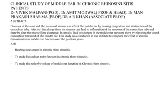
- 1. CLINICAL STUDY OF MIDDLE EAR IN CHRONIC RHINOSINUSITIS PATIENTS Dr VIVEK MALPANI(PG 3) , Dr AMIT MODWAL( PROF & HEAD), Dr MAN PRAKASH SHARMA (PROF),DR A R KHAN (ASSOCIATE PROF) ABSTRACT Diseases of the nose and the paranasal sinuses can affect the middle ear by causing congestion and obstruction of the eustachian tube. Infected discaharge from the sinuses can lead to inflamation of the mucosa of the eustachian tube and there by alter the mucociliary clearance. It can also lead to changes in the middle ear pressure there by elevating the sound conduction threshold of the middle ear. This study was conducted in our institute to compare the effect of chronic rhinosinusitis in middle ear function over the past two years. AIM - Hearing assessment in chronic rhino sinusitis. - To study Eustachian tube function in chronic rhino sinusitis. - To study the path physiology of middle ear function in Chronic rhino sinusitis.
- 2. abstract • Introduction oChronic rhinosinusitis is defined by the presence of at least two out of four cardinal symptoms (i.e., facial pain/pressure, hyposmia/anosmia, nasal drainage, and nasal obstruction) for at least 12 consecutive weeks, in addition to objective evidence. Objective evidence of chronic rhinosinusitis may be obtained on physical examination (anterior rhinoscopy, endoscopy) or radiography, preferably from sinus computed tomography1 oChronic rhinosinusitis (CRS) is phenotypically divided into those cases with nasal polyps (CRSwNP) and those without nasal polyps(CRSNPs) which is based on endoscopic findings.
- 3. oPathological changes which occur in the nasal and sinus mucosa alter the nature of the nasal secretions and also the mucosa of the nasal secretory routes. These infected secretions can cause congestion and obstruction of the eustachian tube orifice by inflammation of the lymphoreticular tissue there by slowing down the mucociliary clearance and may lead to impeded ventilation and/or ascending infection of the middle ear. oMesserlinger was able to demonstrate that there are two major routes for secretions from the paranasal sinuses. The mucus streams from out of the frontal, maxillary and anterior ethmoidal sinuses pass through the frontal recess and the ethmoidal infundibulum.
- 4. oThis mucus is then transported over the posterior free margin of the uncinate process onto the medial surface of the inferior turbinate. oThis stream normally passes anterior and inferior to the tubal orifice. This secretion route is then also joined by the mucus coming from septal mucosa. oThe second route for secretions combines the mucus from the posterior ethmoidal cells and the sphenoid sinuses. These are drained posterior and superior to the tubal orifice. The secretions then pass along the lateral pharyngeal gutter and pyriform fossa. oIn sinusitis the quality and quantity of mucus is altered to either mucopurulent or purulent. Secretions pass over the pharyngeal end of ET and it can lead to inflammation of ET, hypertrophy of lymphoid tissue collection (tubal tonsil hypertrophy). This results in obstruction of ET leading to various middle ear pathologies.
- 5. oDue to the ET dysfunction, negative pressure increases in the middle ear leading to the retraction of the tympanic membrane. o There is host response and liberation of inflammatory mediators like IgA, lysozyme, interleukins and cytokines which increases the vascular permiability and glandular secretions which further causes Inflammation of the middle ear epithelium and there is production of serous or mucosal effusion resulting in OME. oIn acute Eustachian tube blockage, there is collection of transudate and later exudates leading to the acute otitis media (AOM)and even haemorrhage in the middle ear. oIn prolonged Eustachian tube blockage their will be otitis media with effusion( OME), atelectatic ear, retraction pockets, which may lead to erosion of incudostapedial joint,cholesteotoma formation and CSOM.
- 6. Methods and materials • This is a prospective study carried out at All the patients of age group 11 to 60 yrs coming to OPD of the Department of Otorhinolaryngology, National Institute of Medical Sciences & Research, Jaipur; having normal mental & physical status and are willing to participate in the study.
- 7. • Result • Out of 113 patients ,majority(55%) of patients had disease confined to the anterior osteomeatal complex. • The major complaint relating to eustachian tube pathology was fullness of ears about 35%. • In tympanometry 62.5% patient showed Type A curve, while 35% displayed a Type C curve indicative of high negative middle ear pressure, probably eustachian tube dysfunction. • Pure tone audiometry showed normal hearing in 95% cases.
- 8. • Conclusion From this study we are able to conclude that damage to the middle ear becomes more serious with the extend of disease and longer course of the disease in the paranasal sinuses. Though hearing was unaffected, a significantly high (65%) number had elevated air conduction thresholds. Sinusitis involving the posterior group of sinuses were more likely to cause middle ear dysfunction. Also sclerotic mastoids are more likely to adjust poorly to negative pressures.
- 9. • Thanks …