prosthetic joint infection(diagnosis and management ).pptx
•Download as PPTX, PDF•
0 likes•10 views

give details and recent status of prosthetic joint infection, diagnosis , investigation markers ,and management

Recommended
More Related Content
Similar to prosthetic joint infection(diagnosis and management ).pptx
Similar to prosthetic joint infection(diagnosis and management ).pptx (20)
Recently uploaded
Recently uploaded (20)
prosthetic joint infection(diagnosis and management ).pptx
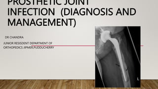
- 1. PROSTHETIC JOINT INFECTION (DIAGNOSIS AND MANAGEMENT) DR CHANDRA JUNIOR RESISDENT DEPARTMENT OF ORTHOPEDICS JIPMER,PUDDUCHERRY
- 2. OBJECTIVES What is prosthetic joint infection(PJI)? Burden of disease How is PJI different from other infections(eg.. FR,ODRI ,OIRI ) Diagnosis Treatment Recent advances Prevention
- 3. WHAT IS PROSTHETIC JOINT INFECTION(PJI)? • Periprosthetic joint infection (PJI) is a unique clinical entity, markedly different from infections involving native bones or joints. • Prosthetic joint infection (PJI), also referred to as periprosthetic infection, is infection involving the joint prosthesis and adjacent tissue • PJI is characterized by a complex interplay between microbes, predominantly bacteria but occasionally fungi, and the host immune response. • Only a minimal microbial burden is required to initiate a PJI.
- 4. • etiologic organisms can adhere to the surfaces of arthroplasty components and form biofilms. • Biofilms notoriously exhibit a marked resistance to a wide array of antimicrobial agents and are adept at avoiding innate immune defenses. • It may occur in the period after the joint replacement or many years late
- 5. • There is no uniformly accepted definition for PJI. • several groups, including the Infectious Diseases Society of America (IDSA) and the Musculoskeletal Infection Society (MSIS), have published proposed or accepted definitions for the diagnosis of PJI . • Although the definitions vary, a recent study showed a high concordance between the IDSA and MSIS definitions of PJI . • Additionally, the European Bone and Joint Infection Society (EBJIS), the American MSIS, and a number of other organizations from around the world recently held an international consensus meeting in an attempt to refine an international consensus definition of PJI .
- 6. MSIS : Definitive evidence of PJI is a sinus tract in communication with the prosthesis or an identical pathogen found in two separate periprosthetic tissue or fluid samples. The presence of four (or more) of six minor criteria can also fulfill a diagnosis of PJI. IDSA : The presence of the sinus tract and two or more sterile site cultures with identical microorganisms to be definitive evidence of PJI. International Consensus Meeting definition of PJI 2018 brought out Diagnosis criteria to overcome short coming of these definitions.
- 7. BURDEN OF DISEASE • Prosthetic joint infection is a difficult complications affecting replacement surgery . • It is painful ,disabling ,often requiring removal of prosthesis . • After the introduction of modern hip arthroplasty ,septic complication threatened the continued viability of procedure . • Charnley reported in 6.8% of 1st 683 procedure. • Experience of Wilson et.al in united states was even more ominous with 11% of 100 arthroplasties becoming infected. • About 1.4-2.5% of all joint replacements worldwide are complicated by PJIs.
- 8. • Currently approximately 1-2% of hip arthroplasty,2-3% of TKR and 4-5 % after revision THR and 5-6% after revision TKR • Infection after Total ankle replacement appears to be relatively infrequent Rate of superficial infection ranges 0-15% ( average 8%) Rate of deeo infection 0-5%( average 1%) • Infection rate after shoulder replacement is low (less than 1%) • Infection rate after elbow arthroplasty is 7-8%.
- 9. Advancesinunderstandingof patientselection, operatingroomenvironment, surgicaltechniqueand useof prophylacticantibioticshavedramatically reducedtheriskofthisdevastingcomplication
- 10. HOW IS PJI DIFFERENT FROM OTHER INFECTIONS(EG.. FR,ODRI ,OIRI ) Common orthopedics Infections and abbreviations : • SSI : surgical site infections • FRI : fracture-related infections • BAI : biomaterial associated infections (or) IAI : implant-associated infections also known ODRI : orthopedic device related infection OIRI : orthopedic implant related infection • PJI : periprosthetic joint infections
- 11. • Although the rate of PJI is low compare to other orthopedic infections , it has dramatic effects on the patients’ health. • Its involved articular surfaces /joints those are inevitable for locomotion • Diagnosis and treatment of PJI challenging • It is difficult to eradicate resulting in a severe complication with a significant patient and socioeconomic burden. • Very close and regular clinical follow ups are crucial to detect and management .
- 12. • Most other bone related infection implant removal and adequate antibiotic therapy may complete the treatment • But prosthesis removal and antibiotic therapy is not a solution for PJI ,Need reintervention . • Biofilm formation more common in PJI compare to other bone related infection. • Biofilm formation on prosthetic surface make eradication of infection more difficult compared to other infections.
- 13. BIOFILM • The most important factor influencing periprosthetic infection treatment • All bacteria make biofilm. • Biofilm consists of approximately 15% cells and 85% polysaccharide matrix and forms on ( All foreign materials ,Devitalized tissues ,Soft tissue and bone) • Biofilm, once established, matures into sophisticated microenvironment. Biofilms typically mature over 4 weeks • Bacteria communicate via signaling molecules and nanowires * Nanowires are very small cell to-cell connections that allow bacteria in a biofilm to communicate with one another
- 14. CLINICAL IMPORTANCE OF BIOFILM STATE • Bacteria become 1000 to 10,000 times more resistant to antibiotics • Essentially, bacteria within a biofilm state cannot be killed with standard dosing regimens of antibiotics • In vivo, biofilm can colonize, grow, and cover a surface within 4–8 days • Effective treatment for established biofilm infection requires: Removal of implants and foreign bodies Removal of all devitalized bone and soft tissue Inadequate debridement of biofilm material is the reason for treatment failure and infection recurrence This includes retained cement and metal left in the medullary canals adjacent to the affected prosthetic joint
- 16. DIAGNOSIS
- 17. CLINICAL HISTORY AND EXAMINATION History : • The clinical manifestations of PJI vary depending upon the virulence of the organism, the mode of initiation of infection, the host immune response, the soft tissue structure surrounding the joint, and the joint involved. • Commonly reported signs or symptoms of PJI include pain(70-90%), joint swelling or effusion, erythema or warmth around the joint, fever, drainage, or the presence of a sinus tract (72%)communicating with the arthroplasty Physical examination : • sinus tract to the joint is a definite infection • warmth, redness, or swelling • low grade fever • Motion limited by pain and swelling
- 18. IMAGING Radiographs : Imaging may support the diagnosis of PJI in certain circumstances but rarely has a definitive role in PJI diagnosis. Plain radiographs are typically obtained in patients undergoing evaluation for possible PJI. They may help identify noninfectious causes for the presenting symptoms, including periprosthetic fracture, fracture of the arthroplasty material, or dislocation .Detection of periprosthetic lucency, loosening of the prosthesis components, effusion, adjacent soft tissue gas or fluid collection, or periosteal new bone formation may suggest infection but is neither sensitive nor specific
- 19. CT scan Computed tomography (CT) and magnetic resonance imaging have the advantages of high spatial resolution and allow evaluation of signs of infection in the periprosthetic tissues. no difference in the evaluation of the bony structures compared to the use of plain radiographs. Furthermore, the use of these techniques is limited by imaging artifacts due to the presence of the metal prosthesis MRI magnetic resonance imaging can be performed only with certain metals, such as titanium or tantalum
- 20. Bone scan Tc-99m (technetium) detects inflammation and In-111 (indium) detects leukocytes triple scan can differentiate infection from fracture or bone remodeling indications if infection is suspected, but cannot be confirmed by aspiration or blood work up 99% sensitivity and 30% to 40% specificity Positron emission tomography (PET) may help to identify areas of high metabolic activity using fluorinated glucose 98% sensitivity and 98% specificity
- 21. LABORATORY • WBC(not specific or sensitive) • CRP( > 3omm/h ),ESR ( 10mg/l) D-dimer (> 850) • Joint aspiration : > One of 3 is elevated with suspicion of infection whenever there is a strong suspicion in order to confirm the diagnosis even of negative blood markers ) Should not undertaken until at least 2weeks after discontinuation of antibiotic (cell count and differential, crystals , gram stain , cultures and specificity ) synovial WBC >10,000 cells/ccl in the first 6 weeks after TKA suggestive of infection WBC >3,000 cells/cc and PMN >80% for hips WBC >1166 cells /cc and PMN >64% for hip antibiotic spacers
- 22. Synovial fluid leukocyte esterase: ( 81% sensitivity and 77% specicity) Leukocyte esterase is an enzyme present in neutrophils. A colorimetric strip measuring leukocyte esterase is widely available as a point-of-care test to determine pyuria for the diagnosis of urinary tract infection. This test strip has recently been proposed as a point-of-care test for synovial fluid from either preoperative or operative aspirates. Gram stain (specificity > sensitivity) positive test would be indicative of infection, however a negative test does not rule out infection repeat aspiration(indicated in cases of inconclusive aspirate and peripheral lab data ) waiting two weeks for a repeat aspiration off antibiotics
- 23. • Preoperative periprosthetic tissue biopsy: sensitivity 73% andspecificity 95% Testing of periprosthetic tissue is one of the most valuable components in the routine microbiological diagnosis of PJI. Samples of periprosthetic tissue are most often obtained at the time of revision surgery but preoperative arthroscopic tissue biopsy may alternatively or additionally be performed cultures obtained by using swabs : • Cultures obtained by using swabs have a limited role in the microbiological detection of PJI. • While the presence of a sinus tract is considered definitive evidence of PJI swab culture of the drainage from the sinus tract is neither sensitive nor specific for the microbiological detection of PJI
- 24. CRITERIA INTERNATIONAL CONSENSUS MEETING (ICM) ON MUSCULOSKELETAL INFECTION :98% SENSITIVITY AND 99.5% SPECIFICITY • .
- 25. CLASSIFIACTION ACUTE INFECTION CHRONIC INFECTION <3WEEKS Considered a non-biofilm state Implants are salvageable Treatment is surgical => 3WEEKS Biofilm state Implants are not salvageable Treatment is surgical and implants are removed
- 26. TSUKAYAMA CLASSIFICATION 1) early postoperative infection : onset within 1st month of surgery 2) Late chronic infection : onset more than 1month of surgery ,insidious onset of symptoms 3) Acute hematogenous infection : onset more than 1month of surgery , acute onset of symptoms in previously well function prosthesis , distant source of infection 4) Positive intraoperative cultures : positive culture obtained at the time of revision for supposedly aseptic conditions.
- 27. TRAMPUZ AND ZIMMERLI CLASSIFICATION Early infection : <3 month Delayed infection : 3-24 month Late infection : > 24 month
- 28. TREATMENT • ACUTE INFECTION : Debridement, Antibiotics and Implant Retention (DAIR) 31 to 82% of success rate Radical debridement surgery is performed, including synovectomy and lavage Modular bearings are exchanged All prosthetic spaces must be accessed and flushed/debrided of bacterial load Intravenous antibiotic therapy used for 4–6 weeks Arthroscopic lavage of an acutely infected joint replacement is not acceptable treatment. The success rate with the DAIR procedure for S. aureus appears to be lower than that for other organisms If infection recurs, treated as a chronic infection. A second debridement surgery attempt should not be made
- 29. CHRONIC INFECTION a)Implant removal : Includes all implant cement (PMMA), nonabsorbable suture material, screws, cables, and metallic fragments b) Radical debridement, including synovectomy, removal of necrotic bone and all devitalized soft tissue, and copious lavage c)Stabilization of joint with methylmethacrylate spacer loaded with high-dose antibiotics d) Intravenous antibiotic therapy for 4–6 weeks e) Second-stage reconstruction
- 31. WOUND COVERAGE MEDIAL GASTROCNEMIUS ROTATIONAL FLAP : The workhorse for deficiencies about the knee Blood supply—medial sural artery Used to cover medial and anterior deficiencies Good excursion LATERAL GASTROCNEMIUS ROTATIONAL FLAP: Blood supply—lateral sural artery Used to cover lateral soft tissue deficiencies Little excursion Risk—peroneal nerve palsy from traction of the flap as it is pulled anteriorly to lateral side of knee
- 32. RECENT ADVANCES
- 33. ONE-STAGE REPLACEMENT ARTHROPLASTY • Indications used more commonly in Europe for infected THA no sinus tract, healthy patient and soft tissue, no prolonged antibiotic use, no bone graft low-virulence organism with good antibiotic sensitivity Technique(use antibiotic-impregnated cement) Advantages • lower cost and convenience with single procedure • earlier mobility Disadvantages • higher risk of continued infection from residual microorganisms • Outcomes : variable success of 75-100%
- 34. TWO-STAGE ARTHROPLASTY EXCHANGE • Success rate 87 to 100% • Two-stage arthroplasty exchange, also referred to as a staged exchange, is considered to be the most definitive strategy in terms of infection eradication and preservation of joint function. • This strategy involves at least two surgeries. In the first surgery, cultures are obtained, all infected tissue is debrided, and the components and PMMA are removed. • An antimicrobial-impregnated PMMA spacer is typically implanted into the joint space prior to closure to deliver local antimicrobial therapy and maintain limb length. • Pathogen-directed antimicrobial therapy is usually given intravenously for 4 to 6 weeks following the first stage. This is then followed by at least a 2- to 6-week antibiotic-free time period.
- 35. ANTIMICROBIAL-LOADED PMMA SPACERS Antimicrobial-loaded PMMA spacers serve two functions in a two-stage arthroplasty exchange. First, both articulating and static spacers provide mechanical support during the time in which the arthroplasty is removed. This preserves proper joint position, prevents muscle contractures, and enhances patient comfort between the first and second stages. The second function of antimicrobial-loaded PMMA spacers is to provide local antimicrobial therapy to augment systemic therapy during the time between the first and second stages. Types static spacer used if joint stability is compromised by soft tissue and/or bone loss Articulated spacer preferred if joint stability preserved. ( Because of better preservation of joint function)
- 37. • ANTIBIOTIC LOADED ACRYLIC CEMENT (ALAC) : Construct consists primarily of methylmethacrylate cement loaded with high dose antibiotics • PROSTHESIS WITH ALAC (PROST ALAC) : Construct that contains temporary metal and plastic prosthesis along with ALAC • Both constructs are acceptable for treatment. There is no proven superiority of ALAC over PROST ALAC. PROST ALAC spacers tend to function better and are generally more stable
- 38. o. Reimplantation of joint arthroplasty: Requires revision/salvage system to accommodate bone and soft tissue loss from infection debridement surgery o.Bony arthrodesis : Used when there is significant loss of functional tissues Bone stock must be adequate for fusion.
- 39. o. Prosthetic end fusion device : Bone defects are spanned by modular rods connected at the knee Device maintains functional leg length unlike classic bony arthrodesis. o. Amputation : Indications: Neuropathy and chronic pain too debilitating for reimplantation to be considered Recurrent infection after resection arthroplasty Patient too medically compromised to be able to combat infection o. Permanent résection : Patient unfit for surgery
- 40. INFECTION (PJI) PREVENTION IN TOTAL JOINT REPLACEMENT Bacterial inoculation of prosthetic joint occurs usually at time of surgery ( majority of cases) • Bioburden ( airborne particles containing bacteria ) : Deposit into the wound or are transferred from contaminated equipment into the surgical wound. • All healthcare personnel shed bacteria into the air from degrading skin cells, which are shed from the body at a rate of 103–104 particles per minute. These particles (called fomites) contain bacteria that are embedded within human skin (called the bacterial biome). These ultrasmall particles float into the air and are carried by vortex air currents and deposited onto the surgical wound or surgical equipment.
- 41. proven infection prevention : >Prophylactic antibiotics: ( first or 2nd generation cephalosporins such as cefazolin or cefuroxime , vancomycin for carriers of resistant s.aureus or high risk of colonization , clindamycin for allergic to cephalosporin) >Administered 30 minutes before skin incision >Continued for 24 hours after surgery >Vertical laminar air flow in operating room >Vertical flow systems are superior to horizontal flow systems (because horizontal flow systems create large vortex currents that circulate unfiltered air into the surgical wound)
- 42. Antibiotic-impregnated cement ; No more than 1 g of antibiotic powder per 40 g packet of cement (so as not to reduce mechanical properties of cement) Indicated for higher-risk patients Use may be associated with increased rates of aseptic loosening (because even 1 g of antibiotic powder may reduce mechanical properties of the cement enough to cause premature cement fatigue in high-load situations)
- 43. REFERENCES • Campbell’s operative orthopedics 14th edition • Miller’s review of orthopaedics 8th edition • www.orthobullets.com • periprosthetic joint infection,(Folusakin Ayoade; Daniel D. Li; Ahmed Mabrouk; John R. Todd.)
- 44. THANKS……