

Recommended
More Related Content
What's hot
What's hot (20)
Similar to Goniometry
Similar to Goniometry (20)
More from Sreeraj S R
More from Sreeraj S R (20)
Recently uploaded
Recently uploaded (20)
Goniometry
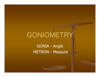
- 1. GONIOMETRY GONIA - Angle METRON - Measure
- 2. BASIC CONCEPTS Goniometry refers to the measurement of angles created at human joints by the bones of the body. Goniometry is used to measure and document the amount of active and passive joint motion.
- 3. USES Goniometric data used in conjunction with other information to: 1. Determine the presence or absence of impairment 2. Establish diagnosis 3. Develop prognosis, treatment goals and plan of care 4. Evaluate progress or lack of progress of treatment 5. Modify treatment 6. Motivate the subject 7. Research the effectiveness of techniques 8. Choose orthoses or adaptive equipment
- 4. JOINT MOTION Arthrokinematics It is a term used to refer to the movement of joint surfaces. They are, 1. Slides/glides: Is a translatory motion 2. Spins/roll : Is a rotary motion Osteokinematics These are movement of shafts of bones.
- 5. JOINT MOTION (cont ) Planes and axis OKs motions are described as taking place in three cardinal Planes and Axis. Planes 1. Sagittal : From ant. to post. of the body. Flexion and extension occurs in this plane in med. Lat. Axis. 2. Frontal : Runs from one side of body to another dividing it to front and back halves. Abduction and adduction occurs in anteroposterior axis. 3. Transverse : Horizontal and divides the body into upper and lower halves. Rotation motion occurs in vertical axis
- 6. RANGE OF MOTION ROM is a motion that occurs at a joint or series of joints. The starting position for ROM is anatomical position except rotations in transverse plane. 3 notation systems have been used to design ROM:0-180˚, 180-0˚, 360˚ 0-180 system of notation is called neutral zero method.
- 7. ACTIVE RANGE OF MOTION AROM is the arc of motion attained by a subject during unassisted voluntary joint motion. This provides the examiner with information about the subject’s willingness to move, coordination, muscle strength and joint range of motion.
- 8. PASSIVE ROM PROM is the arc of motion attained by an examiner without assistance from the subject. Normally PROM is slightly greater than AROM. This provides the examiner with information about the integrity of the articular surfaces and the extensibility of soft tissues around the joint.
- 9. END FEEL The type of structure that limits a ROM has a characteristic feel that may be detected by the examiner. The examiner should be able to detect Normal end feel Abnormal end feel
- 10. NORMAL END FEEL End feel Structure Soft Soft tissue approximation Firm 1. Muscular stretch 2. Capsular stretch 3. Ligamentous stretch Hard Bone to bone
- 11. ABNORMAL END FEEL End feel Examples Soft: Before complete ROM. Feels boggy. Soft tissue edema. Synovitis. Firm: Before complete ROM Hypertone,soft tissue shortening. Hard: Bony grating/bony block #,OA,MO,loose bodies. Empty: No end feel, pain prevents full ROM #,Inflammation.
- 12. HYPOMOBILITY This is decrease in ROM. Cyriax proposed that pathological conditions involving the entire joint capsule cause a particular pattern of restriction involving most of the passive motions of the joint. This pattern is called a capsular pattern. Restriction caused by condition involving structures other than the entire joint capsule is called noncapsular patterns of movement. Noncapsular pattern of movement is not proportioned similar to capsular pattern.
- 13. CAPSULAR PATTERN OF JOINTS Joints Restricted motion glenohumeral Lat.rotn., abd.,med.rotn. elbow Flxn.extn. forearm Supn., pron., flxn., extn. wrist Flxn. extn.=, deviations CMC digit 1 Abd. CMC digit 2-5 All motions = MCP & IP Flxn., extn.
- 14. CAPSULAR PATTERN OF JOINTS joints Restricted motion hip Med.rotn. flxn.abd.extn. knee Flxn. extn ankle Plantar flxn. dorsi flxn. subtalar Inversion midtarsal Add., med.rotn. MTP digit 1 Extn.flxn. MTP digit 2-5 Flxn.extn. IP Extn.flxn.
- 15. HYPERMOBILITY This refers to an increase in passive ROM that exceeds normal values. This is due to the laxity of soft tissues around the joint. Causes are, 1. Injuries 2. Hereditary disorders
- 16. MUSCLE LENGTH TESTING Muscle length is the greatest extensibility of a muscle tendon unit. It is the maximal distance between the proximal and the distal attachments of a muscle to bone. Muscle length is measured indirectly by determining the end of the ROM of the joints crossed by that muscle. Muscle length is tested to ascertain cause of change in ROM.
- 17. MUSCLE LENGTH TESTING (cont ) Muscles are categorized by the number of joints they cross, 1. One joint muscle 2. Two joint muscle 3. Multi joint muscle
- 18. One Joint Muscles Cross and therefore influence the motion of only one joint. No difference exists between the indirect measurement of the length of a one joint muscle and the measurement of joint ROM in the direction opposite to the muscle’s active motion. If there is shortness in muscle length, PROM opposite to the muscles action is decreased. The end feel is firm due to muscle stretch.
- 19. Two joint muscles To asses the length of a two joint muscle, the subject is positioned so that the muscle is lengthened over the proximal or distal joint that the muscle cross. This joint is held in position while the examiner attempts to further lengthen the muscle by moving the second joint through full ROM. The end feel is firm.
- 20. Multi joint muscle The subject is positioned and held so that the muscle is lengthened over all the joints that the muscle crosses except for one last joint. The examiner attempts to further lengthen the muscle by moving the last joint through full ROM. PASSIVE INSUFFICIENCY: The length of two & multi joint muscles are usually not sufficient to allow full passive ROM to occur simultaneously at all joints crossed by these muscles.
- 21. INSTRUMENTS 1. Universal goniometer 2. Gravity dependent goniometer/inclinometers, pendulum, fluid or bubble goniometer. 3. Electrogoniometer
- 22. Alignment Is based on anatomical land marks Stationary arm is in parallel to longitudinal axis of the proximal segment of the joint Moving arm in parallel to the longitudinal axis of the distal segment
- 23. PROCEDURES The examiner must have knowledge of 1. Testing positions 2. Stabilization required 3. Joint structure and function 4. Normal end feel 5. Anatomical bony landmarks 6. Instrument alignment The examiner must have the skill to 1. Position and stabilize correctly 2. Move a body part through proper ROM 3. Determine the end ROM i.e. end feel 4. Palpate bony landmarks 5. Align measuring instrument with landmarks 6. Read measuring instrument 7. Record measurements correctly
- 24. Positioning & stabilization Testing positions are designed to 1. Place the joint in a starting position of 0˚ 2. Permits complete ROM 3. Provide stabilization for the proximal joint segment 4. Positional stabilization may be supplemented by manual stabilization provided by the examiner
- 25. Recording Recordings are done in numerical tables, pictorial charts or within the written text of evaluation. Recordings should include the following 1. Subject’s name age and sex 2. Examiner’s name 3. Date and time 4. Make and type of goniometer 5. Side of body, joint and motion being measured 6. ROM at the beginning of motion and at the end of motion 7. Type of motion i.e. passive or active 8. Subjective information such as pain, discomfort etc 9. Objective information like muscle spasm, crepitus, capsular or noncapsular pattern etc 10. Description on any deviation from recommended testing position
- 26. Factors Affecting ROM Soft tissue tightness Adhesion formation Injuries or inflammation around the joint Muscle bulk Sex Age Nervous system