
Recommended
More Related Content
What's hot
What's hot (20)
Viewers also liked
Viewers also liked (20)
Similar to Slit lamp biomicroscope
Similar to Slit lamp biomicroscope (20)
Recently uploaded
Recently uploaded (20)
Slit lamp biomicroscope
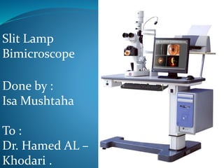
- 1. Slit Lamp Bimicroscope Done by : Isa Mushtaha To : Dr. Hamed AL – Khodari .
- 2. Slit lamp assessment is considered to be the gold standard device for the assessment of the anterior segment of the eye in clinical practice This is because they provide… Excellent image quality Stereoscopic image Flexible illumination Flexible magnification Therefore there are many different uses Even more when attachments are added
- 3. What are the uses of slit lamp ownOn their With accessories Routine examination of anterior segment Adnexa through to anterior vitreous Problem-based examination of anterior segment Gonioscopy Fundoscopy Ocular photography Contact tonometry (Goldmann) Direct contact goniolenses
- 4. • The lamp facilitates an examination of the anterior segment, or frontal structures of the human eye which includes the : 1. Eyelid &eye lashes 2. Cornea 3. Sclera 4. Conjunctiva 5. Iris 6. Aqueous 7. Natural crystalline lens and 8. Anterior vitreous.
- 6. Basic Design 1. Viewing arm Biomicroscope Adjustable focus eyepieces Magnification dial 2. Illumination arm The “slit lamp” Slit size, shape and filter controls Variable size, shape, colour and brightness 3. Biomicroscope and illumination are mechanically coupled around central pivot point (copivotal) Both focus at the same point (parfocal) Both arms can swing independently 180º along horizontal – there is a scale in degrees Both always central regardless of angle (isocentric) 4. Moveable base plate and joystick control
- 7. Types of illumination incorporated in slit lamp 1. Slit width Wide- survey globe/cornea Narrow- depth, width & position of small abnormalities beam as wide as cornea is thick forms a parallelepiped volume: a box of illuminated tissue is seen Thin (slit)- narrowest beam forms an optical section so thin it's just discernible التمييز سهل valuating small changes in clarity & pinpointing الدقة depth of pathology
- 8. 2. Light-source intensity Medium to high: most purposes High: optical section 3. Filters neutral, cobalt blue (for fluorescein), red-free and red free filter (green filter). 4. Magnification low power (~10x) is used for survey medium to high (16-40x) for optic section & parallelepiped Higher than (40x) for specular reflection normally, light is focused at same point as microscope (“parfocal”)
- 10. 1. Start with neutralizing the eye piece at zero if the examiner has no refractive error or wearing his glasses & lower powered objective lens . 2. Use lowest voltage setting on transformer ensure open aperture 3. Select the longest slit length 4. Adjust chin rest : Patient's eyes approximately with level of the marker on head rest . 5. Lamp height of the slit beam centered vertically on Patient's medial canthus 6. Focus by moving joystick How the examiner can adjust and use the slit lamp
- 11. locking nut: loose for free movementOcular focus to 0 adjust beam height for tall, narrow vertical beam adjust width for narrow beam w/ good illumination
- 12. Methods of examination of slit lamp without attachments There are six basic methods of illumination 1. Diffuse illumination 2. Direct focal illumination 3. Indirect illumination 4. Sclerotic scatter 5. Retro-illumination 6. Specular reflection
- 13. 1. Diffuse Illumination : This method is done by using a wide slit which slightly out of focus and this method is used in examination of the iris and the adnexa of the eye . The slit should be wide and the magnification should be low as possible to prevent loss of field and enable large field of view .
- 15. 2. Direct focal illumination : •Illumination and observation are focused in the same plane •slit width narrow to broad •Illumination angle 45° to 60° •Magnification 10x-40x This method is used to examine the cornea in details , the anterior chamber , crystalline lens , anterior part of the vitreous and in determine if there is flare , pus or blood cells in the anterior chamber .
- 17. 3. Indirect illumination : •The beam is focused in an area adjacent to ocular tissue to be observed •Decentered beam •Illumination 2 to 4mm slit •Magnification: Low to medium (depending upon object size). This method is used to determine if there is infiltrates رشح , corneal scars , deposits ترسبات and epithelial and stromal defects .
- 19. 4. Sclerotic scatter : •Light incident on the limbus with 2- 4mm slit at an angle of 45° - 60° •Decentered slit •The microscope focused centrally •Total internal reflection of the incoming light at inner corneal boundaries (endothelium and epithelium) . This method is used in examination of scars, foreign bodies, corneal defects and irregularities in the cornea .
- 20. 5. Retro-Illumination from the Fundus : This technique is used to observe media claritiesصفاءand opacities •The pupil is dilated •the slit beam and microscope are made co-axial and light strikes the fundus and creates a glow behind the opacity in the media •The media opacity creates a shadow in the glow Applications •abnormities in the anterior vitreous, lens, anterior chamber, cornea
- 22. 6. Specular Reflection: it is useful slit lamp technique for scrutinsing the corneal endothelium , the tear film surface , crystalline lens and other surfaces . A bright reflection will be observed in the anterior surface and less bright reflection will be observed in posterior surface of the cornea . The illumination is narrow parallelepiped and the microscope is placed directly in front of the eye with the light source 25degree from the microscope . The endothelium will appear clear and rough surface ( due to individual endothelial cells ) .
- 24. Attachments of the Slit lamp There are many different attachments and accessories,of these : 1. video attachment :used to facilitate teaching of slit lamp . 2. Goldman tonometer :used in measuring the intra ocular pressure of the anterior chamber . 3. Gold 3-mirror lens : used in measuring the angle of the anterior chamber and center of the retina .
- 25. 4. Volk double aspheric lens (+60,+78,+90) :used in examination the fundus os the patient indirectly . 5. Direct contact goniolenses : for examination of the periphrey of the fundus .
- 26. ...