SlideShare a Scribd company logo
1 of 14
Download to read offline
DISCOVERY OF THE PULMONARY
ANGIOTENSIN CONVERTING ENZYME (ACE)
AND ACE INHIBITOR IN 1967: A HISTORICAL
AND PERSONAL PERSPECTIVES
Kevin KF Ng, MD, PhD
Former Associate Professor of Medicine
Division of Clinical Pharmacology
Leonard M. Miller School of Medicine
University of Miami, FL. USA
Email: kevinng68@gmail.com
Kevin KF Ng and John R. Vane
at
DEPARTMENT OF PHARMACOLOGY
ROYAL COLLEGE OF SURGEONS OF ENGLAND
UNIVERSITY OF LONDON, ENGLAND
1967
1
Discovery of pulmonary angiotensin converting
enzyme (ACE) and ACE inhibitor in 1967:
Friday, Sept 22, 2017
A Historical and Personal Perspectives
By
Kevin KF Ng, MD, PhD
Former Associate Professor of Medicine
Division of Clinical Pharmacology
Leonard M. Miller School of Medicine
University of Miami, FL. USA
Email: kevinng68@gmail.com
A Tribute to Sir John R. Vane
2017 marks the 50th
anniversary of the discovery of angiotensin converting enzyme in the lung.
It is an honor and privilege for me to write this as a tribute to Sir John R. Vane (1927-2004) who
was awarded the Nobel Prize for Physiology and Medicine in 1982. It was he who developed the
blood-bathed-organ technique for the continuous in vivo study of the release and disappearance
of pharmacologically active substances in the circulation. He inspired me to use simple
experiments to solve complex biological problems. Little did I realize in 1967 that my
experimental findings from 1967-1968 could contribute to the development and clinical use of
angiotensin converting enzyme (ACE) inhibitors in medicine.
Picture shows Kevin KF Ng using the blood-bathed organ technique to study the “Dynamics of
the Renin-Angiotensin System” in 1967. IV=intravenous infusion. IBB=into bathing blood.
2
History of the Renin-Angiotensin system
The history of the renin-angiotensin system began in 1898 when Robert A. Tigerstedt and Per G.
Bergman reported the pressor effect of renal extracts1
. They named the substance renin because
it came from the kidney. In 1934, Harry Goldblatt induced experimental hypertension in dogs by
clamping a renal artery2
.
Using the Goldblatt kidney to produce experimental hypertension in 1936, two independent
groups of researchers, one led by Eduardo Braun-Mendendez3
in Argentina and the other led by
Irvine Page4
in the United States of America, demonstrated renal secretion of a pressor agent
similar to renin. In the following years, both teams described the presence of yet another new
compound from the blood of ischemic kidneys.
Their subsequent investigations showed that renin acted enzymatically on a plasma protein to
produce a pressor substance. In Buenos Aires, it was called hypertensin. In the United States, it
was called angiotonin. However, in 1958, Braun Menéndez from Argentina and Irvine Page from
the United States agreed to combine these two names together5
. Thus angiotensin was born out
of angiotonin and hypertensin. The substrate in the plasma acted on by renin was named
angiotensinogen.
In 1954, Skeggs et al found that angiotensin existed in two forms: angiotensin I and angiotensin
II. Angiotensin I was a biologically inactive decapeptide which was converted to a highly active
octapeptide by an enzyme in the plasma6
. This two-enzyme pathway of the renin-angiotensin
system is illustrated in the following figure. However, in 1967 Kevin KF Ng using Vane’s
Blood-Bathed-Organ technique showed that the conversion was too slow in the extracorporal
circulating blood in marked contrast to its rapid conversion through the pulmonary circulation.
Substrate Enzyme
Angiotensinogen
Renin
Angiotensin I
Plasma converting enzyme
Angiotensin II
Angiotensinase
Inactive products
Figure shows the concept of renin-angiotensin system prior to 1967.
Fatherly advice from JH Burn
When I was a medical student in Singapore, I was intrigued by a severe fall in the blood
pressure of a patient treated with intramuscular emetine for amoebiasis. My curiosity in
3
this event led to my appointment as Assistant Lecturer in Pharmacology in the University
of Singapore where I discovered that emetine acted like an adrenergic neuron blocking
agent7,8
. In Feb 1966, Joshua H. Burn who was Professor Emeritus from the University of
Oxford came to Singapore as an External Examiner in Pharmacology. He inspired me to
work on the Burn and Rand theory in the mechanism of release of noradrenaline from the
sympathetic nerve endings9,10
.
In June 1966, I was awarded a Merck Sharp and Dohme International Fellowship for
Clinical Pharmacology in the USA. At the same time, I was awarded an Overseas
Scholarship by the University of Singapore to work for a doctorate degree in
Pharmacology. JH Burn who was back in England wrote to me that I should be well trained
in basic pharmacology prior to specializing in clinical pharmacology. He strongly urged me
to work with one of his former staff members John R. Vane who was then a Reader in the
Department of Pharmacology at the Royal College of Surgeons of England, University of
London.
In August 1966, I arrived in London with my wife Gina, two sons Keith and Keiron and my
mother-in-law Helena Goh. After we were settled in, I went to meet John R. Vane at the
Department of Pharmacology in the Royal College of Surgeons of England. He introduced
me to the Chairman of the Department Professor Gustav VR Born, Senior Lecturer Robert
L. Hodge, Lecturer YS Bakhle, his research team: Duncan P. Thomas, Sergio Ferreira,
John Hughes, Norbert Gilmore and other research, technical and administrative staff.
JR Vane led me to his office and asked me what I would like to do. Then he wrote on a
piece of paper "anaphylaxis" and asked me to work on this the next day. As a new comer to
the laboratory, I had to build my own experimental apparatus. Within a few days, I began
to study the effects of acetylcholine, catecholamines (adrenaline, noradrenalin), amines
(histamine, 5-hydroxytryptamine), peptides (bradykinin, angiotensin II, vasopressin),
prostaglandins and other pharmacologically active substances on isolated superfused
organs from various laboratory animals. The goal of these experiments was to look for
tissues that might be suitable for the detection of mediators in anaphylaxis. When Robert
Hodge left for Australia to assume the position of Director of the National Heart
Foundation of Australia, JR Vane asked me to complete the experiments RL Hodge left
behind.
Selection of isolated organs for bioassay.
Isolated organs were removed from the stomach, small intestines, large intestines, colon
and rectum from rats, chickens, guinea pigs, rabbits and cats. These were suspended in
organ baths and tested for sensitivity and specificity. The tissues were deemed sensitive if
they responded to 1-5 nanograms of the substance. To test for specificity, the tissues
arranged in a series of three for parallel bioassay, were superfused with Krebs solution
from a roller pump at a constant rate of 10-15 ml/min. The responses of the tissues were
recorded by auxotonic levers writing on a smoked paper attached to a revolving drum
known as kymogragh. Isolated organs responding to adrenaline, noradrenaline, histamine,
4
5-hydroxytryptamine, angiotensin II, bradykinin and prostaglandins were selected for the
experiments.
Adrenaline relaxed the stomach strip, chick rectum, and rat colon. Noradrenaline relaxed
the rat stomach strip and rat colon without any effect on chick rectum. Angiotensin II
contracted the rat stomach strip and rat colon without effect on the chick rectum. Cat
jejunum was selectively sensitive to bradykinin. Prostaglandin E2 contracted the rat
stomach strip, chick rectum and rat colon. 5-hydroxytrytamine contracted only the stomach
strip.
Figure on the left shows the responses of isolated organs to physiological concentrations of
vasoactive substances. Figure on the right shows the arrangement of the isolated organs in
series and the incubation circuit for the study of half-lives of vasoactive substances in
circulating blood.
Half-life of angiotensin II in circulating blood11
These experiments were done on dogs anesthetized with Halothane and intravenous -
chloralose supplemented with pentobarbital when necessary. The assay organs were a rat
stomach strip and two rat colons; one of the colons was infused intraluminally with
propranolol to block the relaxant effect of catecholamines on the tissue. The trachea was
cannulated and the lungs were ventilated mechanically with room air. Polyethylene
cannulas were tied onto carotid or femoral arteries for blood sampling. Cannulas were also
tied onto carotid or femoral veins for venous return. After the dogs were heparinized,
arterial blood was pumped at a constant rate of 10-15 ml/min (less than 0.5% of cardiac
output of adult dogs) to superfuse the assay organs. The blood was collected in a reservoir
and returned by gravity into the carotid or femoral veins. Thus an extracorporeal circulation
from a major artery to a major vein was created. Mean arterial blood pressure was
measured from the side arm of the arterial cannula with a mercury manometer and recorded
on a kymograph. Synthetic angiotensin II amide (Hypertensin) from CIBA was used in all
experiments.
To study the inactivation of angiotensin II in circulating blood, I lengthened the silicone
tubing and immersed it in a water bath maintained at 37 degrees Fahrenheit. This served as
an incubation circuit which contained 30-45 ml of blood. A bubble of air was injected into
5
the circulating blood at different points of the tubing to determine the incubation time of
angiotensin II. Infusions for 5 minutes were first made into blood before it was pumped to
the assay organs. The transit time was 15 seconds. Infusions of angiotensin II were then
made into the tubing such that angiotensin II would come in contact with circulating blood
for 60 sec, 120 sec and 180 sec. The responses of the assay organs were plotted against
incubation time, a 50% reduction in response relative to incubation time was considered to
be the half-life of angiotensin II. This was found to be 180 seconds.
Disappearance of angiotensin II in vascular beds11
To study the disappearance of angiotensin II in the liver and hind limb, blood was drawn
from carotid artery to superfuse the assay organs and returned via femoral vein. Infusions
of angiotensin II were given into the portal vein and into the femoral artery to study its
disappearance across the vascular beds. To study the disappearance of angiotensin II in the
lungs, blood was drawn from the femoral artery to superfuse the assay organs. It was
returned to the femoral vein by gravity. Intravenous infusions were given into the femoral
vein and intra-aortic infusions was given via cannulas inserted into the ascending aortas
close to the aortic valve. The responses obtained after their passage across the vascular
beds were calibrated by intravenous infusions.
The results of these experiments from October 1966 to early part of 1967 showed that the
half-life of angiotensin II in the circulating blood was 180 seconds and the disappearance
of angiotensin II was 50-75% in the head, liver and hind limbs. The lungs were unique in
that angiotensin II passed through the pulmonary circulation without any loss.
Figure shows schematic diagram of the circulation. Responses of assay organs to Intra-arterial (IA)
infusions were compared with responses to intravenous (IV) infusions. If the IA response 1.0
µg/min was equal to the response to IV 0.5 µg/min, the disappearance was 50%.
While I was doing the experiments on angiotensin II, Sergio Ferreira was doing similar
experiments with bradykinin next to my laboratory. He found that bradykinin was rapidly
inactivated in circulating blood with a half-life of 18 seconds. The disappearance of
bradykinin was 80-95% across the lungs and other vascular beds. This remarkable
6
disappearance of bradykinin in the lungs and a total lack of disappearance of angiotensin II
in the pulmonary circulation came as a big surprise for me.
The lungs had always been considered as an organ for the exchange of carbon dioxide for
oxygen. How polypeptides were handled by the lungs were not known. I presented my
results to JR Vane in his office. Looking at the summary table of my results and the
tracings on smoked papers, he said “Kevin, history is made”. He suggested that angiotensin
II was a systemic hormone, whereas bradykinin was a local hormone and the lung acted
like a filter for the systemic circulation.
Having completed my studies on the half-life of angiotensin II in circulating blood and its
disappearance in vascular beds, I was looking forward to do similar experiments with
angiotensin I. However, angiotensin I was not available commercially. Perhaps I could
make some angiotensin I in vitro with dog renin extract and plasma. JR Vane agreed and in
the summer of 1967, he sent me to Dr. Brown's Blood Pressure Research Unit in Glasgow,
Scotland to extract renin from dog kidneys.
Conversion of angiotensin I to angiotensin II in circulating blood12
On my return from Glasgow, JR Vane told me that he had received five ampoules of
lyophilized horse angiotensin I from Dr. Skeggs. I started the experiments the next day.
Stock solutions of angiotensin I were made with distilled water and kept frozen in the
refrigerator. Working solutions were diluted with normal saline. Rod Flower who joined
the department as a Research technician assisted me daily in my experiments. We
anesthetized the dogs and tied cannulas in the arteries and veins. I prepared the assay
organs and superfused them with Krebs solution at 10 ml/min. Initial experiments were
done to compare the activity of angiotensin I and angiotensin II on the assay organs. After
this, the Krebs solution was replaced by circulating blood from the incubation circuit.
The relative activity of angiotensin I was expressed as a percentage of activity of
angiotensin II. In Krebs solution, angiotensin I had 15% of the activity of angiotensin II.
Incubation in circulating blood for 15 sec increased the activity to 27%. This was further
increased to 40% after 60 sec, 68% after 120 sec and 93% after 180 sec. By plotting these
responses against incubation time, 50% of the increased activity or the half-life of
angiotensin I occurred at 90 sec. Since angiotensin II was inactivated in circulating blood
with a half-life of 180 sec, the increase in activity on the rat colon represented the net result
of conversion of angiotensin I to angiotensin II and the inactivation of angiotensin II.
7
Conversion of angiotensin I to Angiotensin II in the pulmonary circulation12
Angiotensin I had 27% of the activity of angiotensin II on arterial blood-bathed assay
organs. The activity was increased to 80% after intravenous infusion. One circulation time
through the pulmonary circulation of the dog is less than 5 seconds. It was conceivable that
the inactive angiotensin I was converted to the active angiotensin II during its passage
through the lungs. To prove this hypothesis, the following experiments were done.
The first set of experiments were made on dogs in which a fine cannula was inserted into
ascending aorta so that intra-aortic infusions would come into contact with blood after it
has passed through the pulmonary circulation. Blood for the assay organs were sampled
from femoral artery and returned via femoral vein. The responses on the assay organs made
by intra-aortic infusions were compared with those made by intravenous infusions into the
femoral vein. The results showed that the relative activity of intra-aortic angiotensin I was
30% , whereas the relative activity of intravenous angiotensin I was 80%. A net increase
by 50% in angiotensin I activity across the pulmonary circulation must be due to its conversion
from angiotensin I to angiotensin II
The second set of experiments was made by two series of isolated organs arranged in
parallel and the responses of the isolated tissues were recorded on a multichannel
Dynagraph. Synthetic angiotensin I was provided by Dr. M. Weatherall and Dr. S.
Wilkinson of Wellcome Foundation. Under anesthesia, cannulas were inserted into left
ventricle, right ventricle, femoral artery and femoral vein of the dog. Each series of isolated
tissues contained a stomach strip and two rat colons; one colon in each series was infused
intraluminally with propranolol to block the relaxant effect of catecholamines on the
tissues. One series was bathed in venous blood from the right ventricle, the other series was
bathed in arterial blood from the left ventricle. The blood from both series were returned to
the animal via femoral vein.
8
Since angiotensin II passed through the lungs without loss, it was infused into the femoral
vein to determine the relative activity of angiotensin I before and after it had passed
through the pulmonary circulation. The results showed that synthetic angiotensin I had 5%
of the activity of angiotensin II in Krebs solution. Contact with blood for 30 seconds from
the cannula to the assay organs increased the activity to 16%. On transit from femoral vein
to the right ventricle, the relative activity was increased marginally to 18%. In contrast,
blood from left ventricular arterial blood showed a remarkable increase of activity to 36%.
The increase in relative activity from 18% in right ventricle to 36% in left ventricle
represented a 100% change in activity. This showed that angiotensin I was indeed
converted to angiotensin II during its passage through the pulmonary circulation.
The third set of experiments was performed because earlier results were obtained with
horse angiotensin I and synthetic angiotensin I. Both could be different from dog
angiotensin I. To prove this, I used exogenous dog renin which I prepared in Glasgow and
I induced endogenous renin release into the circulation by partial occlusion of blood flow
to the renal arteries of the dog. Blood was sampled from the right and left ventricles for the
assay and returned via the femoral vein by gravity. Two series of isolated tissues were
used; one bathed in venous blood from the right ventricle and the other bathed in arterial
blood from left ventricle.
Exogenous renin had no activity on the rat colons bathed in Krebs solution. It was
impossible to generate angiotensin I in the incubation circuit because of its long half-life in
the circulation and angiotensin I was relatively inactive on the assay tissues. When renin
was infused intravenously for 10 min, the contraction of the rat colons gradually reached a
plateau. When the infusion stopped, the contractions of the rat colons declined
exponentially with a half-life of 15-20 minutes. Calibration of these responses with
angiotensin II showed that the contractions caused by exogenous renin were smaller in
venous blood compared with those in arterial blood. Similar responses were obtained from
endogenous renin when the blood flow to the kidneys was partially reduced by inflating a
balloon above the renal arteries. These results were consistent with the observations made
9
by intravenous horse and synthetic angiotensin I and showed that angiotensin I was indeed
converted to angiotensin II in the pulmonary circulation.
Disappearance of Angiotensin I in peripheral vascular beds13
To discover what happened to angiotensin I in peripheral vascular beds, infusions of
angiotensin I were given into carotid, renal and femora arteries. The effects on the rat
colons bathed in arterial blood were calibrated with intravenous angiotensin I. The
disappearance was 80-90% in the kidneys, 60-90% in hind limbs and 40-50% in the head.
To determine the disappearance of angiotensin I throughout the peripheral vascular beds
excluding the lungs, a coaxial polyethylene tubing was inserted into the aorta close to the
aortic valves. The inner tubing was used for supplying arterial blood to the assay tissues
while the outer tubing which was 2 cm shorter was used to infuse angiotensin I to the
peripheral vascular bed. Calibration by intravenous angiotensin I showed that 50% to 60%
of angiotensin I disappeared in the peripheral vascular beds.
Since angiotensin I and angiotensin II contracted the rat colon, it was not possible to
distinguish the identity of the polypeptides in the arterial blood. To circumvent this
problem, blood was sampled from the right and left ventricles for bioassay. The response of
the assay organs was calibrated with intravenous angiotensin II since it passed through the
pulmonary circulation without loss. The experiments with intra-aortic coaxial catheter
showed that the responses of the rat colons bathed in right ventricle venous blood were
smaller than those bathed in left ventricle arterial blood. I could not determine whether
angiotensin I was converted to angiotensin II in the peripheral vascular beds but the
fraction that passed through appeared to be angiotensin I because its activity was increased
by 100% on transit from right ventricle to left ventricle. The evidence again indicated that
the conversion of angiotensin I to angiotensin II took place in the pulmonary circulation.
Figure shows % disappearance of angiotensin I and angiotensin II in vascular beds¹⁴.
10
Mechanism of conversion of angiotensin I to angiotensin II¹⁵
It has been established that Angiotensin I is a 10 amino acid polypeptide, angiotensin II is a
8 amino acid polypeptide and bradykinin is a 9 amino acid polypeptide as shown below:
1 2 3 4 5 6 7 8 9 10
Angiotensin I: Asp-Arg-Val-Tyr-Ile-His-Pro-Phe-His-Leu
Angiotensin II: Asp-Arg-Val-Tyr-Ile-His-Pro-Phe
Bradykinin: Arg-Pro-Pro-Gly-Phe-Ser-Pro-Phe-Arg
It has also been shown that the plasma converting enzyme split the phenylalanyl-histidine
link in angiotensin I to form angiotensin II and a histidyl-leucine residue. Furthermore, it
has also been shown that bradykinin is inactivated by a carboxypeptidase which removes
the C-terminal arginine from bradykinin. The chemical structure of angiotensin II and that
of bradykinin showed that their last two amino acids were identical. It was possible that
the pulmonary converting enzyme was similar to the enzyme that inactivated bradykinin.
To confirm this, further experiments were done with an angiotensin I analogue and a
bradykinin potenting factor (BPF).
Angiotensin I and a HHL angiotensin I analogue were synthesized by Dr. S. Williams of
Wellcome Foundation. He inserted an extra histidine at position 9 between histidyl-leucine
linkage making it an undecapeptide angiotensin I analogue as follow:
1 2 3 4 5 6 7 8 9 10 11
Angiotensin I: Asp-Arg-Val-Tyr-Ile-His-Pro-Phe-His-Leu
HHLAngiotensin I: Asp-Arg-Val-Tyr-Ile-His-Pro-Phe-His-His-Leu
The pulmonary angiotensin converting enzyme may be an endopeptidase similar to that in
the plasma or a carboxypeptidase which first cleaved histidine and then leucine from
angiotensin I. The availability of HHL angiotensin I analogue made it possible to test this
theory with the blood bathed organ technique.
In anesthetized dogs, blood was sampled from femoral artery to superfuse the assay tissues
and returned via femoral vein. Intravenous infusions were given into femoral vein and
intra-aortic infusions were given into ascending aorta close to the aortic valves. The
contractions of the rat colons in Krebs solution or circulating blood were calibrated with
synthetic angiotensin I or synthetic angiotensin II. In Krebs solution, HHL angiotensin I
had less than 1% of the activity of synthetic angiotensin II. There was no significant change
after incubating in circulating blood for 30 sec. Comparisons of the relative activity of
HHL angiotensin I analogue with angiotensin II after intravenous infusions showed that 50-
60% of the analogue disappeared in the pulmonary circulation. Thus, there was no evidence
of increase in activity after its passage through the pulmonary circulation.
11
The inactivation of HHL angiotensin I analogue in the lungs was consistent with the theory
that the pulmonary converting enzyme was an endopeptidase similar to that in plasma
because stepwise cleavage by a carboxypeptidase would have resulted in angiotensin II
which was resistant to destruction by the lungs.
Inhibition of pulmonary angiotensin converting enzyme by BPF15
In order to prove that the pulmonary converting enzyme was similar to the enzyme that
inactivated bradykinin, I obtained a small quantity of Bradykinin Potentiating Factor (BPF)
from Sergio Ferreira. BPF is a mixture of peptides derived from the venom of Brazilian
snake Bothrops jararaca. It has been shown to potentiate the actions of bradykinin in vitro.
Due to scarcity of BPF, the experiments could only be done in anesthetized rats.
Intravenous injections of angiotensin I and angiotensin II raised the arterial blood pressure
of rats with a potency ratio of 2:1. With intravenous infusion of BPF, the ratio of potency
decreased to 15:1 showing for the first time that the in vivo conversion of angiotensin I to
angiotensin II was inhibited by bradykinin potentiating factor (BPF).
Figure shows the potency ratio of angiotensin I to angiotensin II on blood pressor of a rat. Before
BPF infusion, the potency ration of angiotensin I to angiotensin II was 2:1. During the infusion of
BPF 0.1 mg/min, the potency ration was significantly reduced to 15:1.
In September 1968, I submitted my thesis "The dynamics of the renin-angiotensin system"
for the degree of Doctor of Philosophy to the University of London. The Internal Examiner
Professor JR Vane and the External Examiner Professor WS Peart both congratulated me
for the work well done. The degree of Doctor of Philosophy in Pharmacology was
conferred upon me on October 9th
, 1968. Further experiments on the inhibition of
pulmonary angiotensin converting enzyme by BPF were continued by YS Bakhle and
Sergio Ferreira.
12
As I was making plans to return to Singapore in October 1968, JH Burn went to St. Louis
in Missouri where he met Dr. John C. McGiff. JH Burn told JC McGiff about Vane’s new
technique for the study of vasoactive substances in the circulation. Keen to adapt this new
technique to his research, JC McGiff invited JR Vane to come to the USA. Due to a busy
schedule, JR Vane asked me to stop over USA on my way back to Singapore. This would
give me the opportunity to set up the blood-bathed organ system not only for McGiff in St
Louis, Missouri but also for Dr. Irvine Page in San Francisco, California. My family and I
left London for the USA in late October 1968.
On my return to Singapore from USA, JR Vane introduced me to many of his colleagues
who invited me to give a seminar on the "Dynamics of the renin-angiotensin system" in
the United States. The stop over in USA gave me the opportunity to meet my former
teacher Robert CY Lin in Hong Kong and friends in the Far East. The following is the list
of laboratories and institutes of higher learning where I made my presentations:
• The Wellcome Research Laboratories in New York
• Smith Kline & French Laboratories in Philadelphia
• Department of Pharmacology, State University of New York in Buffalo
• Cleveland Clinic in Ohio
• Department of Internal Medicine, Washington University in Saint Louis
• School of Medicine, Saint Louis University in Missouri
• Cardiology Club of Saint Louis, Missouri
• Lilly Laboratory for Clinical Research in Indianapolis
• Department of Pharmacology, University of Utah in Salt Lake City
• Riker Laboratories in Northridge, California
• Department of Physiology, University of Hawaii in Honolulu
• College of Medicine, National Taiwan University in Taiwan
• Department of Pharmacology, University of Hong Kong
The seminars were well received and the itinerary was memorable. My family and I finally
arrived in Singapore on Christmas day December 25th 1968.
The discovery of the pulmonary converting enzyme and its inhibition by BPF led to the
synthesis of captopril (Capoten), the first ACE inhibitor used clinically in 1982. There are
now ten ACE inhibitors available in the United States for the treatment of hypertension,
congestive heart failure and diabetic kidney disease: captopril, enalapril, fosinopril,
lisinopril, benazepril, moexipril, perindopril, quinapril, ramipril and trandolapril.
References
1 Tigerstedt R and Bergman PG. Niere und Kreislauf. Skand Arch Physiol. 1898; 8: 223–
27
2 Goldblatt H, Lynch J, Hanzal RF and Summerville WW. Studies on experimental
hypertension, I: the production of persistent elevation of systolic blood pressure by means
of renal ischemia. J Exp Med. 1934; 59: 347–379.
13
3 Taquini AC and Braun-Menéndez E. Liberación de substancia vaso-constrictora en el
riñón completamente isquemiado. Rev Soc Arg Biol. 1938; 14: 422–429
4 Page IH. Vasopressor action of extracts of plasma of normal dogs and dogs with
experimentally produced hypertension. Proc Soc Exp Biol. 1937; 35: 112–11
5 Braun-Menéndez E and Page IH. Suggested revision of nomenclature: angiotensin.
Science. 1958; 127: 242.
6 Skeggs LT, Marsh WH, Kahn JR and Shumway, NP. The existense of two forms of
hypertensin. J. Exp. Med. 1954:99:275-280.
7 Ng KKF. A new pharmacological action of emetine. BMJ. 1966;1: 1278-1279.
8 Ng KKF. Blockade of adrenergic and cholinergic transmissions by emetine. Br. J.
Pharmacol. Chemother. 1966; 28:228-237.
9 Burn JH and Ng KKF. The action of pempidine and antiadrenaline substances on
sympathetic post-ganglionic terminations. Br. J. Pharmacol. Chemother. 1965; 24:678-688
10 Ng KKF. The effects of some anti-cholinesterases on the response of the taenia to
sympathetic nerve stimulation. J.Physiol. 1966;182:233-243.
11 Hodge RL, Ng KKF and Vane JR. Disappearance of angiotensin from the circulation of
the dog. Nature. 1967; 215:138-141.
12 Ng KKF and Vane JR. Conversion of angiotensin I to angiotensin II. Nature. 1967;
216:762-766.
13 Ng KKF and Vane JR. Fate of angiotensin I in the circulation. Nature. 1968; 218:144-
150.
14 Ng KKF. The Dynamics of the renin-angiotensin system. PhD thesis, University of
London, 1968.
15 Ng KKF and Vane JR. Some properties of angiotensin converting enzyme in the lung in
vivo. Nature. 1970; 225:1142-1144.

More Related Content

What's hot

ARTEMISININ presentation .pptx
ARTEMISININ presentation .pptxARTEMISININ presentation .pptx
ARTEMISININ presentation .pptx
Himani Kolekar
 

What's hot (20)

Adrenocorticoids and chemistry of Vitamin- D.pptx
Adrenocorticoids and chemistry of Vitamin- D.pptxAdrenocorticoids and chemistry of Vitamin- D.pptx
Adrenocorticoids and chemistry of Vitamin- D.pptx
 
Digoxin & digitoxin
Digoxin & digitoxinDigoxin & digitoxin
Digoxin & digitoxin
 
Phyllanthus niruri in liver dysfunction
Phyllanthus niruri in liver dysfunctionPhyllanthus niruri in liver dysfunction
Phyllanthus niruri in liver dysfunction
 
Pharmacology of Bradykinin , pentagastrin, cholecystokinin and angiotensin
Pharmacology of Bradykinin , pentagastrin, cholecystokinin and angiotensinPharmacology of Bradykinin , pentagastrin, cholecystokinin and angiotensin
Pharmacology of Bradykinin , pentagastrin, cholecystokinin and angiotensin
 
Application of deuterium in drug discovery
Application of deuterium in drug discoveryApplication of deuterium in drug discovery
Application of deuterium in drug discovery
 
Natural products in drug discovery
Natural products in drug discoveryNatural products in drug discovery
Natural products in drug discovery
 
Drug discovery
Drug discoveryDrug discovery
Drug discovery
 
SAR of H1 Receptor Antagonists..
SAR of H1 Receptor Antagonists..SAR of H1 Receptor Antagonists..
SAR of H1 Receptor Antagonists..
 
Digitalis glycoside
Digitalis glycosideDigitalis glycoside
Digitalis glycoside
 
Heterocyclic compounds
Heterocyclic compoundsHeterocyclic compounds
Heterocyclic compounds
 
Herb drug interaction
Herb drug interactionHerb drug interaction
Herb drug interaction
 
Role of natural product in drug discovery
Role of natural product in drug discoveryRole of natural product in drug discovery
Role of natural product in drug discovery
 
Chemistry of prostaglandins, leukotrienes and thromboxanes
Chemistry of prostaglandins, leukotrienes and thromboxanesChemistry of prostaglandins, leukotrienes and thromboxanes
Chemistry of prostaglandins, leukotrienes and thromboxanes
 
Prodrug.pptx
Prodrug.pptxProdrug.pptx
Prodrug.pptx
 
Active constituent of Sweta chirata as Antidibetic
Active constituent of Sweta chirata as AntidibeticActive constituent of Sweta chirata as Antidibetic
Active constituent of Sweta chirata as Antidibetic
 
Quinine (2)
Quinine (2)Quinine (2)
Quinine (2)
 
Utilization of radioactive isotopes in biosynthetic pathway
Utilization of radioactive isotopes in biosynthetic pathwayUtilization of radioactive isotopes in biosynthetic pathway
Utilization of radioactive isotopes in biosynthetic pathway
 
Alkaloids
AlkaloidsAlkaloids
Alkaloids
 
Herbal anti cancer drugs
Herbal anti cancer drugsHerbal anti cancer drugs
Herbal anti cancer drugs
 
ARTEMISININ presentation .pptx
ARTEMISININ presentation .pptxARTEMISININ presentation .pptx
ARTEMISININ presentation .pptx
 

Similar to Discovery of pulmonary angiotensin converting enzyme and ace inhibitor in 1967

History of pharmacology & contributions of various scientists in pharmacology
History of pharmacology & contributions of various scientists in pharmacology History of pharmacology & contributions of various scientists in pharmacology
History of pharmacology & contributions of various scientists in pharmacology
Govt Medical College & Hospital, Sector-32
 
Constituiçõ umidade fleuma genomica suscetibilidade, ajuste e tratamento co...
Constituiçõ umidade fleuma   genomica suscetibilidade, ajuste e tratamento co...Constituiçõ umidade fleuma   genomica suscetibilidade, ajuste e tratamento co...
Constituiçõ umidade fleuma genomica suscetibilidade, ajuste e tratamento co...
valdemar caumo
 
Anaemia and wound healing
Anaemia and wound healingAnaemia and wound healing
Anaemia and wound healing
MNTan1
 
The neuroprotective role of Panax notoginseng saponins in APP/PS1 transgenic ...
The neuroprotective role of Panax notoginseng saponins in APP/PS1 transgenic ...The neuroprotective role of Panax notoginseng saponins in APP/PS1 transgenic ...
The neuroprotective role of Panax notoginseng saponins in APP/PS1 transgenic ...
LucyPi1
 

Similar to Discovery of pulmonary angiotensin converting enzyme and ace inhibitor in 1967 (20)

Natriuretic peptides and oxygen: a narrative review
Natriuretic peptides and oxygen: a narrative reviewNatriuretic peptides and oxygen: a narrative review
Natriuretic peptides and oxygen: a narrative review
 
Discovery of the lung angiotensin converting enzyme 01 22 2016-pdf
Discovery of the lung angiotensin converting enzyme 01 22 2016-pdfDiscovery of the lung angiotensin converting enzyme 01 22 2016-pdf
Discovery of the lung angiotensin converting enzyme 01 22 2016-pdf
 
Autonomic nervous system
Autonomic nervous systemAutonomic nervous system
Autonomic nervous system
 
Basics clinical research
Basics  clinical researchBasics  clinical research
Basics clinical research
 
Mechanism of Rhein Inhibiting Acute Myocardial Infarction–Induced Endoplasmic...
Mechanism of Rhein Inhibiting Acute Myocardial Infarction–Induced Endoplasmic...Mechanism of Rhein Inhibiting Acute Myocardial Infarction–Induced Endoplasmic...
Mechanism of Rhein Inhibiting Acute Myocardial Infarction–Induced Endoplasmic...
 
Serendipitous drug discovery in the field of pharmacy
Serendipitous drug discovery in the field of pharmacySerendipitous drug discovery in the field of pharmacy
Serendipitous drug discovery in the field of pharmacy
 
History of pharmacology & contributions of various scientists in pharmacology
History of pharmacology & contributions of various scientists in pharmacology History of pharmacology & contributions of various scientists in pharmacology
History of pharmacology & contributions of various scientists in pharmacology
 
Update on organon detailed
Update on organon detailedUpdate on organon detailed
Update on organon detailed
 
Introduction to Pharmacognosy
Introduction to PharmacognosyIntroduction to Pharmacognosy
Introduction to Pharmacognosy
 
ANTI ASTHMATIC DRUG SCREENING MODEL.pptx
ANTI ASTHMATIC DRUG SCREENING MODEL.pptxANTI ASTHMATIC DRUG SCREENING MODEL.pptx
ANTI ASTHMATIC DRUG SCREENING MODEL.pptx
 
Constituiçõ umidade fleuma genomica suscetibilidade, ajuste e tratamento co...
Constituiçõ umidade fleuma   genomica suscetibilidade, ajuste e tratamento co...Constituiçõ umidade fleuma   genomica suscetibilidade, ajuste e tratamento co...
Constituiçõ umidade fleuma genomica suscetibilidade, ajuste e tratamento co...
 
A new conept of topical pain relief by phytochemicals
A new conept of topical pain relief by phytochemicalsA new conept of topical pain relief by phytochemicals
A new conept of topical pain relief by phytochemicals
 
A brief history of pharmacology
A brief history of pharmacologyA brief history of pharmacology
A brief history of pharmacology
 
Anaemia and wound healing
Anaemia and wound healingAnaemia and wound healing
Anaemia and wound healing
 
History of Pharmcology
History of PharmcologyHistory of Pharmcology
History of Pharmcology
 
Insulin therapy: An Old Story
Insulin therapy: An Old StoryInsulin therapy: An Old Story
Insulin therapy: An Old Story
 
Innis
InnisInnis
Innis
 
The Evolution of the Clinical Trials Process – A Brief History Lesson
The Evolution of the Clinical Trials Process – A Brief History LessonThe Evolution of the Clinical Trials Process – A Brief History Lesson
The Evolution of the Clinical Trials Process – A Brief History Lesson
 
The neuroprotective role of Panax notoginseng saponins in APP/PS1 transgenic ...
The neuroprotective role of Panax notoginseng saponins in APP/PS1 transgenic ...The neuroprotective role of Panax notoginseng saponins in APP/PS1 transgenic ...
The neuroprotective role of Panax notoginseng saponins in APP/PS1 transgenic ...
 
00.toxicological assessment-of-venlafaxine-acute-and-subchronic-toxicity-stud...
00.toxicological assessment-of-venlafaxine-acute-and-subchronic-toxicity-stud...00.toxicological assessment-of-venlafaxine-acute-and-subchronic-toxicity-stud...
00.toxicological assessment-of-venlafaxine-acute-and-subchronic-toxicity-stud...
 

More from Kevin KF Ng

More from Kevin KF Ng (20)

The Use of Vitamin D in Chronic Diseases
The Use of Vitamin D in Chronic DiseasesThe Use of Vitamin D in Chronic Diseases
The Use of Vitamin D in Chronic Diseases
 
Synergistic actions of vitamin D3 and vitamin K2
Synergistic actions of vitamin D3  and vitamin K2Synergistic actions of vitamin D3  and vitamin K2
Synergistic actions of vitamin D3 and vitamin K2
 
Target pain receptors with essential oils
Target pain receptors with essential oilsTarget pain receptors with essential oils
Target pain receptors with essential oils
 
Beta-glucans as immunostimulants
Beta-glucans as immunostimulants Beta-glucans as immunostimulants
Beta-glucans as immunostimulants
 
Solution for chronic inflammatory diseases
Solution for chronic inflammatory diseasesSolution for chronic inflammatory diseases
Solution for chronic inflammatory diseases
 
CBD pain relief_Final.pdf
CBD pain relief_Final.pdfCBD pain relief_Final.pdf
CBD pain relief_Final.pdf
 
Shoulder pain relief based on new science
Shoulder pain relief based on new scienceShoulder pain relief based on new science
Shoulder pain relief based on new science
 
Clove is a versatile spice and medicine
Clove is a versatile spice and medicineClove is a versatile spice and medicine
Clove is a versatile spice and medicine
 
Answers to a poll on how many cells are replaced every day.
Answers to a poll on how many cells are replaced every day.Answers to a poll on how many cells are replaced every day.
Answers to a poll on how many cells are replaced every day.
 
Black pepper update 2023
Black pepper update 2023Black pepper update 2023
Black pepper update 2023
 
Scientific studies on watermelon as medicine
Scientific studies on watermelon as medicineScientific studies on watermelon as medicine
Scientific studies on watermelon as medicine
 
A Super Chicken Soup Recipe
A Super Chicken Soup RecipeA Super Chicken Soup Recipe
A Super Chicken Soup Recipe
 
How chicken soup relieves common cold symptoms
How chicken soup relieves common cold symptomsHow chicken soup relieves common cold symptoms
How chicken soup relieves common cold symptoms
 
Soy sauce is a flavor enhancer
Soy sauce is a flavor enhancerSoy sauce is a flavor enhancer
Soy sauce is a flavor enhancer
 
Kumquat is a symbol of prosperity with health benefits
Kumquat is a symbol of prosperity with health benefitsKumquat is a symbol of prosperity with health benefits
Kumquat is a symbol of prosperity with health benefits
 
You are what you eat and its effect on obesity
You are what you eat and its  effect on obesityYou are what you eat and its  effect on obesity
You are what you eat and its effect on obesity
 
Top flavor enhancer
Top flavor enhancerTop flavor enhancer
Top flavor enhancer
 
Chinese philosophy and the applications of Yin Yang and the Five Elements
Chinese philosophy and the applications of Yin Yang and the Five Elements Chinese philosophy and the applications of Yin Yang and the Five Elements
Chinese philosophy and the applications of Yin Yang and the Five Elements
 
Saffron, the spice of happiness
Saffron, the spice of happinessSaffron, the spice of happiness
Saffron, the spice of happiness
 
Persimmon_fruit of the gods
Persimmon_fruit of the godsPersimmon_fruit of the gods
Persimmon_fruit of the gods
 

Recently uploaded

Call Girls Bhubaneswar Just Call 9907093804 Top Class Call Girl Service Avail...
Call Girls Bhubaneswar Just Call 9907093804 Top Class Call Girl Service Avail...Call Girls Bhubaneswar Just Call 9907093804 Top Class Call Girl Service Avail...
Call Girls Bhubaneswar Just Call 9907093804 Top Class Call Girl Service Avail...
Dipal Arora
 
Call Girls in Gagan Vihar (delhi) call me [🔝 9953056974 🔝] escort service 24X7
Call Girls in Gagan Vihar (delhi) call me [🔝  9953056974 🔝] escort service 24X7Call Girls in Gagan Vihar (delhi) call me [🔝  9953056974 🔝] escort service 24X7
Call Girls in Gagan Vihar (delhi) call me [🔝 9953056974 🔝] escort service 24X7
9953056974 Low Rate Call Girls In Saket, Delhi NCR
 

Recently uploaded (20)

Top Rated Hyderabad Call Girls Erragadda ⟟ 9332606886 ⟟ Call Me For Genuine ...
Top Rated  Hyderabad Call Girls Erragadda ⟟ 9332606886 ⟟ Call Me For Genuine ...Top Rated  Hyderabad Call Girls Erragadda ⟟ 9332606886 ⟟ Call Me For Genuine ...
Top Rated Hyderabad Call Girls Erragadda ⟟ 9332606886 ⟟ Call Me For Genuine ...
 
Pondicherry Call Girls Book Now 9630942363 Top Class Pondicherry Escort Servi...
Pondicherry Call Girls Book Now 9630942363 Top Class Pondicherry Escort Servi...Pondicherry Call Girls Book Now 9630942363 Top Class Pondicherry Escort Servi...
Pondicherry Call Girls Book Now 9630942363 Top Class Pondicherry Escort Servi...
 
Night 7k to 12k Navi Mumbai Call Girl Photo 👉 BOOK NOW 9833363713 👈 ♀️ night ...
Night 7k to 12k Navi Mumbai Call Girl Photo 👉 BOOK NOW 9833363713 👈 ♀️ night ...Night 7k to 12k Navi Mumbai Call Girl Photo 👉 BOOK NOW 9833363713 👈 ♀️ night ...
Night 7k to 12k Navi Mumbai Call Girl Photo 👉 BOOK NOW 9833363713 👈 ♀️ night ...
 
Call Girls Bangalore Just Call 8250077686 Top Class Call Girl Service Available
Call Girls Bangalore Just Call 8250077686 Top Class Call Girl Service AvailableCall Girls Bangalore Just Call 8250077686 Top Class Call Girl Service Available
Call Girls Bangalore Just Call 8250077686 Top Class Call Girl Service Available
 
Best Rate (Hyderabad) Call Girls Jahanuma ⟟ 8250192130 ⟟ High Class Call Girl...
Best Rate (Hyderabad) Call Girls Jahanuma ⟟ 8250192130 ⟟ High Class Call Girl...Best Rate (Hyderabad) Call Girls Jahanuma ⟟ 8250192130 ⟟ High Class Call Girl...
Best Rate (Hyderabad) Call Girls Jahanuma ⟟ 8250192130 ⟟ High Class Call Girl...
 
Call Girls Bareilly Just Call 8250077686 Top Class Call Girl Service Available
Call Girls Bareilly Just Call 8250077686 Top Class Call Girl Service AvailableCall Girls Bareilly Just Call 8250077686 Top Class Call Girl Service Available
Call Girls Bareilly Just Call 8250077686 Top Class Call Girl Service Available
 
Call Girls Haridwar Just Call 8250077686 Top Class Call Girl Service Available
Call Girls Haridwar Just Call 8250077686 Top Class Call Girl Service AvailableCall Girls Haridwar Just Call 8250077686 Top Class Call Girl Service Available
Call Girls Haridwar Just Call 8250077686 Top Class Call Girl Service Available
 
Best Rate (Guwahati ) Call Girls Guwahati ⟟ 8617370543 ⟟ High Class Call Girl...
Best Rate (Guwahati ) Call Girls Guwahati ⟟ 8617370543 ⟟ High Class Call Girl...Best Rate (Guwahati ) Call Girls Guwahati ⟟ 8617370543 ⟟ High Class Call Girl...
Best Rate (Guwahati ) Call Girls Guwahati ⟟ 8617370543 ⟟ High Class Call Girl...
 
Mumbai ] (Call Girls) in Mumbai 10k @ I'm VIP Independent Escorts Girls 98333...
Mumbai ] (Call Girls) in Mumbai 10k @ I'm VIP Independent Escorts Girls 98333...Mumbai ] (Call Girls) in Mumbai 10k @ I'm VIP Independent Escorts Girls 98333...
Mumbai ] (Call Girls) in Mumbai 10k @ I'm VIP Independent Escorts Girls 98333...
 
Call Girls Bhubaneswar Just Call 9907093804 Top Class Call Girl Service Avail...
Call Girls Bhubaneswar Just Call 9907093804 Top Class Call Girl Service Avail...Call Girls Bhubaneswar Just Call 9907093804 Top Class Call Girl Service Avail...
Call Girls Bhubaneswar Just Call 9907093804 Top Class Call Girl Service Avail...
 
(Low Rate RASHMI ) Rate Of Call Girls Jaipur ❣ 8445551418 ❣ Elite Models & Ce...
(Low Rate RASHMI ) Rate Of Call Girls Jaipur ❣ 8445551418 ❣ Elite Models & Ce...(Low Rate RASHMI ) Rate Of Call Girls Jaipur ❣ 8445551418 ❣ Elite Models & Ce...
(Low Rate RASHMI ) Rate Of Call Girls Jaipur ❣ 8445551418 ❣ Elite Models & Ce...
 
Call Girls Nagpur Just Call 9907093804 Top Class Call Girl Service Available
Call Girls Nagpur Just Call 9907093804 Top Class Call Girl Service AvailableCall Girls Nagpur Just Call 9907093804 Top Class Call Girl Service Available
Call Girls Nagpur Just Call 9907093804 Top Class Call Girl Service Available
 
Top Rated Bangalore Call Girls Mg Road ⟟ 9332606886 ⟟ Call Me For Genuine S...
Top Rated Bangalore Call Girls Mg Road ⟟   9332606886 ⟟ Call Me For Genuine S...Top Rated Bangalore Call Girls Mg Road ⟟   9332606886 ⟟ Call Me For Genuine S...
Top Rated Bangalore Call Girls Mg Road ⟟ 9332606886 ⟟ Call Me For Genuine S...
 
Top Quality Call Girl Service Kalyanpur 6378878445 Available Call Girls Any Time
Top Quality Call Girl Service Kalyanpur 6378878445 Available Call Girls Any TimeTop Quality Call Girl Service Kalyanpur 6378878445 Available Call Girls Any Time
Top Quality Call Girl Service Kalyanpur 6378878445 Available Call Girls Any Time
 
Call Girls Service Jaipur {9521753030} ❤️VVIP RIDDHI Call Girl in Jaipur Raja...
Call Girls Service Jaipur {9521753030} ❤️VVIP RIDDHI Call Girl in Jaipur Raja...Call Girls Service Jaipur {9521753030} ❤️VVIP RIDDHI Call Girl in Jaipur Raja...
Call Girls Service Jaipur {9521753030} ❤️VVIP RIDDHI Call Girl in Jaipur Raja...
 
Call Girls Ludhiana Just Call 9907093804 Top Class Call Girl Service Available
Call Girls Ludhiana Just Call 9907093804 Top Class Call Girl Service AvailableCall Girls Ludhiana Just Call 9907093804 Top Class Call Girl Service Available
Call Girls Ludhiana Just Call 9907093804 Top Class Call Girl Service Available
 
Call Girls in Gagan Vihar (delhi) call me [🔝 9953056974 🔝] escort service 24X7
Call Girls in Gagan Vihar (delhi) call me [🔝  9953056974 🔝] escort service 24X7Call Girls in Gagan Vihar (delhi) call me [🔝  9953056974 🔝] escort service 24X7
Call Girls in Gagan Vihar (delhi) call me [🔝 9953056974 🔝] escort service 24X7
 
Russian Call Girls Service Jaipur {8445551418} ❤️PALLAVI VIP Jaipur Call Gir...
Russian Call Girls Service  Jaipur {8445551418} ❤️PALLAVI VIP Jaipur Call Gir...Russian Call Girls Service  Jaipur {8445551418} ❤️PALLAVI VIP Jaipur Call Gir...
Russian Call Girls Service Jaipur {8445551418} ❤️PALLAVI VIP Jaipur Call Gir...
 
Call Girls Ooty Just Call 8250077686 Top Class Call Girl Service Available
Call Girls Ooty Just Call 8250077686 Top Class Call Girl Service AvailableCall Girls Ooty Just Call 8250077686 Top Class Call Girl Service Available
Call Girls Ooty Just Call 8250077686 Top Class Call Girl Service Available
 
Call Girls Siliguri Just Call 8250077686 Top Class Call Girl Service Available
Call Girls Siliguri Just Call 8250077686 Top Class Call Girl Service AvailableCall Girls Siliguri Just Call 8250077686 Top Class Call Girl Service Available
Call Girls Siliguri Just Call 8250077686 Top Class Call Girl Service Available
 

Discovery of pulmonary angiotensin converting enzyme and ace inhibitor in 1967

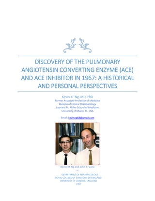
  • 1. DISCOVERY OF THE PULMONARY ANGIOTENSIN CONVERTING ENZYME (ACE) AND ACE INHIBITOR IN 1967: A HISTORICAL AND PERSONAL PERSPECTIVES Kevin KF Ng, MD, PhD Former Associate Professor of Medicine Division of Clinical Pharmacology Leonard M. Miller School of Medicine University of Miami, FL. USA Email: kevinng68@gmail.com Kevin KF Ng and John R. Vane at DEPARTMENT OF PHARMACOLOGY ROYAL COLLEGE OF SURGEONS OF ENGLAND UNIVERSITY OF LONDON, ENGLAND 1967
  • 2. 1 Discovery of pulmonary angiotensin converting enzyme (ACE) and ACE inhibitor in 1967: Friday, Sept 22, 2017 A Historical and Personal Perspectives By Kevin KF Ng, MD, PhD Former Associate Professor of Medicine Division of Clinical Pharmacology Leonard M. Miller School of Medicine University of Miami, FL. USA Email: kevinng68@gmail.com A Tribute to Sir John R. Vane 2017 marks the 50th anniversary of the discovery of angiotensin converting enzyme in the lung. It is an honor and privilege for me to write this as a tribute to Sir John R. Vane (1927-2004) who was awarded the Nobel Prize for Physiology and Medicine in 1982. It was he who developed the blood-bathed-organ technique for the continuous in vivo study of the release and disappearance of pharmacologically active substances in the circulation. He inspired me to use simple experiments to solve complex biological problems. Little did I realize in 1967 that my experimental findings from 1967-1968 could contribute to the development and clinical use of angiotensin converting enzyme (ACE) inhibitors in medicine. Picture shows Kevin KF Ng using the blood-bathed organ technique to study the “Dynamics of the Renin-Angiotensin System” in 1967. IV=intravenous infusion. IBB=into bathing blood.
  • 3. 2 History of the Renin-Angiotensin system The history of the renin-angiotensin system began in 1898 when Robert A. Tigerstedt and Per G. Bergman reported the pressor effect of renal extracts1 . They named the substance renin because it came from the kidney. In 1934, Harry Goldblatt induced experimental hypertension in dogs by clamping a renal artery2 . Using the Goldblatt kidney to produce experimental hypertension in 1936, two independent groups of researchers, one led by Eduardo Braun-Mendendez3 in Argentina and the other led by Irvine Page4 in the United States of America, demonstrated renal secretion of a pressor agent similar to renin. In the following years, both teams described the presence of yet another new compound from the blood of ischemic kidneys. Their subsequent investigations showed that renin acted enzymatically on a plasma protein to produce a pressor substance. In Buenos Aires, it was called hypertensin. In the United States, it was called angiotonin. However, in 1958, Braun Menéndez from Argentina and Irvine Page from the United States agreed to combine these two names together5 . Thus angiotensin was born out of angiotonin and hypertensin. The substrate in the plasma acted on by renin was named angiotensinogen. In 1954, Skeggs et al found that angiotensin existed in two forms: angiotensin I and angiotensin II. Angiotensin I was a biologically inactive decapeptide which was converted to a highly active octapeptide by an enzyme in the plasma6 . This two-enzyme pathway of the renin-angiotensin system is illustrated in the following figure. However, in 1967 Kevin KF Ng using Vane’s Blood-Bathed-Organ technique showed that the conversion was too slow in the extracorporal circulating blood in marked contrast to its rapid conversion through the pulmonary circulation. Substrate Enzyme Angiotensinogen Renin Angiotensin I Plasma converting enzyme Angiotensin II Angiotensinase Inactive products Figure shows the concept of renin-angiotensin system prior to 1967. Fatherly advice from JH Burn When I was a medical student in Singapore, I was intrigued by a severe fall in the blood pressure of a patient treated with intramuscular emetine for amoebiasis. My curiosity in
  • 4. 3 this event led to my appointment as Assistant Lecturer in Pharmacology in the University of Singapore where I discovered that emetine acted like an adrenergic neuron blocking agent7,8 . In Feb 1966, Joshua H. Burn who was Professor Emeritus from the University of Oxford came to Singapore as an External Examiner in Pharmacology. He inspired me to work on the Burn and Rand theory in the mechanism of release of noradrenaline from the sympathetic nerve endings9,10 . In June 1966, I was awarded a Merck Sharp and Dohme International Fellowship for Clinical Pharmacology in the USA. At the same time, I was awarded an Overseas Scholarship by the University of Singapore to work for a doctorate degree in Pharmacology. JH Burn who was back in England wrote to me that I should be well trained in basic pharmacology prior to specializing in clinical pharmacology. He strongly urged me to work with one of his former staff members John R. Vane who was then a Reader in the Department of Pharmacology at the Royal College of Surgeons of England, University of London. In August 1966, I arrived in London with my wife Gina, two sons Keith and Keiron and my mother-in-law Helena Goh. After we were settled in, I went to meet John R. Vane at the Department of Pharmacology in the Royal College of Surgeons of England. He introduced me to the Chairman of the Department Professor Gustav VR Born, Senior Lecturer Robert L. Hodge, Lecturer YS Bakhle, his research team: Duncan P. Thomas, Sergio Ferreira, John Hughes, Norbert Gilmore and other research, technical and administrative staff. JR Vane led me to his office and asked me what I would like to do. Then he wrote on a piece of paper "anaphylaxis" and asked me to work on this the next day. As a new comer to the laboratory, I had to build my own experimental apparatus. Within a few days, I began to study the effects of acetylcholine, catecholamines (adrenaline, noradrenalin), amines (histamine, 5-hydroxytryptamine), peptides (bradykinin, angiotensin II, vasopressin), prostaglandins and other pharmacologically active substances on isolated superfused organs from various laboratory animals. The goal of these experiments was to look for tissues that might be suitable for the detection of mediators in anaphylaxis. When Robert Hodge left for Australia to assume the position of Director of the National Heart Foundation of Australia, JR Vane asked me to complete the experiments RL Hodge left behind. Selection of isolated organs for bioassay. Isolated organs were removed from the stomach, small intestines, large intestines, colon and rectum from rats, chickens, guinea pigs, rabbits and cats. These were suspended in organ baths and tested for sensitivity and specificity. The tissues were deemed sensitive if they responded to 1-5 nanograms of the substance. To test for specificity, the tissues arranged in a series of three for parallel bioassay, were superfused with Krebs solution from a roller pump at a constant rate of 10-15 ml/min. The responses of the tissues were recorded by auxotonic levers writing on a smoked paper attached to a revolving drum known as kymogragh. Isolated organs responding to adrenaline, noradrenaline, histamine,
  • 5. 4 5-hydroxytryptamine, angiotensin II, bradykinin and prostaglandins were selected for the experiments. Adrenaline relaxed the stomach strip, chick rectum, and rat colon. Noradrenaline relaxed the rat stomach strip and rat colon without any effect on chick rectum. Angiotensin II contracted the rat stomach strip and rat colon without effect on the chick rectum. Cat jejunum was selectively sensitive to bradykinin. Prostaglandin E2 contracted the rat stomach strip, chick rectum and rat colon. 5-hydroxytrytamine contracted only the stomach strip. Figure on the left shows the responses of isolated organs to physiological concentrations of vasoactive substances. Figure on the right shows the arrangement of the isolated organs in series and the incubation circuit for the study of half-lives of vasoactive substances in circulating blood. Half-life of angiotensin II in circulating blood11 These experiments were done on dogs anesthetized with Halothane and intravenous - chloralose supplemented with pentobarbital when necessary. The assay organs were a rat stomach strip and two rat colons; one of the colons was infused intraluminally with propranolol to block the relaxant effect of catecholamines on the tissue. The trachea was cannulated and the lungs were ventilated mechanically with room air. Polyethylene cannulas were tied onto carotid or femoral arteries for blood sampling. Cannulas were also tied onto carotid or femoral veins for venous return. After the dogs were heparinized, arterial blood was pumped at a constant rate of 10-15 ml/min (less than 0.5% of cardiac output of adult dogs) to superfuse the assay organs. The blood was collected in a reservoir and returned by gravity into the carotid or femoral veins. Thus an extracorporeal circulation from a major artery to a major vein was created. Mean arterial blood pressure was measured from the side arm of the arterial cannula with a mercury manometer and recorded on a kymograph. Synthetic angiotensin II amide (Hypertensin) from CIBA was used in all experiments. To study the inactivation of angiotensin II in circulating blood, I lengthened the silicone tubing and immersed it in a water bath maintained at 37 degrees Fahrenheit. This served as an incubation circuit which contained 30-45 ml of blood. A bubble of air was injected into
  • 6. 5 the circulating blood at different points of the tubing to determine the incubation time of angiotensin II. Infusions for 5 minutes were first made into blood before it was pumped to the assay organs. The transit time was 15 seconds. Infusions of angiotensin II were then made into the tubing such that angiotensin II would come in contact with circulating blood for 60 sec, 120 sec and 180 sec. The responses of the assay organs were plotted against incubation time, a 50% reduction in response relative to incubation time was considered to be the half-life of angiotensin II. This was found to be 180 seconds. Disappearance of angiotensin II in vascular beds11 To study the disappearance of angiotensin II in the liver and hind limb, blood was drawn from carotid artery to superfuse the assay organs and returned via femoral vein. Infusions of angiotensin II were given into the portal vein and into the femoral artery to study its disappearance across the vascular beds. To study the disappearance of angiotensin II in the lungs, blood was drawn from the femoral artery to superfuse the assay organs. It was returned to the femoral vein by gravity. Intravenous infusions were given into the femoral vein and intra-aortic infusions was given via cannulas inserted into the ascending aortas close to the aortic valve. The responses obtained after their passage across the vascular beds were calibrated by intravenous infusions. The results of these experiments from October 1966 to early part of 1967 showed that the half-life of angiotensin II in the circulating blood was 180 seconds and the disappearance of angiotensin II was 50-75% in the head, liver and hind limbs. The lungs were unique in that angiotensin II passed through the pulmonary circulation without any loss. Figure shows schematic diagram of the circulation. Responses of assay organs to Intra-arterial (IA) infusions were compared with responses to intravenous (IV) infusions. If the IA response 1.0 µg/min was equal to the response to IV 0.5 µg/min, the disappearance was 50%. While I was doing the experiments on angiotensin II, Sergio Ferreira was doing similar experiments with bradykinin next to my laboratory. He found that bradykinin was rapidly inactivated in circulating blood with a half-life of 18 seconds. The disappearance of bradykinin was 80-95% across the lungs and other vascular beds. This remarkable
  • 7. 6 disappearance of bradykinin in the lungs and a total lack of disappearance of angiotensin II in the pulmonary circulation came as a big surprise for me. The lungs had always been considered as an organ for the exchange of carbon dioxide for oxygen. How polypeptides were handled by the lungs were not known. I presented my results to JR Vane in his office. Looking at the summary table of my results and the tracings on smoked papers, he said “Kevin, history is made”. He suggested that angiotensin II was a systemic hormone, whereas bradykinin was a local hormone and the lung acted like a filter for the systemic circulation. Having completed my studies on the half-life of angiotensin II in circulating blood and its disappearance in vascular beds, I was looking forward to do similar experiments with angiotensin I. However, angiotensin I was not available commercially. Perhaps I could make some angiotensin I in vitro with dog renin extract and plasma. JR Vane agreed and in the summer of 1967, he sent me to Dr. Brown's Blood Pressure Research Unit in Glasgow, Scotland to extract renin from dog kidneys. Conversion of angiotensin I to angiotensin II in circulating blood12 On my return from Glasgow, JR Vane told me that he had received five ampoules of lyophilized horse angiotensin I from Dr. Skeggs. I started the experiments the next day. Stock solutions of angiotensin I were made with distilled water and kept frozen in the refrigerator. Working solutions were diluted with normal saline. Rod Flower who joined the department as a Research technician assisted me daily in my experiments. We anesthetized the dogs and tied cannulas in the arteries and veins. I prepared the assay organs and superfused them with Krebs solution at 10 ml/min. Initial experiments were done to compare the activity of angiotensin I and angiotensin II on the assay organs. After this, the Krebs solution was replaced by circulating blood from the incubation circuit. The relative activity of angiotensin I was expressed as a percentage of activity of angiotensin II. In Krebs solution, angiotensin I had 15% of the activity of angiotensin II. Incubation in circulating blood for 15 sec increased the activity to 27%. This was further increased to 40% after 60 sec, 68% after 120 sec and 93% after 180 sec. By plotting these responses against incubation time, 50% of the increased activity or the half-life of angiotensin I occurred at 90 sec. Since angiotensin II was inactivated in circulating blood with a half-life of 180 sec, the increase in activity on the rat colon represented the net result of conversion of angiotensin I to angiotensin II and the inactivation of angiotensin II.
  • 8. 7 Conversion of angiotensin I to Angiotensin II in the pulmonary circulation12 Angiotensin I had 27% of the activity of angiotensin II on arterial blood-bathed assay organs. The activity was increased to 80% after intravenous infusion. One circulation time through the pulmonary circulation of the dog is less than 5 seconds. It was conceivable that the inactive angiotensin I was converted to the active angiotensin II during its passage through the lungs. To prove this hypothesis, the following experiments were done. The first set of experiments were made on dogs in which a fine cannula was inserted into ascending aorta so that intra-aortic infusions would come into contact with blood after it has passed through the pulmonary circulation. Blood for the assay organs were sampled from femoral artery and returned via femoral vein. The responses on the assay organs made by intra-aortic infusions were compared with those made by intravenous infusions into the femoral vein. The results showed that the relative activity of intra-aortic angiotensin I was 30% , whereas the relative activity of intravenous angiotensin I was 80%. A net increase by 50% in angiotensin I activity across the pulmonary circulation must be due to its conversion from angiotensin I to angiotensin II The second set of experiments was made by two series of isolated organs arranged in parallel and the responses of the isolated tissues were recorded on a multichannel Dynagraph. Synthetic angiotensin I was provided by Dr. M. Weatherall and Dr. S. Wilkinson of Wellcome Foundation. Under anesthesia, cannulas were inserted into left ventricle, right ventricle, femoral artery and femoral vein of the dog. Each series of isolated tissues contained a stomach strip and two rat colons; one colon in each series was infused intraluminally with propranolol to block the relaxant effect of catecholamines on the tissues. One series was bathed in venous blood from the right ventricle, the other series was bathed in arterial blood from the left ventricle. The blood from both series were returned to the animal via femoral vein.
  • 9. 8 Since angiotensin II passed through the lungs without loss, it was infused into the femoral vein to determine the relative activity of angiotensin I before and after it had passed through the pulmonary circulation. The results showed that synthetic angiotensin I had 5% of the activity of angiotensin II in Krebs solution. Contact with blood for 30 seconds from the cannula to the assay organs increased the activity to 16%. On transit from femoral vein to the right ventricle, the relative activity was increased marginally to 18%. In contrast, blood from left ventricular arterial blood showed a remarkable increase of activity to 36%. The increase in relative activity from 18% in right ventricle to 36% in left ventricle represented a 100% change in activity. This showed that angiotensin I was indeed converted to angiotensin II during its passage through the pulmonary circulation. The third set of experiments was performed because earlier results were obtained with horse angiotensin I and synthetic angiotensin I. Both could be different from dog angiotensin I. To prove this, I used exogenous dog renin which I prepared in Glasgow and I induced endogenous renin release into the circulation by partial occlusion of blood flow to the renal arteries of the dog. Blood was sampled from the right and left ventricles for the assay and returned via the femoral vein by gravity. Two series of isolated tissues were used; one bathed in venous blood from the right ventricle and the other bathed in arterial blood from left ventricle. Exogenous renin had no activity on the rat colons bathed in Krebs solution. It was impossible to generate angiotensin I in the incubation circuit because of its long half-life in the circulation and angiotensin I was relatively inactive on the assay tissues. When renin was infused intravenously for 10 min, the contraction of the rat colons gradually reached a plateau. When the infusion stopped, the contractions of the rat colons declined exponentially with a half-life of 15-20 minutes. Calibration of these responses with angiotensin II showed that the contractions caused by exogenous renin were smaller in venous blood compared with those in arterial blood. Similar responses were obtained from endogenous renin when the blood flow to the kidneys was partially reduced by inflating a balloon above the renal arteries. These results were consistent with the observations made
  • 10. 9 by intravenous horse and synthetic angiotensin I and showed that angiotensin I was indeed converted to angiotensin II in the pulmonary circulation. Disappearance of Angiotensin I in peripheral vascular beds13 To discover what happened to angiotensin I in peripheral vascular beds, infusions of angiotensin I were given into carotid, renal and femora arteries. The effects on the rat colons bathed in arterial blood were calibrated with intravenous angiotensin I. The disappearance was 80-90% in the kidneys, 60-90% in hind limbs and 40-50% in the head. To determine the disappearance of angiotensin I throughout the peripheral vascular beds excluding the lungs, a coaxial polyethylene tubing was inserted into the aorta close to the aortic valves. The inner tubing was used for supplying arterial blood to the assay tissues while the outer tubing which was 2 cm shorter was used to infuse angiotensin I to the peripheral vascular bed. Calibration by intravenous angiotensin I showed that 50% to 60% of angiotensin I disappeared in the peripheral vascular beds. Since angiotensin I and angiotensin II contracted the rat colon, it was not possible to distinguish the identity of the polypeptides in the arterial blood. To circumvent this problem, blood was sampled from the right and left ventricles for bioassay. The response of the assay organs was calibrated with intravenous angiotensin II since it passed through the pulmonary circulation without loss. The experiments with intra-aortic coaxial catheter showed that the responses of the rat colons bathed in right ventricle venous blood were smaller than those bathed in left ventricle arterial blood. I could not determine whether angiotensin I was converted to angiotensin II in the peripheral vascular beds but the fraction that passed through appeared to be angiotensin I because its activity was increased by 100% on transit from right ventricle to left ventricle. The evidence again indicated that the conversion of angiotensin I to angiotensin II took place in the pulmonary circulation. Figure shows % disappearance of angiotensin I and angiotensin II in vascular beds¹⁴.
  • 11. 10 Mechanism of conversion of angiotensin I to angiotensin II¹⁵ It has been established that Angiotensin I is a 10 amino acid polypeptide, angiotensin II is a 8 amino acid polypeptide and bradykinin is a 9 amino acid polypeptide as shown below: 1 2 3 4 5 6 7 8 9 10 Angiotensin I: Asp-Arg-Val-Tyr-Ile-His-Pro-Phe-His-Leu Angiotensin II: Asp-Arg-Val-Tyr-Ile-His-Pro-Phe Bradykinin: Arg-Pro-Pro-Gly-Phe-Ser-Pro-Phe-Arg It has also been shown that the plasma converting enzyme split the phenylalanyl-histidine link in angiotensin I to form angiotensin II and a histidyl-leucine residue. Furthermore, it has also been shown that bradykinin is inactivated by a carboxypeptidase which removes the C-terminal arginine from bradykinin. The chemical structure of angiotensin II and that of bradykinin showed that their last two amino acids were identical. It was possible that the pulmonary converting enzyme was similar to the enzyme that inactivated bradykinin. To confirm this, further experiments were done with an angiotensin I analogue and a bradykinin potenting factor (BPF). Angiotensin I and a HHL angiotensin I analogue were synthesized by Dr. S. Williams of Wellcome Foundation. He inserted an extra histidine at position 9 between histidyl-leucine linkage making it an undecapeptide angiotensin I analogue as follow: 1 2 3 4 5 6 7 8 9 10 11 Angiotensin I: Asp-Arg-Val-Tyr-Ile-His-Pro-Phe-His-Leu HHLAngiotensin I: Asp-Arg-Val-Tyr-Ile-His-Pro-Phe-His-His-Leu The pulmonary angiotensin converting enzyme may be an endopeptidase similar to that in the plasma or a carboxypeptidase which first cleaved histidine and then leucine from angiotensin I. The availability of HHL angiotensin I analogue made it possible to test this theory with the blood bathed organ technique. In anesthetized dogs, blood was sampled from femoral artery to superfuse the assay tissues and returned via femoral vein. Intravenous infusions were given into femoral vein and intra-aortic infusions were given into ascending aorta close to the aortic valves. The contractions of the rat colons in Krebs solution or circulating blood were calibrated with synthetic angiotensin I or synthetic angiotensin II. In Krebs solution, HHL angiotensin I had less than 1% of the activity of synthetic angiotensin II. There was no significant change after incubating in circulating blood for 30 sec. Comparisons of the relative activity of HHL angiotensin I analogue with angiotensin II after intravenous infusions showed that 50- 60% of the analogue disappeared in the pulmonary circulation. Thus, there was no evidence of increase in activity after its passage through the pulmonary circulation.
  • 12. 11 The inactivation of HHL angiotensin I analogue in the lungs was consistent with the theory that the pulmonary converting enzyme was an endopeptidase similar to that in plasma because stepwise cleavage by a carboxypeptidase would have resulted in angiotensin II which was resistant to destruction by the lungs. Inhibition of pulmonary angiotensin converting enzyme by BPF15 In order to prove that the pulmonary converting enzyme was similar to the enzyme that inactivated bradykinin, I obtained a small quantity of Bradykinin Potentiating Factor (BPF) from Sergio Ferreira. BPF is a mixture of peptides derived from the venom of Brazilian snake Bothrops jararaca. It has been shown to potentiate the actions of bradykinin in vitro. Due to scarcity of BPF, the experiments could only be done in anesthetized rats. Intravenous injections of angiotensin I and angiotensin II raised the arterial blood pressure of rats with a potency ratio of 2:1. With intravenous infusion of BPF, the ratio of potency decreased to 15:1 showing for the first time that the in vivo conversion of angiotensin I to angiotensin II was inhibited by bradykinin potentiating factor (BPF). Figure shows the potency ratio of angiotensin I to angiotensin II on blood pressor of a rat. Before BPF infusion, the potency ration of angiotensin I to angiotensin II was 2:1. During the infusion of BPF 0.1 mg/min, the potency ration was significantly reduced to 15:1. In September 1968, I submitted my thesis "The dynamics of the renin-angiotensin system" for the degree of Doctor of Philosophy to the University of London. The Internal Examiner Professor JR Vane and the External Examiner Professor WS Peart both congratulated me for the work well done. The degree of Doctor of Philosophy in Pharmacology was conferred upon me on October 9th , 1968. Further experiments on the inhibition of pulmonary angiotensin converting enzyme by BPF were continued by YS Bakhle and Sergio Ferreira.
  • 13. 12 As I was making plans to return to Singapore in October 1968, JH Burn went to St. Louis in Missouri where he met Dr. John C. McGiff. JH Burn told JC McGiff about Vane’s new technique for the study of vasoactive substances in the circulation. Keen to adapt this new technique to his research, JC McGiff invited JR Vane to come to the USA. Due to a busy schedule, JR Vane asked me to stop over USA on my way back to Singapore. This would give me the opportunity to set up the blood-bathed organ system not only for McGiff in St Louis, Missouri but also for Dr. Irvine Page in San Francisco, California. My family and I left London for the USA in late October 1968. On my return to Singapore from USA, JR Vane introduced me to many of his colleagues who invited me to give a seminar on the "Dynamics of the renin-angiotensin system" in the United States. The stop over in USA gave me the opportunity to meet my former teacher Robert CY Lin in Hong Kong and friends in the Far East. The following is the list of laboratories and institutes of higher learning where I made my presentations: • The Wellcome Research Laboratories in New York • Smith Kline & French Laboratories in Philadelphia • Department of Pharmacology, State University of New York in Buffalo • Cleveland Clinic in Ohio • Department of Internal Medicine, Washington University in Saint Louis • School of Medicine, Saint Louis University in Missouri • Cardiology Club of Saint Louis, Missouri • Lilly Laboratory for Clinical Research in Indianapolis • Department of Pharmacology, University of Utah in Salt Lake City • Riker Laboratories in Northridge, California • Department of Physiology, University of Hawaii in Honolulu • College of Medicine, National Taiwan University in Taiwan • Department of Pharmacology, University of Hong Kong The seminars were well received and the itinerary was memorable. My family and I finally arrived in Singapore on Christmas day December 25th 1968. The discovery of the pulmonary converting enzyme and its inhibition by BPF led to the synthesis of captopril (Capoten), the first ACE inhibitor used clinically in 1982. There are now ten ACE inhibitors available in the United States for the treatment of hypertension, congestive heart failure and diabetic kidney disease: captopril, enalapril, fosinopril, lisinopril, benazepril, moexipril, perindopril, quinapril, ramipril and trandolapril. References 1 Tigerstedt R and Bergman PG. Niere und Kreislauf. Skand Arch Physiol. 1898; 8: 223– 27 2 Goldblatt H, Lynch J, Hanzal RF and Summerville WW. Studies on experimental hypertension, I: the production of persistent elevation of systolic blood pressure by means of renal ischemia. J Exp Med. 1934; 59: 347–379.
  • 14. 13 3 Taquini AC and Braun-Menéndez E. Liberación de substancia vaso-constrictora en el riñón completamente isquemiado. Rev Soc Arg Biol. 1938; 14: 422–429 4 Page IH. Vasopressor action of extracts of plasma of normal dogs and dogs with experimentally produced hypertension. Proc Soc Exp Biol. 1937; 35: 112–11 5 Braun-Menéndez E and Page IH. Suggested revision of nomenclature: angiotensin. Science. 1958; 127: 242. 6 Skeggs LT, Marsh WH, Kahn JR and Shumway, NP. The existense of two forms of hypertensin. J. Exp. Med. 1954:99:275-280. 7 Ng KKF. A new pharmacological action of emetine. BMJ. 1966;1: 1278-1279. 8 Ng KKF. Blockade of adrenergic and cholinergic transmissions by emetine. Br. J. Pharmacol. Chemother. 1966; 28:228-237. 9 Burn JH and Ng KKF. The action of pempidine and antiadrenaline substances on sympathetic post-ganglionic terminations. Br. J. Pharmacol. Chemother. 1965; 24:678-688 10 Ng KKF. The effects of some anti-cholinesterases on the response of the taenia to sympathetic nerve stimulation. J.Physiol. 1966;182:233-243. 11 Hodge RL, Ng KKF and Vane JR. Disappearance of angiotensin from the circulation of the dog. Nature. 1967; 215:138-141. 12 Ng KKF and Vane JR. Conversion of angiotensin I to angiotensin II. Nature. 1967; 216:762-766. 13 Ng KKF and Vane JR. Fate of angiotensin I in the circulation. Nature. 1968; 218:144- 150. 14 Ng KKF. The Dynamics of the renin-angiotensin system. PhD thesis, University of London, 1968. 15 Ng KKF and Vane JR. Some properties of angiotensin converting enzyme in the lung in vivo. Nature. 1970; 225:1142-1144.