Angiography
•Download as PPTX, PDF•
101 likes•55,607 views

Angiography is a medical imaging technique that uses radiography to visualize blood vessels and organs after injecting a contrast medium. It can detect diseases of the arteries and veins like atherosclerosis, aneurysms, and internal bleeding. The procedure involves a team inserting a catheter into the blood vessels and injecting iodinated contrast dye before capturing x-ray images. Risks are generally low but can include minor bleeding, vessel damage, and allergic reactions to the contrast medium. Advances in digital subtraction angiography now make angiography the gold standard for assessing vascular diseases when other imaging modalities are inconclusive.

Recommended
More Related Content
What's hot
What's hot (20)
Similar to Angiography
Similar to Angiography (20)
Recently uploaded
Recently uploaded (20)
Angiography
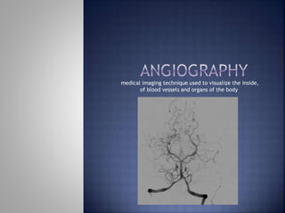
- 1. medical imaging technique used to visualize the inside, of blood vessels and organs of the body
- 2. What do we visualize with an angiographic procedure?
- 3. Is the general term that describes the radiologic examination of vascular structures within the body after the introduction of an iodinated contrast medium or gas
- 4. The first angiogram was performed only months after Roentgen's discovery Which was when? Two physicians injected chalk or mercury salts into an amputated hand and created an image of the arteries
- 5. It is used to assess for diseases of the: Arteries (these take blood to the brain, limbs andabdominal organs) Veins (these carry blood back to the heart) These diseases may include: 1. Atherosclerosis: 'furring' of the arteries causing them to narrow. 2. Aneurysms: blood vessels that become enlarged with a risk of bursting. 3. Conditions causing internal bleeding, amongst many others.
- 8. which demonstrate the vasculature to a greater or less degree CT MRI (MRA) Ultrasound (particularly Doppler) Nuclear Medicine are all used to image vessels and each has its advantages and disadvantages Vessel imaging is a constantly evolving area.
- 9. Radiologist/ Specialist Nurse 2-3 Radiologic Technologists (CV) Sometimes Anesthesiologist
- 10. Verify the presence of tumors Blood supply to tumors Internal bleeding Possible anemia Stenosis Can be caused form atherosclerosis Occlusions Clots Thrombus Embolus Aneurysms Heart disease
- 11. Previous severe reaction to contrast Impaired renal function Impaired blood clotting factors Inability to undergo surgical procedure
- 12. Iodinated contrast media is used Can produce nausea & an uncomfortable burning sensation Allergic reactions Severe: anaphylactic shock Shock, rapid shallow breathing, high pulse rate & ALOC Mild: Hives or slight difficulty breathing
- 13. Once you are home you should drink plenty of fluids (avoiding alcohol) for the first 24 hours, and take things easy for 48 hours. You should avoid driving, strenuous activity and sexual intercourse. This allows time for the artery to heal. A more detailed booklet will be given to you called "Discharge advice following a Vascular Radiology Procedure" when you go home.
- 14. Conventional angiography is usually a very safe procedure but because it is more invasive than MRA or CTA, the risks of complications are greater. The risks include: minor bleeding/bruising and a small risk of damage to the vessels, but the risk of serious complications is rare. It is possible to suffer an allergic reaction due to the contrast medium required during the test. Thankfully, they are uncommon and usually minor (mild rash or itching). More severe reactions are possible (1 in 2500 patients) and very rarely can be life threatening (1 in 25,000). It is important to tell your Doctor or radiographer if you have had a previous reaction to contrast medium before your test commences.
- 15. Maintain flow rate Includes heating device ( To reduce the viscosity of the contrast media by keeping it near body temperature )
- 16. 16 In most angiographic studies contrast must be administered at a consistent speed either faster as in abdominal angiography or slower as in lymphangiography SAFETY MEASURES: P 638 LIGHT / ALARM /
- 18. Now primarily uses: DSA: Digital subtraction angiography This is still considered the gold standard of vessel imaging when other modalities are inconclusive Now common practice to be considered as an area needing advanced training for: 1. Radiologist: Interventional 2. R. T. (CIT, CV) etc ANGIO tech
- 19. Technical innovations image intensification three-phase generators rapid film changers automatic pressure injectors advanced catheter technology all helped to establish angiography as an essential diagnostic tool by the 1960s
- 20. An important offshoot of angiographic imaging have created a therapeutic technology Embolization intra-arterial drug therapy transluminal angioplasty are among the procedures that have radically changed and broadened the scope of the diagnostic imaging department
- 21. Biplane C-arm digital imaging Autoinjector --syringes, a heating device, a high-pressure mechanism a control panel Image Intensifying screen Sliding table Rapid film changer (NOW DIGITAL*) Cut film 6 & Cassette changer /magazine
- 22. 1. Puncture Needle Stylet and Cannula large cannula size (1.6mm) 2. Guide Wire --Soft flexible wire with the strength to pass through curved vessels (.6 – 1.0)
- 23. Vascular access needles Size based on external diameter of needle Allows for appropriate Guidewires matching So internal diameter must also be known
- 24. Used as a platform over which a catheter is to be advanced Once positioned guidewire is fixed and catheter is advanced until it meets the tip of the guidwire Mostly constructed on stainless steel & coated with Teflon
- 25. Short catheters used when multiple catheters will be used Placed in lieu of a catheter
- 28. A subtraction mask is taken before contrast injected Each of digitized image is from the mask Images acquired form 1 image every 2-3 sec Up to 30 images per sec
- 43. 1. www.sth.nhs.uk/clientfiles/File/pd6831_Angiography.pdf 2. http://www.elcamino.edu/faculty/mcolunga/RT%20255/A ngiography%20and%20Arteriography.ppt 3. https://en.wikipedia.org/wiki/Angiography 4. https://www.slideshare.net/jdtomines/angiography-basics