An approach to a Floppy infant - Dr Sujit
•
232 likes•40,925 views

One of the Most discussed topic in every residency

Recommended
More Related Content
What's hot
What's hot (20)
Similar to An approach to a Floppy infant - Dr Sujit
Similar to An approach to a Floppy infant - Dr Sujit (20)
More from Sujit Shrestha
Recently uploaded
Recently uploaded (20)
An approach to a Floppy infant - Dr Sujit
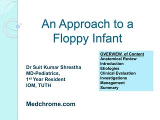
- 1. An Approach to a Floppy Infant Dr Suit Kumar Shrestha MD-Pediatrics, 1st Year Resident IOM, TUTH Medchrome.com OVERVIEW of Content Anatomical Review Introduction Etiologies Clinical Evaluation Investigations Management Summary
- 2. What is Tone? Muscle tone is the resistance offered by a muscle against stretch in resting condition due to state of partial contraction of the extrafusal fibers resulting from asynchronous discharge of the motor neurons. Types- 1. Passive tone 2. Active tone Structures responsible- Muscle Muscle Spindle (Receptor) Group I and II sensory fibers Spinal Cord ( Center) Alpha and Gamma Motor Neurons ( Nerve paths) Supraspinal Control ( Brainstem, Cerebellum, Corex, RF )
- 3. Mechanism The myotactic reflex is the basis of normal tone in a muscle. Muscle tone is maintained at the peripheral level by the participation of the fusimotor system: pathways involving the muscle spindles that promote muscle contraction in response to stretch And the inverse myotactic reflex involving the golgi tendon organ that provides a braking mechanism to the contraction of muscle. A lesion interrupting the stretch reflexes at any level in the lower motor neuron (LMN) will result in a loss of muscle tone and stretch reflexes i.e. flaccidity .
- 4. The output of gamma motor neurons to the muscle spindle is influenced by supraspinal influences predominantly inhibitory. Thus lesions affecting the UMN reduction of these inhibitory influences increase in excitatory output of the gamma motor neurons to the muscle spindle In early infancy, contrary to expected increase in muscle tone, the response to an upper motor neuron lesion in the early stages is flaccidity and loss of muscle tone . This pattern- hypotonia is associated with preserved or hyperactive reflexes later evolves into spasticity. Clinical distinction between an UMN and LMN lesion provides a rationale for investigations based on the localization of lesion in the pathway of motor control (central vs. peripheral hypotonia)
- 5. Hypotonia is characterized by reduced resistance to passive range of motion in joints versus weakness, which is a reduction in the maximum muscle power that can be generated. (Dubowitz, 1985; Crawford, 1992; Martin, 2005) Weak infants always have hypotonia, but hypotonia may exist without weakness. A hypotonic newborn should be considered septic until proven otherwise
- 6. Introduction “Floppy Infant” o A well-recognized entity o An organized approach is essential for Evaluation o Problems like frequent infections, feeding problems, ptosis, ophthalmoplegia and dislocated hips. The word ‘floppy’ can be used to mean: ◦ decrease in muscle tone (hypotonia) ◦ decrease in muscle power (weakness) ◦ ligamentous laxity and increased range of joint mobility. But strictly speaking - hypotonia. Floppy infant refers to those children presenting with generalized hypotonia, most often arising out of an insult incurred during fetal or neonatal period.
- 7. Useful indicators of weakness are: Ability to cough and clear airway secretions (‘cough test’)- Apply pressure to the trachea and wait for a single cough that clears secretions. If more than one cough is needed to clear secretions, this is indicative of weakness. Poor swallowing ability as indicated by drooling and oropharyngeal pooling of secretions. The character of the cry — infants with consistent respiratory weakness have a weak cry. Paradoxical breathing pattern — intercostal muscles paralysed with intact diaphragm.
- 8. Causes of Floppy Infant Syndrome Neurology Chapter of IAP Central nervous system – Perinatal asphyxia Neonatal encephalopathy Kernicterus, Cerebral palsy (atonic type) Intracranial hemorrhage Chromosomal anomalies- Down syndrome Inborn errors of metabolism e.g., aminocidurias, mucopolysaccharidosis and cerebral lipidosis. .
- 9. Causes… Spinal cord lesions Anterior horn cell disease – Werdnig Hoffman spinal muscular atrophy, SMA II and III, Myelopathies. Poliomyelitis Peripheral nervous Acute polyneuropathy Familial dysautonomia Congenital sensory neuropathy. Myoneural junction Neonatal myasthenia gravis Infantile botulism Following antibiotic therapy
- 10. Causes of Floppy Infant Syndrome (Contd.) Neurology Chapter of IAP Muscles Muscular dystrophies, Congenital myotonic dystrophies, Congenital myopathies (including central core disease and nemalin myopathy), Polymyositis, Glycogen storage disease (pompe’s), and Arthrogryposis multiplex congenital. Miscellaneous Protein energy malnutrition, rickets, Prader willi syndrome, Malabsorption syndromes, Ehler-danlos syndrome, Cutis laxa, cretinism.
- 11. The Floppy Infant : Evaluation of Hypotonia DOI: 10.1542/pir.30-9-e66 Pediatrics in Review 2009;30;e66 Dawn E. Peredo and Mark C. Hannibal Causes of Hypotonia Percentage (n=277) HIE 19% Chromosomal /Genetic Syndromes Down syndrome 13% Prader-Willi syndrome 5% 31% Brain Anomalies 13% Myopathies 5% Congenital Myotonic Dystrophy 4% Metabolic disorders 3% Benign Neonatal Hypotonia 3% Spinomuscular Arophy 2% Muscular Dystrophy 2% Others 5% Unknown 13%
- 12. Approach to a Floppy Infant History Presenting Complaints: ◦ Decreased Tone ◦ Difficulty sucking, chewing; Weak cry ◦ Decreased movement ◦ Delayed motor milestones ◦ Complications of muscle weakness: Recurrent respiratory infections, Respiratory difficulty Age of Onset: The onset -important to distinguish between congenital and aquired aetiologies. Eg ◦ SMA Type I -: < 6 months; ◦ SMA Type II : 3mo- 15 mon ◦ SMA Type III : at or after 12 mon ◦ Neonatal myasthenia – soon after birth; ◦ Juvenile myasthenia - >6 mo
- 13. History: Sudden onset: IVH in Premature infants Course- progressive,static or fluctuating Distribution of weakness: ◦ Proximal (Unable to stand from sitting) – Myopathies ◦ Distal (Unable to hold things) – Neuropathy Associated Atrophy of muscles: ◦ Myopathies, ◦ neuropathies Muscle pain: ◦ Acute polyneuropathies, ◦ myositis, ◦ Metabolic disease, ◦ Ischemic myopathies
- 14. History: Joint deformities ◦ Arthrogryposis Zellweger Syndrome, Chromosomal disorder Congenital Muscular dystrophy, Myotonic dystrophy, Transitory Neonatal myasthenia Congenital dislocation of hip- frequent finding CNS causes: ◦ Seizures , Abnormal movement, ◦ Mental retardation/Learning disabilities, ataxia ◦ Poor state of alertness ◦ Lack of response to visual and auditory stimuli ◦ Dysmorphic features
- 15. Dysmorphic Features: ◦ Cerebral dysgenesis, ◦ Zellweger syndrome, ◦ Prader-Willi Syndrome ◦ Fiber type Disproportion myopathies, ◦ Neonatal myotonic Dystrophies Severity: ◦ Respiratory difficulty (Involvement of respiratory muscles) ◦ Apnea, Aspiration (Gag reflex lost) H/o Fatigue on continuous sucking – Myasthenia H/o Constipation – Botulism H/o easy bruising, poor wound healing – Ehler- Danlos
- 16. Relevant History: The pre-, peri- and postnatal history is important. Enquire about the quality and quantity of fetal movements, breech presentation and the presence of either poly or oligohydramnios. Neonatal seizures and an encephalopathic state ( Central) Documentation of birth trauma, birth anoxia, delivery complications, low cord pH and Apgar scores are crucial as HIE remains an important cause. H/O consanguinity and identify other affected family members in order to reach a definitive diagnosis, using a detailed family pedigree to assist future genetic counselling
- 17. Development h/o: ◦ Delayed milestones Family h/o: A family history of neuromuscular abnormalities may be informative because many disorders are inherited. Examples of familial neuromuscular diseases include congenital myotonic dystrophy, spinal muscular atrophy, metabolic disorders (e.g., mitochondrial disease, acid maltase deficiency, defects of creatine synthesis).
- 18. Examination: Detection of Hypotonia: Main Features: ◦ Bizarre or Unusual postures ◦ Decreased resistance of joints to passive movement ◦ Increase in range of movement of joints ◦ Decreased Spontaneous movements
- 19. Clinical signs in a floppy infant 1. ‘frog-leg’ posture- generally implies reduced spontaneous movement, with the legs fully abducted and arms lying beside the body either extended or flexed 2. Significant head lag on traction or pull-to-sit manoeuvre and excessively rounded back when sitting (>33 weeks) 3. Rag-doll posture on ventral suspension 4. Vertical suspension test – feeling of ‘slipping through the hands’ when the infant is held under the arms 5. Various associated examination findings such as flat occiput or congenital dislocation of the hips, arthrogryposis.
- 20. Appearance of the Floppy Infant: Supine position: ◦ Paucity of spontaneous movement ◦ Fully abducted at hip joint ◦ Thighs externally rotated (Frog-leg posture) ◦ Arms extended or flexed at elbow with hands beside the head Traction response Head lag when pulled to sit Normally, Flexion at the elbow, knee and ankle (To counter traction) 4wks: Head in plane of body momentarily 20 wks: No head lag
- 21. Examination: Sitting Posture: ◦ Head falls forward ◦ Trunk control poor ◦ Unable to sit unsupported Vertical suspension: ◦ Hold at axilla ◦ Head falls forward ◦ Legs dangle or scissoring (Normally flex) ◦ Tendency to slip through one’s hands Horizontal suspension: Normally, head erect, back straight, limbs flexed Hypotonic – head and legs hang limply, Rag doll Prone: Unable to lift head and trunk
- 22. Examination: Palpation of muscles: Flabby Adductor angle: Angle between thighs when hips maximally abducted with extension at knees Popliteal angle: Hips flexed onto abdomen by holding at the knees Dorsiflexion angle of the foot: By gentle pressure on the sole Heel to ear manoeuvre: Both extended legs lifted towards the ears without lifting the pelvis Scarf sign: Flexed at elbow and pulled across the chest by holding at the hand and wrist Adductor angle
- 24. Diagnostic approach The initial approach to a floppy infant is to determine whether the problem is of central or peripheral origin. This is of crucial importance when forming a plan for diagnostic investigation.
- 26. Floppy Strong Floppy Weak
- 27. Differentiating Features of a Floppy Infant according to Site of Involvement Site of involvement Extent of weakness Proximal vs. distal weakness Face Arms Legs Central - + + > or = Anterior horn cell + ++++ ++++ > or = Peripheral nerve - +++ +++ < Neuromuscular junction +++ +++ +++ = Muscle Variable ++ + >
- 28. Differentiating Features of a Floppy Infant according to Site of Involvement (Contd.) Site of involvement Deep tendon reflexes EMG Muscle biopsy Central Normal or increased Normal Normal Anterior horn cell Absent Fasciculation / fibrillation Denervation pattern Peripheral nerve Decreased Fibrillation Denervation pattern Neuromuscular junction Normal Decremental / incremental Normal Muscle Decreased Short duration small amplitude potential Characteristic
- 29. A distinct pattern of weakness • Axial weakness is a significant feature in central hypotonia. • Generalised weakness with sparing of the diaphragm, facial muscles, pelvis and sphincters suggests anterior horn cell involvement. • With myasthenic syndromes, the bulbar and oculomotor muscles exhibit a greater degree of involvement. • Progressive proximal symmetrical weakness suggests a dystrophinopathy. Signs of proximal weakness in the older infant include a lordotic posture, Trendelenburg gait and Gower sign. • A striking distribution of weakness of the face, upper arms and shoulders suggests fascioscapulohumeralmuscular dystrophy. • Distal muscle groups are predominantly affected with peripheral neuropathies.
- 33. Clinical features - Hypotonia of central origin Social and cognitive impairment in addition to motor delay Dysmorphic features implying a syndrome or other organ malformations sometimes implying a syndrome Fisting of hands Normal or brisk tendon reflexes Features of pseudobulbar palsy, brisk jaw jerk, crossed adductor response or scissoring on vertical suspension Features that may suggest an underlying spinal dysraphism History suggestive of hypoxic-ischaemic encephalopathy, birth trauma or symptomatic hypoglycaemia Seizures
- 34. Indicators of peripheral hypotonia Delay in motor milestones with relative normality of social and cognitive development Family history of neuromuscular disorders/maternal myotonia Reduced or absent spontaneous antigravity movements, reduced or absent deep tendon jerks and increased range of joint mobility Frog-leg posture or ‘jug-handle’ posture of arms in association with marked paucity of spontaneous movement Myopathic facies (open mouth with tented upper lip, poor lip seal when sucking, lack of facial expression, ptosis and restricted ocular movements) Muscle fasciculation- rare but diagnostic Other corroborative evidence including muscle atrophy, muscle hypertrophy and absent or depressed deep tendonreflexes
- 35. Investigations: I. Motor Unit disorder: Serum Creatine Kinase: Prior to EMG or Biopsy ◦ ↑↑ in rapidly progressive myopathies ◦ Maybe normal in Fiber type Disproportion myopathies and some metabolic myopathies ◦ May ↑ in rapidly progressive neuronopathies (SMA) EMG: ◦ Myopathies – Brief, small amplitude, Polyphasic potentials (BSAPPs) Muscular Dystrophies, Myotonic Dystrophies ◦ Neuropathies- Denervation potentials at rest (Fibrillations, Fasciculations, Sharp waves) and Motor unit potentials large in size, prolonged and polyphasic
- 36. Inv: Nerve Conduction Test: ◦ Demyelinating Conditions – Slower velocity ◦ Helps localise site of traumatic nerve injury ◦ Repetitive nerve stimulation: Increase in size of motor unit potentials – Infantile botulism Decremental response - Myasthenia Gravis • Muscle Biopsy: immunohistochemical staining and electron microscopy – The method of choice for differentiating myopathies and muscular dystrophies – more invasive – If biopsy shows specific abnormalites, could be an essential part of the diagnostic evaluation in the newborn to guide subsequent DNA molecular diagnostic studies
- 37. Nerve Biopsy: Sural nerve ◦ Demyelination ◦ Multifocal endoneurial edema, Mononuclear infiltrates – CIDP ◦ Metabolic products Tensilon Test: ◦ Edrophonium 0.15mg/kg sc in neonates, response in 10 mins ◦ 0.2mg/kg iv in infants, response in 1 min Serum Acetylcholine receptor Protein Antibody Stool – Clostridium botulinum
- 38. Inv: CSF examination: ◦ Increased protein (Albumino-cytological Dissociation) – GBS, Congenital hypomyelinating neuropathy ECG: ◦ For associated Cardiomyopathy ◦ Acid maltase deficiency: Short PR, High QRS in all leads Chest X-Ray: ◦ Pneumonia ◦ CHF: Acid maltase deficiency, Carnitine deficiency
- 39. Investigations - CNS causes of Hypotonia: CT Scan of head/MRI: ◦ Intraventricular/parenchymal hemorrhage ◦ CNS malformations ◦ Ischemic changes EEG: Epileptiform activities Chromosome analysis: Trisomy 21- Down’s, Chromosome 15 translocation – Prader Willi X-Ray/MRI of Spine OTHERS- ◦ TFT ◦ TORCH ◦ Investigations for liver, renal involvement Look for Sepsis Looks Like Sepsis without Sepsis
- 40. Investigations in cases where a central cause for hypotonia is suspected Investigations of peripheral hypotonia Serum electrolytes, including ca,phosphate, serum ALP, venous blood gas, TFT Plasma copper/ ceruloplasmin assay (as screening test for Menkes syndrome) Chromosomal analysis (trisomy), testing for Prader-Willi syndrome(15q11–13) Plasma amino acids and urine organic acids, Urine mucopolysaccharide screen (GAG) Molecular/biochemical diagnosis of pro-collagen disorders Very long chain fatty acids Medical genetics opinion Ophthalmology opinion Brain imaging (CT/MRI) Creatinine kinase Lactate EMG /NCS/repetitive nerve stimulation test M uscle biopsy (histology, immunohistochemistry, electron microscopy, respiratory chain enzyme analysis) Genetic testing (SMN gene deletion present in 95% of cases of spinal muscular atrophy type 1, myotonic dystrophy, congenital myasthenic syndromes) Nerve biopsy (rarely) Tensilon test
- 41. USEFUL EMG FEATURES IN PERIPHERAL HYPOTONIA EMG /NCS studies may distinguish between neurogenic, myopathic and myasthenic aetiologies for hypotonia Neurogenic – large amplitude action potentials, reduced interference pattern, increased internal instability Myopathic – small amplitude action potentials with increased interference pattern Myotonic – increased insertional activity Myasthenic – abnormal repetitive and single fibre studies
- 42. Diagnostic Yield Method of Diagnosis % Successfully Diagnosed History and Physical Examination (Step 1) 50% ◦ Family history, Pregnancy and delivery ◦ Clinical and neurologic examination Imaging Study (CT or MRI/MRS) (Step 2) 13% Clinical Genetic Evaluation (Step 3) 9% Genetic Testing (Step 4) 6% ◦ Karyotype, FISH, CGH Biochemical Evaluation (Step 5) 6% ◦ Amino acids, organic acids, peroxisomes, ◦ carnitine, CDG test Neuromuscular Testing (Step 6) 6% ◦ CK, EMG, NCV, DNA for SMA and CMD, muscle biopsy Follow-up Testing 7% ◦ Some tests repeated/Further tests Adapted from Paro-Panjan D, Neubauer D. Congenital hypotonia: is there an algorithm? J Child Neurol. 2004;19:439–44
- 43. Management: Most have no cure and have a progressive course Aim: ◦ Provide Life support: Intubation and mechanical ventilation, Feeding support ◦ Prevent and relieve contractures- Physiotherapy, Casts, Surgical management ◦ Prevent and treat Infections (Pneumonia)
- 44. Principles of management Physiotherapy - stretches aimed at prevention of contractures Occupational therapy - appliances, improvement of posture and function, facilitating activities of daily living Prevention and correction of scoliosis Evaluation and treatment of associated cardiac dysfunction Respiratory support - assessment of requirement for invasive or non-invasive ventilation and/or tracheostomy
- 45. Cont… management. Feeding - nasogastric feeding, caloric supplementation,gastrostomy Management of gastro-oesophageal reflux - medical or fundoplication Orthopaedic intervention in setting of established or evolving joint contractures Encouragement of overall development and stimulation of learning Prevention (influenza and pneumococcal vaccination) and prompt treatment of respiratory infections
- 46. Mx- Specific Treatment of Cause: ◦ Gabapentin, Riluzole, Caspase inhibitors in SMA ◦ Prednisolone for CIDP, Inflammatory Myopathies ◦ High Protein diet in Pompe’s ◦ L-carnitine replacement in carnitine deficiency Genetic Counseling Psychological support, Counselling of parents
- 47. Recent developments Recent advances in genetics have uncovered new conditions causing hypotonia and weakness such as congenital myasthenic syndromes and spinal muscular atrophy variants Advances in immunohistochemistry, electron microscopy and genetics have led to a more specific diagnosis of myopathies Some of these advances have allowed for specific therapeutic interventions, e.g. use of acetylcholinesterase inhibitors in some congenital myasthenic syndromes
- 48. Neurology Chapter of IAP Common causes of floppy infant Cerebral Palsy Many hypotonic children due to causes in central nervous system are mentally retarded. In atonic or hypotonic cerebral palsy, reflexes are brisk in spite of generalized flaccidity. Floppy infant due to cerebral causes is associated with lethargy, poor feeding, and lack of alertness, poor Moro’s reflex, and seizures during the neonatal period.
- 49. Neurology Chapter of IAP Werdnig Hoffman disease It is characterized by marked hypotonia, sluggish fetal movement, and fasciculation of tongue. The child is alert. Feeding behaviour and cry are poor. Deep tendon reflexes are absent. Muscle biopsy shows neurogenic type of atrophy or that the muscle spindles are atrophied in groups. Disease is inherited as an autosomal may be available. Death occurs by 2-4 years of age.
- 50. Neurology Chapter of IAP Myasthenia gravis Occurs in about 12 percent of the babies born to mothers wih MG. Characterized by marked hypotonia, pooling of oral secretions, poor feeding, feeble cry and generalized muscle weakness appearing within 2-3 days after the birth. Baby is alert. Facial weakness manifests by mask-like facies, open mouth and staring look. External opthalmoplegia and ptosis are rare. Deep tendon reflexes are normal. The prognosis is substantiated by improvement in the muscle functions following intramuscular injection of edrophonium chloride 1 mg or neostigmine methyl sulfate 0.1 mg. the condition lasts for 3 to 4 weeks. The child is treated with neostigmine methyl sulphate 0.1 to 0.5 mg IM 10 minutes before each feel for 1 or 2 days followed by neostigmine bromide, 1 to 4 mg orally half an hour before each feed.
- 51. Neurology Chapter of IAP Congenital myopathies These are rare inherited disorders resulting in a benign congenital hypotonia, with generally good outlook for normal life span. Nemaline myopathy is the most common variant. Other disorders of this group include the central core disease, myotubular myopathy and congenital fiber type disproportion.
- 52. Neurology Chapter of IAP Others In polyneuritis there is symmetrical weakness of the limbs with sensory changes. The diagnosis of Pompe’s disease is suspected when the child has macroglossia, cardiomegaly and generalized hypotonia. Babies with prader-willi syndrome are mentally retarded and obese; deep tendon reflexes are diminished. Diabetes mellitus occurs later in life. Testes may be undescended. Ehlers-danlos syndrome is characterized by hyperelasticity of the skin, hyperflexibility of joints and extreme, fragility of skin. Wound healing is delayed and there are freely movable subcutaneous nodules. In cutis laxa, the child has loose skin hanging in baggy folds.
- 53. references Nelson Text Book of Pediatrics 18th Edition Essential Pediatrics- OP Ghai 6th Edition The Floppy Infant : Evaluation of Hypotonia-Pediatrics in Review 2009;30;e66 Dawn E. Peredo and Mark C. Hannibal The floppy infant: contribution of genetic and metabolic disorders- Asuri N. Prasada,*, Chitra Prasad- Received 4 October 2002; received in revised form 29 January 2003; accepted 19 March 2003 IAP Neurology Evaluation of the floppy infant Vasantha Gowda ,Jeremy Parr, Sandeep Jayawant Neurology Symposium From Volpe J: Neurology of the Newborn, 4th ed. Philadelphia, WB Saunders, 2001, p 645