

Recommended
More Related Content
What's hot
What's hot (20)
Viewers also liked
Viewers also liked (20)
Similar to Imaging and pathology of larynx (2)
Similar to Imaging and pathology of larynx (2) (20)
More from Sunil Kumar
Recently uploaded
Recently uploaded (20)
Imaging and pathology of larynx (2)
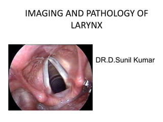
- 1. IMAGING AND PATHOLOGY OF LARYNX DR.D.Sunil Kumar
- 2. • Imaging of the larynx can be an extremely difficult endeavor. The anatomy is complex. The target moves with every breath and every swallow. In certain situations, however, imaging addresses questions that the clinician has difficulty in answering and can make a considerable difference in treatment planning. • The mucosa can be examined thoroughly. To be relevant, imaging must provide information that cannot be obtained by direct visualization. Thus, the usual intent of the radiologist is to evaluate the deeper tissues. • In some cases, a bulky lesion of the upper larynx may block the view of the lower larynx, and imaging may assist in defining caudal extent of disease.
- 3. Anatomy • The larynx extends from the tip of epiglottis to the inferior margin of the cricoid cartilage. • Laryngeal Cartilages – The laryngeal skeleton provides the framework by which the supporting soft tissues function. The laryngeal skeleton consists of three singular cartilages the thyroid, epiglottic, and cricoid cartilages and three paired small cartilages: the arytenoid, cuneiform, and corniculate cartilages. – Understanding of the normal cross-sectional CT anatomy of the larynx is aided by familiarity with these cartilagenous structures at different anatomic levels.
- 4. • Thyroid Cartilage. – The thyroid cartilage is a shield-shaped structure consisting of paired Iaminae fused in the midline, forming a more acute angle in males than in females. – In addition, the thyroid cartilage has two superior projections (cornu), which extend toward the hyoid, and two similar projections interiorly, which articulate with the cricoid. – The broad thyroid cartilage provides some protection from trauma for the intrinsic soft tissues of the larynx.
- 5. • The cricoid cartilage is the foundation of the larynx and is the only complete ring. • The posterior part is larger than the anterior, giving the cartilage the appearance of a signet ring facing posteriorly. • The lower margin of the cricoid cartilage represents the lower margin of the larynx.
- 6. • Epiglottis. – This cartilaginous structure is situated behind the body of the hyoid. – Its widest part is cephalad; it then tapers to a point at its inferior tip where it attaches to the thyroid laminae by the thyroepiglottic ligament. – At its cephalad, free margin the epiglottis is attached to the lateral pharyngeal wall by the two lateral glossoepiglottic folds and in its midline anchored by the medial glossoepiglottic fold extending toward the base of the tongue
- 7. Normal anatomy of larynx on axial contrast CT images - supraglottis. Axial contrast CT image shows the tip of the epiglottis in the midline (thin arrow) Paired valleculae (curved arrows) on either sides of hyoepiglottic ligament (elbow arrow). Epiglottis is seen in the midline (thin arrow)
- 8. • Arytenoids. – The paired pyramidal structures lie on top of the cricoid cartilage . – Anteriorly projecting vocal processes extend from the base of the arytenoids to the origin of the true vocal cord. – Laterally projecting muscular processes project toward the thyroid lamina. – During phonation, the arytenoid cartilages slide medially allowing the true vocal cords to oppose each other.
- 9. • The vertical height of the arytenoid spans the laryngeal ventricle. Because of this and its characteristic shape and position, the arytenoid cartilage can help localize the ventricle on axial scanning.
- 10. • The imaging appearance of these cartilages depends on whether or not they are ossified. • The epiglottis and the vocal process of arytenoids are fibrocartilages that do not ossify. • Nonossified cartilages have soft tissue attenuation on CT and intermediate signal intensity on T1-weighted (T1W) and T2-weighted (T2W) images. • The thyroid, cricoid and arytenoid are hyaline cartilages that show progressive ossification with age. • On CT, the ossified cartilages have hyperattenuating inner and outer margins with low attenuation of the medullary cavity. • On MRI, the ossified cortical margins are of low signal and the fat-filled medullary cavity is of high signal on T1W and T2W images.
- 11. Laryngeal Soft Tissues • The soft-tissue structures can be separated into the major functional intrinsic structures of the larynx and the surrounding deep laryngeal spaces. • The intrinsic structures consist of the epiglottis, true vocal cords, false vocal cords, and the aryepiglottic folds. • The deep laryngeal spaces consist of those areas that cannot be evaluated by clinical examination or even laryngography because they lie beneath the mucosal surface. These spaces consist of the preepiglottic space and the paralaryngeal spaces as well as the space deep to the cricothyroid membrane.
- 12. • The inner mucosal surface of the larynx, or endolarynx, is the working part of the organ. • Two prominent parallel folds stretch from front to back along the lateral aspect of each side of the airway • These are the true and false vocal cords or folds, and they are in the horizontal plane. • The true cord (the glottis) is the key functional component in the generation of voice and has a fine edge at the medial margin. The thyroarytenoid muscle makes up the bulk of the true cord. • The more superiorly placed false vocal fold has a more blunted medial edge.
- 13. • Laryngeal Ventricular Complex (LVC) • This is the key component in organizing the larynx into the supraglottis, glottis and subglottis. • It comprises the false cords, true cords and intervening laryngeal ventricle. • The LVC is best identified on coronal images; the ventricle itself is seen as a small air-filled outpouching between the false and true cords.
- 15. • The three parallel structures-the true cord, the false cord, and the ventricle organize the larynx 'into three regions: the supraglottis, the glottis, and the subglottis. • The true cord is the glottis. The glottic region of the larynx extends from the upper surface of the true cord to a line somewhat arbitrarily chosen as being 1 cm below the level of the ventricle. • The subglottic region is between this arbitrary line and the inferior edge of the cricoid cartilage, the lower margin of the larynx. • There is no defined mucosal structure representing the exact boundary or separation of the glottic and subglottic regions. • The supraglottic region is the part of the larynx that is above the ventricle. This region includes the false cords, the epiglottis, and the AE folds.
- 16. • Above the false cord the mucosa sweeps upward and outward to the aryepiglottic (AE) folds, which are mucosal folds extending from epiglottis to arytenoid cartilage. • Inferiorly, the mucosa covering of the true cord sweeps outward into the subglottic area, eventually merging smoothly with the mucosa of the trachea.
- 17. Anterior and Posterior Commissure • The anterior commissure is the midline anterior meeting point of the true vocal cords. • It comprises of the anterior cord, the anterior junction of the two vocal cords, the thyroid cartilage and the Broyle’s ligament, a fibrous structure connecting the vocal ligaments to the cartilage. There is no perichondrium at that point, so the fibers extend directly from the vocal ligament into the cartilage. • The posterior commissure is the mucosal surface on the anterior surface of the cricoid cartilage between the arytenoid cartilages. • Both the commissures are seen very well on the axial images The thyroarytenoid muscle forms the bulk of the true vocal cords (block arrows) at this level. Note the anterior commissure (elbow arrow) and the posterior commissure (curved arrow)
- 18. The Paraglottic Space (PLS) • The PGS is located deep to the mucosal surfaces of the true and false cords and bound laterally by the thyroid and cricoid cartilages and is best seen on axial CT and MR sections through the supraglottis, where it is entirely composed of fat. • At the level of the supraglottic larynx, the paraglottic space is filled mostly with fat. Below the ventricle, the thyroarytenoid muscle fills the paraglottic region. • It extends caudally upto the undersurface of the true vocal cords. The entire extent is clearly demonstrated on coronal images . • The PGS is continuous with the extralaryngeal soft tissues between the thyroid and cricoid cartilages antero-laterally; an important pathway for extralaryngeal tumor spread.
- 19. Pre-Epiglottic Space (PES) • The pre-epiglottic space (PES) is a fat-filled space, rich in lymphatics. • It is bound superiorly by the hyoepiglottic ligament, anteriorly by the thyrohyoid membrane, inferiorly by the thyroepiglottic ligament and posteriorly by the epiglottis. • The PES and PGS communicate with each other superiorly. Sagittal images are best suited to delineate the entire extent of the PES
- 22. MR imaging, larynx. Preepiglottic space (P) is hyperintense secondary to fat.
- 23. Imaging Considerations • CT – CT has the advantage of fast imaging speed. – The scan is faster and thus allows the entire larynx to be covered in a single breathhold. – The thinner-slice thickness allows multiplanar reformatted images comparable in quality to direct imaging. – Motion artifact is a significant problem in imaging of the larynx. CT scanning tries to avoid the problem by its use of fast imaging. The spiral acquisition may cover the larynx in 10 to 20 seconds. The examination can be performed during slow, shallow respiration, or the scan can be done with the breath held.
- 24. • The patient is in the supine position, and he is instructed to resist swallowing or coughing. • Axial slices are obtained from the base of the skull to the trachea with a scan orientation parallel to the true vocal cords. • Iodinated contrast material (total dose, 35–40 g iodine) is given intravenously. • Images are obtained during quiet breathing rather than during apnea because the abducted position of the true vocal cords facilitates evaluation of the anterior and posterior commissures
- 25. • At present, magnetic resonance imaging (MRI) is better at differentiating various soft tissues compared with CT. MRI may allow better analysis of potential cartilage invasion or improved definition of the tumor muscle interface. • An additional examination for better assessment of the tumor in laryngeal ventricle, anterior commisure and aryepiglottic folds may be done with e-phonation
- 26. • MRI – A high field MRI scanner using a dedicated neck coil is preferred. – A combination of multiplanar noncontrast T1-weighted, T2-weighted and T2-weighted fat saturation images with postcontrast T1 fat-suppressed images are routinely used. – The entire examination takes about 30 minutes, and the patient is asked to refrain from coughing and swallowing during the acquisition. – The choice of imaging modality is subject to the availability of the CT or MR scanner and the expertise in interpretation of the scans, as also the ability of the patient to tolerate an MR examination. – In most institutions, CT is the preferred imaging method for evaluating laryngeal SCC and MRI is used as a complementary problem-solving tool when CT does not provide all the information prior to therapy.
- 27. Carcinoma Larynx • Cancers of the larynx constitute about 25% of all head and neck malignancies. • They commonly present in adults between 50 and 70 years and show a strong male predominance • Tobacco smoking and alcohol consumption are important risk factors for laryngeal SCC.
- 28. • Clinical examination followed by endoscopy is always the first step in T staging of laryngeal SCC. • CT and MRI are performed to define the submucosal extent and deeper margins of the tumor. • Small and superficial mucosal tumors may not be appreciated at CT or MRI and hence, it is mandatory that an endoscopy is done prior to any imaging study. • Integration of cross-sectional imaging with endoscopy findings significantly improves the accuracy of T staging.
- 29. Tumor Origin and Characteristic Patterns of Spread (T staging) • Supraglottic SCC – Approximately 30% of all laryngeal cancers arise in the supraglottis. – They often present in advanced stages, because symptoms (hoarseness, due to vocal cord involvement) do not occur until late. – Due to the rich lymphatic network of the supraglottis, nodal disease (levelII and III) is a frequent finding in these patients. – Supraglottic SCC may arise in the anterior compartment (epiglottis) or the postero-lateral compartment (aryepiglottic fold and false cords).
- 30. Supraglottic SCC – epiglottis. Axial contrast CT image shows a lobulated enhancing epiglottic mass filling the preepiglottic space (black asterisk) Supraglottic SCC – epiglottis. Axial contrast CT image in another patient shows the epiglottic mass (arrowheads) filling the right vallecula (white asterisk). Enlarged necrotic deep cervical node level II on the right side (elbow arrow)
- 31. Supraglottic SCC – aryepiglottic fold. A right aryepiglottic fold mass (thin white arrows) is seen invading into preepiglottic (white asterisk) and right paraglottic space (black asterisk) and narrowing the right piriform sinus (curved white arrow). Note sclerosis of thyroid lamina (thin black arrow) with extralaryngeal tumor (white curved elbow arrows)
- 32. • Glottic SCCs represent about 65% of all laryngeal cancers. • Hoarseness of voice due to vocal cord involvement is the primary presenting symptom in these patients. • Metastatic nodal disease is rare in glottic carcinomas due to the sparse lymphatic drainage of the glottis. • Glottic SCCs commonly arise from the anterior half of the vocal cord and spread into the anterior commissure. • Anterior commissural disease is seen on CT or MRI as soft tissue thickening of more than 1-2 mm. The accuracy of CT in predicting anterior commissure involvement is about 75%.
- 33. Glottic SCC. Axial contrast CT image shows a glottis mass in the left true cord reaching the anterior commissure (black asterisk). Mild thickening of posterior commissure is noted (thick black arrow) with sclerosis of left arytenoid and left lamina of thyroid cartilage Advanced glottic SCC. Axial contrast CT image at a caudal level shows the mass (thin white arrows) with disease in the posterior commissure (curved black arrow) and cricoid erosion (thick black arrow)
- 34. • While vocal cord mobility is best assessed at endoscopy, disease in the cricoarytenoid joint and interarytenoid region have been described as imaging correlates for vocal cord fixation Advanced glottic SCC. Axial contrast CT image shows aleft vocal cord mass (thin white arrows) reaching anterior commissure (asterisk). Note the sclerosis of left thyroid lamina and left cricoarytenoid joint (thin black arrows)
- 35. • Subglottic SCC • These cancers are rare, accounting for only 5% of all laryngeal cancers, clinically silent and present late in the course of the disease and have a poor prognosis with a 5-year survival rate of 40%. • Lymph node metastases are common and affect the pre and paratracheal nodes. Hence, the neck CT should be extended to include the superior mediastinum in patients with primary subglottic cancer.
- 36. Advanced subglottic SCC. Axial CT image through the subglottis in another patient shows a circumferential subglottic mass with destruction of the cricoid and the thyroid cartilages (curved black elbow arrows) and extralaryngeal spread of tumor (thin white arrows)
- 37. • Transglottic SCC • Laryngeal SCC encroaching on both, the glottis and supraglottis, with or without subglottic component and when the site of origin is unclear, is termed as transglottic tumor.
- 38. Large transglottic SCC. Coronal CT shows the entire extent of transglottic mass spreading along the right paraglottic space (thin arrows).
- 40. Cartilage invasion • MRI has a high sensitivity (89%-95%) but lower specificity (74%-84%) as compared to CT for the detection of cartilage invasion. • Nonossified cartilages(epiglottis) have soft tissue attenuation on CT and intermediate signal intensity on T1-weighted (T1W) and T2-weighted (T2W) images. • On MRI, the ossified cortical margins(thyroid, cricoid, arytenoid) are of low signal and the fat-filled medullary cavity is of high signal on T1W and T2W images.
- 41. • Presence of tumor invasion can be readily identified on the T1-weighted images if the cartilage is ossified . • Tumor is seen as abnormal soft tissue intensity within the bright signal of the medullary fat of the cartilage. Cartilage invasion on MRI. T1 axial image shows a large mass destroying left thyroid lamina with extralaryngeal spread encasing left carotid artery (curved arrow). See the normal ossified right thyroid lamina (thick arrow
- 42. • High intracartilaginous signal on fat-suppressed T2 weighted and cartilaginous enhancement on postcontrast fat-suppressedT1-weigh ted sequences have been the accepted criteria for positive identification of neoplastic cartilaginous invasion. Cartilage invasion on MRI. T2 axial image shows the large mass with cartilage destruction. The intracartilage signal is similar to the adjacent mass
- 43. • However, peritumoral inflammation may mimic neoplastic invasion if these criteria are used, especially in the thyroid cartilage, thereby leading to false positive assessments. • Recently, Becker, et al, reported that in assessing neoplastic infiltration of laryngeal cartilage at MR imaging, the T2-weighted and gadolinium-enhanced T1-weighted signal intensity should be compared with the signal intensity of the adjacent tumor on the corresponding sequences. • If the cartilage displays higher signal intensity than tumor, a diagnosis of peritumoral inflammation within the cartilage is suggested; if, however, the cartilage displays a similar signal intensity to tumor, neoplastic cartilage invasion is suggested
- 44. T1-weighted MR image obtained at supraglottic level shows left-sided laryngeal tumor (T) with intermediate to low signal intensity. Adjacent anterior right and left thyroid lamina also show an area of intermediate to low signal intensity (arrows). (b) T2-weighted fast spin- echoMRimage obtained at same level as a shows that the tumor (T) has intermediately high signal intensity, whereas adjacent thyroid cartilage (arrows) has similar signal intensity.
- 45. T1-weightedMRimage obtained after intravenous administration of contrast material shows enhancement of the tumor mass (T) and similar enhancement of the adjacent thyroid lamina (arrows). This suggests— according to the old and the newMRimaging criteria—that the thyroid cartilage is invaded by tumor anteriorly. Corresponding axial slice from surgical specimen at same level confirms that anterior thyroid cartilage is invaded by tumor (long arrows).
- 46. (a) T1-weighted spin-echoMRimage obtained at glottic level shows left-sided piriform sinus tumor (T) with intermediate to low signal intensity. Adjacent left thyroid lamina (arrow) shows similar intermediate to low signal intensity. (b) T2-weighted fast spin- echoMRimage obtained at same level as a shows that the tumor (T) has moderately high signal intensity, while the adjacent thyroid lamina (arrow) has much higher signal intensity than the soft-tissue component of the tumor itself.
- 47. (c) T1-weighted spin-echoMRimage obtained after intravenous administration of contrast material shows enhancement of the tumor mass (T) and stronger enhancement of the thyroid lamina (arrow) relative to the adjacent tumor mass. According to the old diagnosticMRimaging criteria, neoplastic invasion of the thyroid cartilage should be suspected . According to the new criteria, the most likely diagnosis is inflammation of the thyroid cartilage without neoplastic invasion because the signal intensity on b is higher than that of the adjacent tumor and the enhancement of the thyroid cartilage is stronger than the tumor enhancement. Corresponding axial slice from surgical specimen at same level shows that the left thyroid lamina is not invaded by tumor (arrow). The tumor (T) arises from the lateral wall of the left piriform sinus. Black dotslateral tumor borders
- 48. • The CT criteria for reporting cartilage invasion include sclerosis, erosion, lysis and the presence of extralaryngeal tumor.
- 49. Post-Treatment Evaluation • Surveillance is especially crucial in the first 2-3 years because two-thirds of local recurrence and nodal metastases occur in this period. • PET-CT has superior diagnostic accuracy in detecting tumor recurrence. • A baseline pretreatment FDG PET CT has been recommended to use for comparison at subsequent post-treatment follow-up in patients with laryngeal SCC. • A decreased FDG activity in the early phase of combined chemoradiation is associated with greater tumor response, survival and local control. • A pretreatment SUV(standardised uptake value) less than 9 in the primary tumor has been found to be predictive of a lower rate of local recurrence and improved disease free survival compared with a primary tumor SUV of 9 or more
- 50. • While endoscopy remains the preferred method to diagnose mucosal recurrences, imaging contributes to the detection of deep recurrence. • Recurrence following surgery is generally reported on CT, when focal areas of nodularity or soft tissue are noted in the surgical bed. • Radiation for laryngeal SCC is followed by significant edema, thickening and abnormal enhancement in the laryngeal tissues.
- 51. Laryngeal cyst •Laryngeal cysts originate from the minor salivary glands within the mucosa of the larynx. •They occur in any part of larynx, but are more frequent on supraglottic locations, such as epiglottis and vallecula. •On CT, cysts demonstrate a low attenuation values (0-20 HU) and they show no enhancement after injection of contrast material. •On MR, the cysts display a high signal intensity on T2weighted images and variable signal intensities on TI- weighted images owing to the variable protein content.
- 53. LARYNGOCELE • It refers to a dilatation of the laryngeal ventricular saccule. • Laryngocoeles are usually acquired rather than congenital. • Laryngoceles may contain air or fluid. In the latter case they are also referred to as saccular cyst or laryngeal mucocele.
- 54. • Risk factors • Raised intralaryngeal pressure secondary to excessive cough • playing woodwind/brass instruments • Lesion Obstructing the laryngeal saccule(laryngeal appendix) where it opens into the laryngeal ventricle., e.g. tumour – The finding of a laryngocoele should prompt a search for an underlying laryngeal carcinoma obstructing the orifice of the laryngeal ventricle
- 55. • Classification • Three laryngocoele subtypes are described – internal: the dilated ventricular saccule is confined to the paralaryngeal space (~40%) – external: the saccule herniates through the thyrohyoid membrane, and the superficial portion is dilated (~25%) – mixed: with dilated internal and external components (~45%)
- 56. • CT • Typically seen as a well defined, air or fluid density lesion related to the paraglottic space, which has continuity with the laryngeal ventricle.
- 57. Infection of laryngomucocele may occur which results in laryngopyocele formation. On CT it appears as Fluid density lesion with thick enhancing walls
- 58. ACUTE LARYNGOTRACHEOBRONCHITIS(Croup) • It is due to viral infection of the upper airway by parainfluenza virus or respiratory syncytial virus (RSV). • It is commonly seen between 6 months to 3 years with peak at 18 months. • It is the most common cause of upper respiratory distress in infants and young children.
- 59. Radiographic features • Plain film • The typical radiographic findings: – steeple sign: seen on AP radiographs of the neck or chest and neck demonstrates uniform narrowing of the subglottic airway due to mucosal edema; also referred to as a wine bottle sign
- 60. • A corresponding lateral x-ray would show narrowing of the subglottic trachea and ballooning of the hypopharynx, due to the patient's attempt at decreasing airway resistance. • Epiglottis appears normal which helps in differentiating epiglottitis.
- 61. Epiglottitis • Epiglottitis is a medical emergency caused by inflammation of the epiglottis and aryepiglottic folds. • It is a life threatening condition which can lead to acute airway obstruction. • The commonest age of presentation is in children of 3 to 6 years. It is exceedingly rare in the adult population. • Epiglottitis is almost always caused by infection Haemophilus influenzae type B (HIB). However, with the increasing availability of the HIB vaccine, the incidence of epiglottitis has changed.
- 62. Radiographic features • Plain Film • Lateral radiograph demonstrates thickening and enlargement of epiglottis and aryepiglottic folds due to edema which is called thumb sign . • Hypopharynx may be over-distended.
- 63. • CT • CT is only rarely obtained, and usually when the diagnosis is unclear. Indeed, placing the child in the supine position can actually precipitate respiratory arrest. If a scan is obtained, marked oedema and thickening of epiglottis and aryepiglottic folds may be seen with narrowing of the airway. • The entire supraglottic larynx, tongue base, and tonsils often are edematous, and a phlegmonous collection may be seen within the adjacent soft tissues. • CT may be used to assess the extent of disease and possible complications such as necrotizing epiglottitis and epiglottic or deep neck abscess.
- 64. Acute epiglottitis caused by a gas-forming organism. Axial (a) and sagittal (b) contrast-enhanced CT images show the epiglottis, which is enlarged and contains multiple foci of air (arrow in b).
- 65. VOCAL CORD PALSY • Vocal cord paralysis (VCP) can be caused by any process that interferes with the normal function of the vagal nerves or recurrent laryngeal nerve. • It is important to recognise the imaging findings of VCP and know the course of the vagal and recurrent laryngeal nerves, imaging evaluation of these nerves and thereby the possible sites for pathology, the imaging characteristics and imaging mimics of VCP.
- 66. • The vocal cords play a crucial role in phonation. The muscles that are responsible for vocal cord movement are mainly innervated by the recurrent laryngeal nerves. The recurrent laryngeal nerves are branches of the vagal nerves. • Vocal cord paralysis (VCP) can therefore be caused by any lesion along the course of the vagal nerves above the branching of the recurrent laryngeal nerves or of the recurrent laryngeal nerves itself. • An offending lesion located in the brainstem or the skull base usually results in multiple cranial nerve deficits because at this level the vagal nerve is intimately related to other cranial nerves. • Pathology involving the recurrent laryngeal nerves and/or the extracranial vagal nerves frequently results in isolated laryngeal symptoms. VCP most frequently affects one side but can be bilateral.
- 67. • Iatrogenic injury by mediastinal and neck surgery is the most important cause of VCP. • Bilateral VCP was more often caused by thyroid surgery, while unilateral VCP was more often caused by other surgery, like carotid endarterectomy, anterior approaches to the cervical spine, and heart or great vessel surgery. • Idiopathic and malignancy are second and third common causes of VCP.
- 68. • The movement of the vocal cords is controlled by the intrinsic laryngeal muscles. All intrinsic laryngeal muscles except the cricothyroid muscle are innervated by the recurrent laryngeal nerves, which are branches of the vagal nerves. • The cricothyroid is supplied by superior laryngeal nerve, the paralysis of which is rare.
- 69. Imaging strategy • Brainstem and skull base. • Bilateral VCP is indicative of a central cause in the medulla oblongata. Acute onset of symptoms also points toward a central cause. • The area of the medullary nuclei of the vagal nerve can best be evaluated with magnetic resonance imaging(MRI). • If multiple cranial nerves are involved, the jugular foramen should be scrutinised.
- 70. • Extracranial vagal nerves and recurrent laryngeal nerves • To depict pathology in the course of the extracranial vagal nerves and the recurrent laryngeal nerves, a contrast- enhanced computed tomography (CT) from the midbrain to the aortic arch including the AP window is preferred.
- 71. • The CT should be acquired during quiet breathing, in order to bring the vocal cords to an abducted position. • Axial reconstructions should always be obtained parallel to the true vocal cords for optimal diagnostic accuracy.
- 72. • During quiet respiration the cords are in a relaxed, abducted state. • Breath-holding brings the cords together in an adducted midline position.
- 73. Imaging characteristics of VCP • The most specific findings of unilateral VCP are – (1) widening of the laryngeal ventricle(passive result of atrophy of the ipsilateral cord).
- 74. –(2) medial deviation and thickening of the aryepiglottic fold –(3) dilatation of the piriform sinus. • The changes in the aryepiglottic fold and the piriform sinus are a result of paralysis of the posterior cricoarytenoid muscle. This muscle is the only muscle that abducts the vocal cords. Atrophy of this muscle and the inability to abduct the cord results in anteromedial rotation of the arytenoid, medial deviation of the aryepiglottic fold, and (passive) dilatation of the piriform sinus.
- 76. Bilateral VCP. a Axial CT reformat showing bilateral medial deviation and thickening of the aryepiglottic fold (arrows) and bilateral dilatation of the piriform sinuses (*). b Coronal CT reformat showing bilateral widening of the laryngeal ventricles (arrows)
- 77. • In addition to these imaging findings, several other less specific supportive signs have been described in the literature. • On coronal reformats, the atrophied cord can have a pointed appearance due to reduced bulk.
- 78. • On axial images, the subglottic area on the affected side can appear full due to sagging of the paralysed cord. • Also the paralysed cord often has a paramedian position due to the inability to abduct
- 79. Mimics of VCP • To avoid misdiagnosis, first of all, the axial CT reformats have to be angulated parallel to the true vocal cords with both crycoarytenoid joints within the same plane. • Tilted axial reformats may lead to subglottic air projecting anterior to the angulated normal vocal cord. This imaging appearance may mimic a widened laryngeal ventricle and thereby VCP.
- 80. • Second, malignant infiltration or fibrosis of the vocal cord can result in immobilisation or thickening, mimicking a paramedian position of the cord. • Laryngoscopy helps in differentiating focal lesions of the vocal cord from VCP. Small squamous cell carcinoma of the left vocal cord (*) mimicking the paramedian position seen in VCP
- 81. • Third, traumatic injury to the arytenoid cartilage with medial dislocation may mimic VCP. • This dislocation can be either the result of direct blunt trauma to the larynx or intubation. Especially after intubation the differentiation between traumatic dislocation and paralysis can be difficult since intubation can cause both. • In addition, intubation can result in fibrosis of the vocal cord, which can also mimic VCP.
- 82. CT scan obtained following traumatic intubation shows arytenoid cartilage dislocation mimicking right VCP. The right arytenoid cartilage (arrow) lies anterior to the cricoid cartilage, slightly below the expected level of the true vocal cords.
- 83. • A confusing imaging finding, which the radiologist should be aware of, can be seen on 18Ffluorodeoxyglucose positron emission tomography (FDG-PET) exams of patients with VCP. • In such cases, the PET may show increased metabolism in the unaffected vocal cord due to compensatory hypertrophy. • This finding mimics malignancy on the unaffected side.
- 84. FDG-PET findings in VCP. a Non-contrast CT image showing medial deviation and thickening of the left aryepiglottic fold (*) indicative of left VCP. b Fused PET-CTshowing FDG uptake in the right vocal cord (arrow) due to compensatory hypertrophy. This should not be confused with disease. Note the paramedian position of the left true vocal cord (T)
- 85. Hemangioma Larynx • Laryngeal hemangiomas have been subdivided into two groups: the infantile type and the adult type. • Infantile Hemangioma is the most common benign tumor of the head and neck in infants, constituting about 1.6% of all congenital laryngeal anomalies. • Although present at birth, hemangiomas of the airway usually become symptomatic between 1 and 6 months of age, when their rapid growth causes increasing obstruction of the airway • Clinically, hemangiomas present with sudden onset of acute respiratory difficulty that varies in severity.
- 86. • The location of the hemangioma along the airway is variable. Hemangiomas can be present in the supraglottic area, subglottic region, trachea, and in the mainstem bronchus. • In each of these locations, the hemangioma has the potential complication of causing significant, sometimes life-threatening airway obstruction.
- 87. • At CT, hemangiomas enhance strongly after administration of contrast material, and phleboliths are pathognomonic for the cavernous type. • At MR imaging, hemangiomas have very high signal intensity on T2-weighted images and enhance strongly after administration of gadolinium chelates, which allows a correct diagnosis in most cases.
- 88. Unenhanced axial CT image shows supraglottic hemangioma (arrow). B, Contrast-enhanced axial image (B) show soft-tissue mass in right aryepiglottic fold (arrows) with marked enhancement after contrast administration.
- 89. LARYNGEAL TUBERCULOSIS • Laryngeal tuberculosis is a rare form of extrapulmonary tuberculosis (TB). It was considered in the preantibiotic era to be the most common disease of the larynx, affecting 35--83% of patients with tuberculosis. • It is usually a complication of pulmonary TB, and most patients with laryngeal TB have coexisting active pulmonary TB, sputum-positive rate being 90-95%. • Currently its incidence is estimated to be less than 1% of all TB cases.Due to its vague clinical presentation and because of a lack of clinical suspicion, laryngeal TB is frequently confused with entities like laryngeal carcinoma and chronic laryngitis.
- 90. • The radiological appearance of laryngeal TB depends on the stage of the disease. • In the acute phase, the lesions are of exudative nature and have a diffuse distribution within the larynx. • On CT scan, patients with acute TB laryngitis show bilateral diffuse thickening of the vocal cords, epiglottis, and paralaryngeal spaces, whereas those with chronic TB laryngitis show focal or asymmetric thickening or a mass.
- 91. Axial contrast-enhanced CT scans. At the supraglottic level (A) there is soft tissue thickening and enhancement of both aryepiglottic folds and pyriform sinuses , with an enlarged left deep cervical group lymph node (black arrow) the paralaryngeal soft tissues bilaterally (arrows). At the level of the glottis (C) there is soft tissue thickening that extends to the anterior commissure (arrow)
- 92. • The most important differential diagnosis to be considered in acute and chronic TB larynx is laryngeal carcinoma – diffuse circumglottic in the former and localized in the latter. • Imaging features that favor a diagnosis of TB laryngitis over laryngeal carcinoma include a bilateral diffuse pattern of involvement ; thickening of the free edge of the epiglottis; intact laryngeal architecture (i.e., no sclerosis or destruction of the cartilages); lack of subglottic or hypopharyngeal extension; and focal low- attenuation areas in the lesion, which are probably indicative of caseation necrosis
- 93. • Ultrasound has a relatively good view of the different structures of the larynx (and even, pharynx), allowing an excellent assessment of cervical soft tissue remaining. • A possible exception to this rule involves access to older men, where the thyroid cartilage morphology and calcification may impede an easy access of the larynx from an echographic point of view. • In these cases, a side or lateral access of the vocal cords allows a better assessment of them. • The use of color or power Doppler allows the detection of vascularization and movement of both focal lesions and functionality of the vocal cords. High-resolution ultrasound of the larynx
- 94. • VOCAL NODULES • Vocal nodules are located between the anterior and middle third of the vocal cords and are usually bilateral. • Often occur in individuals who abuse of the voice (singers, teachers). • Valsalva maneuver (adduction of the vocal cords) may help for identifying them
- 96. VOCAL CORD POLYP • It is the most common benign neoplasm and occurs mainly in men aged 30 to 40 years. • This is usually unilateral nodular lesions in the free edge of the vocal cord. • They usually have a larger size than nodules, so they are more accessible by ultrasound
- 97. Vocal cord polyp. A) CT showing a small exophytic lesion in the middle third of left vocal cord (arrows). B) B- mode ultrasound correlation demonstrating a hyperechoic nodular lesion in the same location observed in CT (B).
- 98. Vocal cord polyp. A) Axial CT section showing a small exophytic lesion in the anterior third of left vocal cord (arrow). B) Angle and calcification of the thyroid cartilage does not allow a proper assessment of the vocal cords. C) A lateral access allows the identification of a hyperechoic nodular lesion corresponding to the vocal cord polyp identified in CT scan
- 99. Vocal cord palsy • Ultrasound, particularly if there is good ultrasound access, allows a dynamic assessment of the vocal cords movement. • It is possible to identify an alteration in the mobility of vocal cords, with quite breathing, as with the Valsalva maneuver and phonation
- 100. Left vocal cord paralysis in a patient with a history of breast cancer. Axial Bmode ultrasound at the level of vocal cords showing positional asymmetry. Absence of left vocal cord mobility is demostrated in dynamic evaluation, which is located in paramedian position (recurrent nerve injury).
- 101. Right vocal cord paralysis after thyroidectomy. A y B) Axial B-mode ultrasound at the level of vocal cords in time sequence showing the setting of right vocal cord and normal mobility of the left. C) In this case, color Doppler assessment objective absence of Doppler signal in paralyzed cord.
- 102. Laryngoceles • Ultrasonography laryngoceles are identified as nodular structures with aerodigestive walls
- 103. Right laryngocele. A) CT scan in an elderly patient with a suggestive image of right external laryngocele with air content (arrow). B) Corresponding ultrasound image, medial to the great cervical vessels with well defined wall layers (aerodigestive) (arrow).
- 104. MALIGNANT TUMOURS • On ultrasound laryngeal tumors shows as hypoechoic lesions with relatively well-defined edges and presenting hypervascularity at color Doppler study.
- 105. Hypopharyngeal carcinoma. A) Axial contrast-enhanced CT. Lobulated mass affecting the right pyriform sinus, lateral wall of the hypopharynx with involvement of soft tissue lateral to the thyroid cartilage (arrow). B) Color Doppler ultrasound showing hypervascularity and identifying the soft-tissue perilaryngeal involment more clearly than CT (arrow).