SlideShare a Scribd company logo
1 of 60
PRINCIPLES AND APPLICATION OF SPECTROPHOTOMETRY IN DISEASE DIAGNOSIS
                          Absorption : UV/Visible/IR


• Certain molecules absorb light in a characteristic way: helps
  to identify and quantify biological molecules
• Absorption occurs when the energy contained in a photon is
  absorbed by an electron resulting in a transition to an excited
  state
• The absorption efficiency of an analyte is affected by: The
  nature of the analyte, number of available microstates, The
  solvent
• Absorption spectroscopy: Bioanalytical methods;          signal
  intensity is directly proportional to the concentration
• Pigment Chlorophyll- which absorbs light;
  in the blue and red region of the visible light
  spectrum.
• For this reason, leaves are- green (because
  they reflect green).

• If Leaf is extracted in an organic solvent, the
  leaf extract (containing the solute chlorophyll)
  with a high chlorophyll content will produce:
  dark green colour
• A leaf extract with a low chlorophyll content
  will yield a pale green extract.
Spectrophotometry is
• a mean of measuring how densely green
  the solution is.(concentration)
SPECTROSCOPY / SPECTROCHEMICAL ANALYSIS.


The study how the chemical compound interacts with
different wavelenghts in a given region of electromagnetic
radiation
Spectrophotometry : Quantitative measurement of the
reflection or transmission properties of a material as a
function of wavelength.;         Involves the use of a
spectrophotometer.
SPECTROPHOTOMETER : The combination of two devices, a
spectrometer and a photometer.
•A device that is used to measure intensity of light as a function
of the wavelength of light.
• An instrument that measures the amount of light of a specified
wavelength that passes through (is transmitted through) a sample
(medium)
BLOCK DIAGRAM OF SPECTROPHOTOMETER
The Spectrophotometer
            Single-beam


            Double-beam
SPECTROPHOTOMETE
     Spectrometer: for producing light of any selected wavelength or color
    Photometer:         used    for    measuring      the      intensity    of  light.


        Rx i t s l i t
    The two devices are placed at either side of a cuvette filled with a liquid

         E

       E n tr a n c e s lit                                                          D e te c to r

                                          Red                     I0           I
                                                                                                     R eadout
                                                                                                      d e v ic e

   I0= radiant power arriving at the cuvette           a = absorptivity of the sample (extinction coefficient)
                            P r is m                   L = length of the path through the sample C
   I = radiant power leaving the cuvette
                                             V io le   tc = concentration vof the absorbing substance
                                                                       C u e tte


L ig h t s o u r c e          M o n o c h ro m a to r
PRINCIPLES : Spectroscopy:

 Deals with the production, measurement, and
  interpretation of spectra arising from the
  interaction of electromagnetic radiation with
  matter.
 Electromagnetic spectrum of energy: the gamma
  rays (wavelengths < 0.1 nanometres) to radio
  waves (wavelengths > 250 millimetres.)
 Spectroscopy deals with :
 the ultraviolet (180 to 380nm)
 the visible (380 to 800nm)
 the infrared (0.8 to 50 micrometres).
Colors & Wavelengths
                     COLOR    WAVELENGTH (λ in nm)
       Ultraviolet           < 380
       Violet                380 – 435
       Blue                  436 – 480
       Greenish-blue         481 – 490
       Bluish-green          491 – 500
       Green                 501 – 560
       Yellowish-green       561 – 580
       Yellow                581 – 595
       Orange                596 – 650
si V


       Red                   651 – 780
       Near Infrared         > 780
SPECTROPHOTOMETRY                           COLORIMETRY

•    A photometer (a device for        •   The measurement of color
    measuring light intensity)         •   Any technique used to evaluate an
•   Measure intensity as a                 unknown color in reference to
    function of the color, or more         known colors
    specifically, the wavelength of
    light                              •   It determines color based on the red,
•     Tungsten or xenon flashlamp          blue, and green components of light
                                           absorbed by the object or sample,
    as the source of white light
•   Tungsten          lamp       for
                                       •   Colored light beam through an
    measurements in visible
                                           optical filter, which transmits only
    region(360-900nm)
                                           one particular color / band of
•     Hydrogen /deuterium lamp             wavelengths of light to the
    for UV region(200-380nm)               photodectector
Spectroscopy and Spectrophotometry

• Light can either be transmitted or absorbed by dissolved
  substances

• Presence & concentration of dissolved substances is analyzed
  by passing light through the sample

• Spectroscopes measure electromagnetic emission

• Spectrophotometers measure electromagnetic absorption
• Principle: Based on Beer Lambert’s LAW
 Spectrometer produces the light of desired wavelength and it
passes through the tube and reaches photometer that measures its
intensity.

 Then the photometer produces a voltage signal to a display
device, usually a galvanometer.

     As the amount of light absorbed by the liquid changes; the
signal also changes.

 The concentration of a substance in solution can be measured by
calculating the amount of absorption of light at the appropriate
wavelength or a particular colour
 Reading of Spectrophotometer: (Number)- Absorbance that is
directly proportional to the color intensity, and also the
concentration of the species responsible for the color.
• To use absorbance for analytical purposes, a calibration
  curve must be generated by measuring the absorbance
  of several solutions that contain known concentrations of
  analyte.
If development of color is linked to the concentration of
 a substance in solution then: That concentration can be
 measured by determining the extent of absorption of
 light at the appropriate wavelength.

 For example : Hemoglobin appears red
• Hemoglobin absorbs blue and green light rays much
  more effectively than red.)
• Thus, The degree of absorbance of blue or green light is
  proportional to the concentration of hemoglobin.
Terms:/Parameters
Transmittance : The passing of light through a sample
Absorbance: Amount of light absorbed by a sample (the
  amount of light that does not pass through or reflect off
  a sample)
   %Transmittance: The manner in which a
  spectrophotometer reports the amount of light that
  passes through a sample
Absorbance units: A unit of light absorbance
  determined by the decrease in the amount of light in a
  light beam
Absorbance spectrum: A graph of a sample’s
  absorbance at different wavelengths
Lambdamax: The wavelength that gives the highest
  absorbance value for a sample
Absorption: The Beer-Lambert Law

                                                   August Beer (1825-1863): Added
                                                   absorption co-efficient and related to
                                                   conc. in solution.


  Pierre Bouguer
                          Johan Lambert
 Astronomer: Light
                     Mathematician, first to
 is diminished as it
                     prove that π is irrational.
 passes through the
                     No absorption coefficient
     atmosphere.


A = − log( I1 / I 0 ) = εcl
                         €: Extinction coefficient

                                          c: Concentration         l : Path length
BEER–LAMBERT’S LAW
 (Beer–Lambert–Bouguer law)

• Relates the absorption of light to the properties of the material
  through which the light is travelling.
BEER'S LAW
• When monochromatic light (light of a specific wavelength) passes
  through a solution there is usually a quantitative relationship
  between the solute concentration and the intensity of the
  transmitted light
• The amount of light absorbed by the a medium ( solution/ sample)
  is proportional to the concentration of the absorbing material or
  solute present.
• Thus the concentration of a coloured solute in a solution may
  be determined in the lab by measuring the ABSORBANCY OF
  LIGHT AT A GIVEN WAVELENGTH
BEER–LAMBERT’S LAW (Beer–Lambert–Bouguer law)
                    ….contd


LAMBERT'S LAW
o Lambert described how intensity changes
  with distance in an absorbing medium.
o The amount of light absorbed by the a
  medium ( solution/ sample) at a given
  wavelength is proportional to thickness of the
  absorbing layer: path length of the light
Beer – Lambert Law
States that the Absorbance (O.D) of a solution is
directly proportional to the concentration of the
absorbing species in the solution and the path
length.


The fraction of the incident light absorbed by a solution at a given wavelength is
related to
          a. thickness of the absorbing layer (path length) and
          b. concentration of the absorbing species
Transmittance
  Defined as the ratio of the intensity of light emerging from the
  solution (I) to that of incident light entering (Io)
  There is a logarithmic dependence between the transmission (or
  transmissivity), T, of light through a substance and
  The product of : the absorption coefficient of the substance, α,
  and the distance the light travels through the material (i.e. the
  path length), ℓ.
• The ABSORPTION COEFFICIENT: (α ) =
   Molar absorptivity (extinction coefficient) of the absorber, (c)
                   X the concentration (c) of absorbing species in the material


  I0 : intensity(power) of the incident light I : intensity(power) of the
  transmitted light ; ℓ : . thickness of the absorbing layer (path length) and
  cross section of light absorption by a single particle;
 
                            T- Transmittance
             I
      T=                    I0 - Original light intensity
             I0
                            I- Transmitted light intensity
 
                                   I
% Transmittance (T)=    100 x
                                   I0

                     1
Absorbance (A) = Log
                     T
(OPTICAL DENSITY)
                               I0
                    = Log      I = KCL
       I0
Log I is proportional to : C (concentration of
solution) and L (length of light path through the
solution).
By definition of the Beer - Lambert Law.

                        α = εc
                       A=α ℓ

                      A = ECL
  A = Transmission/Transmissivity ; expressed in terms
of Absorbance (numerical number only)- (OPTICAL
DENSITY)
E = Molar Extinction Coefficient of the absorber (ε)-
Extinction Coefficient of a solution containing 1g
molecule of solute per 1 liter of solution
C = concentration of solution ( C; moles per unit
vol) L= length of light path through the solution (ℓ ; )
IMPLICATIONS OF BEER-LAMBERTS LAW
• The absorbance (A) becomes linear with the
  concentration ( C; number density of absorbers)
• Thus, if the path length and the Molar absorptivity ae
  known; & the         absorbance is measured: The
  concentration of the substance (or the number
  density of absorbers) can be obtained.
• As Concentration (C) increases, light Absorption
  (A) increases, LINEARLY
• As Concentration (C) increases, light
  Transmission (T) decreases: EXPONENTIALLY
  (INVERSLY)
As Concentration (C) increases, light Absorption (A) increases,
LINEARLY                                                          .
· As Concentration (C) increases, light Transmission (T) decreases,
EXPONENTIALLY
COMPONENTS OF SPECTROPHOTOMETER


1. Light source(UV and visible)
2. Optical system/Wavelength selector
   (Monochromator)
3. Sample containers
4. Detector
5. Output: Signal processor and readout
SPECTROPHOTOMETER
                      E x it s lit

       E n tr a n c e s lit                                                            D e te c to r

                                          Red                   I0                 I
                                                                                                       R eadout
                                                                                                        d e v ic e


                              P r is m
                                             V io le t               C u v e tte
I0= radiant power arriving at the cuvette            a = absorptivity of the sample (extinction coefficient)
                                                     L = length of the path through the sample C
I = radiant power leaving the cuvette                c = concentration of the absorbing substance

L ig h t s o u r c e          M o n o c h ro m a to r
WORKING OF SPECTROPHOTOMETER

•   White light radiation source that passes through a MONOCHROMATOR ( prism
    or a diffraction grating that separates the white light into all colors of the
    visible spectrum) .
•   After the light is separated, it passes through a FILTER (to block out unwanted
    light, sometimes light of a different color) and a SLIT (to narrow the beam of
    light).
•     Next the beam of light passes through the SAMPLE that is in the sample
    holder.(cuvette)
•    The light passes through the sample and the unabsorbed portion (reflected)
    strikes a PHOTODETECTOR that produces an electrical signal which is
    proportional to the intensity of the light.
•     The signal is then converted to A READABLE OUTPUT (absorbance )that is
    used in the analysis of the sample.
•     Calibration curve : generated by measuring the absorbance of several
    solutions that contain known concentrations of analyte.


•
COMPONENTS OF SPECTROPHOTOMETER
                          1. LIGHT SOURCE


•   Deuterium Lamps - Continuous spectrum in the ultraviolet region is
    produced by electrical excitation of deuterium at low pressure. (160nm-
    375nm)
•   Tungsten Filament Lamps - the most common source of visible and
    near infrared radiation ( at wavelength 320 to 2500 nm)
•   Hydrogen Gas Lamp and Mercury Lamp, Xenon (wavelengths from
    200 to 800 nm)- in UV Spectrophotometer
•   Silicon Carbide (SiC) Rod : Radiation at wavelengths:1200 -40000
    nm

•   NiChrome wire (750 nm to 20000 nm); ZrO2 (400 nm to 20000 nm)
    – for IR Region:
•   Laser: Used when high intensity line source is required
OPTICAL SYSTEM/WAVELENGTH SELECTOR
                               MONOCHROMATOR

•   Optical device
•   Disperses a beam of
    light into its                                                                               E x it s lit
    component
    wavelengths                                E n t r a n c e s lit                                                 D e te c to r
•    Allows only a narrow
    band of wavelengths to                                                         Red                      I0   I
    pass                                                                                                                             R eadout
•   Blocks all other                                                                                                                  d e v ic e
    wavelengths
1. An entrance slit
2.I =A     collimating     lens                                         P r is m
                                                              a = absorptivity of the sample (extinction coefficient)
   0 radiant power arriving at the cuvette                                                     C u v e tte
     (concave)                                                L = length of the patht through the sample C
                                                                                 V io le
  I = radiant power leaving the cuvette                       c = concentration of the absorbing substance
3. A dispersing device
     (usually a prism or a
     grating)                         L ig h t s o   u rc e            M o n o c h ro m a to r
4. A focusing lens
5. An exit slit
MONOCHROMATOR
•Czerny-Turner setup
• AS A FILTER: It will select a narrow portion of the spectrum
(the bandpass) of a given source.
• IN ANALYSIS: the monochromator will sequentially select
for the detector to record the different components
(spectrum) of any source or sample emitting light.
• Mirror collimates light (parallel rays)
• Gating disperses light ( Prisms were formerly used)
• Light coming through entrance slit is polychromatic
• Light out of exit slit is monochromatic
CUVETTES ( SAMPLE CONTAINERS)
• The containers for the sample- usually plastic or quartz:
• Reference solution must be transparent to the radiation which will
pass through them.
• Quartz or fused crystalline silica cuvettes for UV spectroscopy .
• Glass cuvettes for Visible Spectrophotometer
• NaCl and KBr Crystals for IR wavelengths
Cell Types I                    AND II


      Open-topped rectangular standard cell (a)
• The photomultiplier tube (In UV-Vis spectroscopy)
Consists of :
                     Detectors
• A photoemissive cathode (a cathode which emits electrons
  when struck by photons )
• Several dynodes (which emit several electrons for each
  electron striking them)
• An anode.
• Produces an electric signal proportional to the radiation
  intensity
• Signal is amplified and made available for direct display
• A sensitivity control amplifies the signal
• Examples: Phototube (UV); Photomultiplier tube (UV-Vis);
  Thermocouple (IR); Thermister (IR)
Photomultiplier Detector
5.   OUTPUT: SIGNAL PROCESSOR AND READOUT
                (DISPLAY DEVICE)
        DISPLAY DEVICE (Output device)
• Consist of a moving–coil meter or a pen
  recorder displaying % transmission (%T).
• At present: Instrument control, operation,
  standardization and data processing or
  storage: carried out by a microcomputer or
  microprocessor built in or interfaced to it.
Steps in working with spectrophotomoter

 When warming up the spectrophotometer, there should be no cuvettes in the
  machine
 Preparation of samples
 A series of standard solutions of known concentration
 Set spectrophotometer to wavelength of maximum light absorption
• Measure light absorbance of standards
 Set the % transmittance of light as 0%
 In the sample space, lodge a cuvette, filled with solvent and close the sample
  space.
 Set the transmittance at 100%
 For comparing, fill the cuvette with sample and place it in sample space and
  close the sample space.
 Note down the reading on the Photometer for calculations.
 Plot standard curve: Absorbance vs. Concentration
 Calculating the concentration of sample using Beer Lambert Equation:
                                     A = ECL
Measuring the Absorbance
MEASURING THE CONCENTRATION USING STANDARD
DIFFERENT TYPES OF SPECTROPHOTOMETERS


Classification Based on:
 Different measurement techniques Differ with respect to the
species to be analysed (such as molecular or atomic spectroscopy)
 The sources of intensity variation: Type of radiation-matter
interaction to be monitored (such as absorption, emission, or
diffraction)
 The region of the electromagnetic spectrum (The wavelengths
they work with )used in the analysis
· Based on the absorption or emission of radiation, in the
ultraviolet (UV), visible (Vis), infrared (IR), and radio (nuclear
magnetic resonance, NMR) frequency ranges are most commonly
encountered
TYPES AND APPLICATIONS OF SPECTROPHOTOMETER


• Primarily used for QUANTITATIVE Analysis
  of Known Compounds
Tissue absorption


Major tissue absorbers include: Hemoglobin, lipids (beta carotene), melanin, water,
proteins, blood components, body fluids
Oxy and deoxy hemoglobin have distinct spectra. Optical measurements can provide
information on tissue oxygenation, oxygen consumption, blood hemodynamics
APPLICATIONS OF SPECTROPHOTOMETER


 Forensic sciences.
 Molecular biology: in measuring the growth of micro
  organisms like bacteria.
 UV-Vis : Most Popular in Pharmaceutical, Foods and
  Paints Industries, Water Laboratories
 In Disease diagnosis/ Pathological states (changes):
  detected by the analysis of various samples.,taken from
  the body : are analyzed in three different areas –
  Chemistry, Hematology and Microbiology section
 Blood (the blood plasma, and the formed elements – the
  blood cells )- The most common substance for analysis
TYPES AND APPLICATION OF SPECTROSCOPY…contd
                    Types of Spectroscopy
Absorption Spectroscopy :

 The power of a beam of light measured before and after
interaction with a sample is compared.

 Specific absorption techniques tend to be referred to by the
wavelength of radiation measured such as ultraviolet, infrared or
microwave absorption spectroscopy

 Absorption occurs when the energy of the photons matches the
energy difference between two states of the material.
 The absorption of ultraviolet radiation by molecules is
dependent upon the electronic structure of the molecule. So the
ultraviolet spectrum is called electronic spectrum
Ultraviolet Spectroscopy
  All atoms absorb in the Ultraviolet (UV) region because these
 photons are energetic enough to excite outer electrons.
 Used in quantifying protein and DNA concentration, the ratio of
 protein to DNA concentration in a solution; Amino Acids
 (aromatic), Pantothenic Acid, Glucose Determination and Enzyme
 Activity (Hexokinase)

 Several amino acids usually found in protein, such as tryptophan,
  absorb light in the 280 nm range and DNA absorbs light in the
  260 nm range. (Ratio of 260/280 nm absorbance- general indicator
  of the relative purity of a solution)
 Used as a detector for high performance liquid chromatography
  (HPLC). The presence of an analyte gives a response which can be
  assumed to be proportional to the concentration
Visible Spectroscopy

 Many atoms emit or absorb visible light.
 In order to obtain a fine line spectrum, the atoms must be in
  a gas phase.
 This means that the substance has to be vaporised.
 The spectrum is studied in absorption or emission.
 Often combined : UV absorption spectroscopy in UV/Vis
  spectroscopy.
  spectroscopy
 Applications- Estimation of : Niacin, Pyridoxine, Vitamin
  B12, Metal Determination (Fe), Fat-quality Determination
  (TBA) and Enzyme Activity (glucose oxidase)
Infrared Spectroscopy
• The IR spectral region Further subdivided into ; near-infrared
  (NIR), mid-infrared (MIR), and far-infrared (FIR) based on
  wavelength.
• The MIR region : most familiar to the organic chemist as
  offers the possibility to measure different types of
  interatomic bond vibrations at different frequencies.
• In organic chemistry the analysis of IR absorption spectra
  shows types of bonds are present in the sample.

•    IR-based methods: Most common clinical analytical tests,
    those involving serum, whole blood, and urine.; fluids that
    are less commonly assayed (e.g. saliva and amniotic fluid)
Near /Mid Infrared Spectroscopy
• Near Infrared Spectroscopy : NIRange, immediately beyond the
  visible wavelength range, -Much greater penetration depth into the
  sample than in the case of mid IR spectroscopy range.
• Allows large samples to be measured in each scan
•      Practical     applications      :    Medical       diagnosis,,
  pharmaceuticals/medicines, biotechnology, genomics analysis,
  proteomic analysis, interatomics research, inline textile monitoring,
  food analysis and chemical imaging/hyperspectral imaging of intact
  organisms, agricultural: rapid grain analysis; insect detection
• Forensic lab application, crime detection and various military
  applications.
• To identify changes in biofluid metabolite concentrations reflecting
  site and mechanism-specific toxicity, to define novel indices of toxic
  insult, to evaluate control data, to monitor disease progression and
  response to therapeutic intervention and to track progression and
  regression of toxin-induced lesions over a time period
X-Ray Spectroscopy
• When X-rays of sufficient frequency (energy) interact with a
  substance, inner shell electrons in the atom are excited to
  outer empty orbitals, or they may be removed completely,
  ionizing the atom.
 The inner shell "hole" will then be filled by electrons from
  outer orbitals.
 The energy available in this de-excitation process is emitted
  as radiation (fluorescence) or will remove other less-bound
  electrons from the atom (Auger effect).
 The absorption or emission frequencies (energies) are
  characteristic of the specific atom.
 Used in chemistry and material sciences to determine
  elemental composition and chemical bonding.
Atomic Absorption Spectroscopy -

 Uses a pre-burner nebulizer (or nebulizing chamber) to
  create a sample mist and a slot-shaped burner that
  gives a longer path length flame.
 The nebulizer and flame are used to desolvate and
  atomize the sample, but the excitation of the analyte
  atoms is done by the use of lamps shining through the
  flame at various wavelengths for each type of analyte.
 The amount of light absorbed after going through the
  flame determines the amount of analyte in the sample.
 A graphite furnace for heating the sample to desolvate
  and atomize is commonly used for greater sensitivity.
 Good sensitivity and selectivity: Used for trace
  elements in aqueous (and other liquid) samples.
Photo Emission Spectroscopy
 Photoelectron spectroscopy

 Refers to energy measurement of electrons emitted
 from solids, gases or liquids by the photoelectric effect,
 in order to determine the binding energies of electrons in
 a substance.

     Various techniques, depending on whether the
    ionization energy is provided by an X-ray photon or an
    ultraviolet photon.
Mass Spectroscopy
• Unique among the various techniques
•    Mass spectrometry: Highly sensitive detection and
  identification technique, allowing determination of molecular
  structures, and thus of a sample’s composition
• Weigh atoms, molecules, cluster, nano-particle, virus, cell
  and etc. In general, it can only determine mass (mass-to-
  charge ratio (M/Z) for a particle in gas phase.)
• .For most mass spectrometers, Z is equal to 1 so that mass
  can be determined
• Involves the interaction of electromagnetic radiation or some
  form of energy with molecules.
•   The molecules absorb the radiation and produce a spectrum : during
    absorption process or as the excited molecules return to the ground state.
Mass Spectrometry
 The Components of a Mass Spectrometer

         1.   Ion Source
         2.   Analyser
         3.   Detector
         4.   Data
Mass Spectrometry


            Provides Information on

1.    Molecular Mass
2.    Molecular Structure (fragmentation)
3.    Elemental composition
MS Applications
Non-biomedical
• Pollutant Analysis
• Trace Metal Analysis
• Explosive Analysis
• Illegal Drug Detection
• Alcohol Analysis
• Organic Chemical Analysis
• Inorganic Chemical Analysis
Biomedical
• Proteomic Analysis
• DNA sequencing
• DNA fingerprinting for Forensic Applications
• Biomolecule structure analysis
• Polysaccharide Analysis
• Metabolomic Analysis and Pharmacological Applications
Mass Spectrometry
        Hyphenated techniques; GC-MS
                              GC (Gas Chromatograph)


                               Excellent in separation and quantitation
                               Poor in identification


                                      MS (Mass Spectrometer)


                            Excellent in identification and quantitation
                                         Poor in separation


GC-MS         Excellent in separation, identification and
                            quantitation!
Raman Spectroscopy
•     Interactions between matter and electromagnetic radiation also
    give rise to scattering processes, such as elastic scattering, and
    inelastic scattering
•   It relies on inelastic scattering, or Raman scattering, of
    monochromatic light, usually from a laser in the visible, near
    infrared, or near ultraviolet range.
•   The laser light interacts with molecular vibrations, phonons or
    other excitations in the system, resulting in the energy of the
    laser photons being shifted up or down.
•   The shift in energy gives information about the phonon modes
    in the system.
•   This process,it takes place with no change in frequency
•   for the radiation forming the beam involved.
•   To study vibrational, rotational, and other low-frequency
Nuclear Magnetic Resonance Spectroscopy


• Analyses the magnetic properties of
  certain atomic nuclei to determine different
  electronic local environments of hydrogen,
  carbon, or other atoms in an organic
  compound or other compound

•    Used to determine the structure of the
    compound.
APPLICATIONS OF NMR IN MEDICINE
BRAIN
         Distinguishing gray matter & white matter
         Imaging posterior fossae, brain stem, spinal      cord
         Detect demyelinating lesions, tumors, hemorrhages, infarctions
ABDOMEN
    1. Metabolic liver disease
    2. Measures liver iron over load in hemochromatosis
    3. Focal areas of inflammation in chronic active hepatisis
KIDNEYS
     Distinguishing renal cortex & medulla
       To evaluate transplanted kidney
PELVIS
     Differentiates between BPH & prostatic carcinoma
     Detects bladder tumours
HEART
o Tomographic images of heart muscle, chambers, valvular structures
o Discrimination between infarcted, ischemic & normal myocardium


    o

More Related Content

What's hot

What's hot (20)

Spectrophotometry
SpectrophotometrySpectrophotometry
Spectrophotometry
 
Spectrophoto meter
Spectrophoto meterSpectrophoto meter
Spectrophoto meter
 
Spectrophotometry
SpectrophotometrySpectrophotometry
Spectrophotometry
 
colorimeter
colorimetercolorimeter
colorimeter
 
Spectrophotometry
Spectrophotometry Spectrophotometry
Spectrophotometry
 
Turbidimetry
TurbidimetryTurbidimetry
Turbidimetry
 
pH meter
pH meterpH meter
pH meter
 
Colorimeter pdf
Colorimeter pdfColorimeter pdf
Colorimeter pdf
 
Radioisotope technique and methods
Radioisotope technique and methodsRadioisotope technique and methods
Radioisotope technique and methods
 
Chemiluminescence
ChemiluminescenceChemiluminescence
Chemiluminescence
 
Photometry, colorimetry, spectophotometry
Photometry, colorimetry, spectophotometryPhotometry, colorimetry, spectophotometry
Photometry, colorimetry, spectophotometry
 
Atomic absorption Spectrophotometry
Atomic absorption Spectrophotometry Atomic absorption Spectrophotometry
Atomic absorption Spectrophotometry
 
Spetrophotometer
SpetrophotometerSpetrophotometer
Spetrophotometer
 
Colorimeter and spectrophotometer, Mass Spectrometer
Colorimeter and spectrophotometer, Mass SpectrometerColorimeter and spectrophotometer, Mass Spectrometer
Colorimeter and spectrophotometer, Mass Spectrometer
 
Colorimetery
ColorimeteryColorimetery
Colorimetery
 
Photometry by Dr. Anurag Yadav
Photometry by Dr. Anurag YadavPhotometry by Dr. Anurag Yadav
Photometry by Dr. Anurag Yadav
 
Types of centrifuges
Types of centrifugesTypes of centrifuges
Types of centrifuges
 
Nephlerometry and turbidimetry
Nephlerometry and turbidimetryNephlerometry and turbidimetry
Nephlerometry and turbidimetry
 
Spectrophotometry
Spectrophotometry Spectrophotometry
Spectrophotometry
 
Colorimetry & spectrophotometry
Colorimetry & spectrophotometryColorimetry & spectrophotometry
Colorimetry & spectrophotometry
 

Viewers also liked

Spectrophotometry
SpectrophotometrySpectrophotometry
Spectrophotometrysushma93
 
ATOMIC ABSORPTION SPECTROPHOTOMETRY
ATOMIC ABSORPTION SPECTROPHOTOMETRYATOMIC ABSORPTION SPECTROPHOTOMETRY
ATOMIC ABSORPTION SPECTROPHOTOMETRYRaju Sanghvi
 
uv -visible spectroscopy
 uv -visible spectroscopy uv -visible spectroscopy
uv -visible spectroscopykeshav pai
 
Introduction, Principle, Instrumentation and Applications of SDS-PAGE
Introduction, Principle, Instrumentation and Applications of SDS-PAGEIntroduction, Principle, Instrumentation and Applications of SDS-PAGE
Introduction, Principle, Instrumentation and Applications of SDS-PAGEMohammed Mubeen
 
Go ions v2_021312
Go ions v2_021312Go ions v2_021312
Go ions v2_021312Femi Prince
 
backward wave oscillator
backward wave oscillatorbackward wave oscillator
backward wave oscillatorAJAL A J
 
UV Visible Spectroscopy
UV Visible SpectroscopyUV Visible Spectroscopy
UV Visible SpectroscopyM.T.H Group
 
New ppt of uv visible
New ppt of uv visibleNew ppt of uv visible
New ppt of uv visiblekeshav pai
 
Seminar review 1
Seminar review 1Seminar review 1
Seminar review 1AJAL A J
 
Optical Emission Spectrometry OES
Optical Emission Spectrometry  OESOptical Emission Spectrometry  OES
Optical Emission Spectrometry OESGamal Abdel Hamid
 
Uv-visible spectroscopy
Uv-visible spectroscopyUv-visible spectroscopy
Uv-visible spectroscopyMzgin Mohammed
 
Uv visible spectroscopy- madan
Uv visible spectroscopy- madanUv visible spectroscopy- madan
Uv visible spectroscopy- madanMadan Sigdel
 
Application of u.v. spectroscopy
Application of u.v. spectroscopyApplication of u.v. spectroscopy
Application of u.v. spectroscopyManish Kumar
 
Microwave Spectroscopy
Microwave SpectroscopyMicrowave Spectroscopy
Microwave Spectroscopykrishslide
 

Viewers also liked (20)

Spectrophotometry
SpectrophotometrySpectrophotometry
Spectrophotometry
 
ATOMIC ABSORPTION SPECTROPHOTOMETRY
ATOMIC ABSORPTION SPECTROPHOTOMETRYATOMIC ABSORPTION SPECTROPHOTOMETRY
ATOMIC ABSORPTION SPECTROPHOTOMETRY
 
uv -visible spectroscopy
 uv -visible spectroscopy uv -visible spectroscopy
uv -visible spectroscopy
 
Spectrophotometer
SpectrophotometerSpectrophotometer
Spectrophotometer
 
Introduction, Principle, Instrumentation and Applications of SDS-PAGE
Introduction, Principle, Instrumentation and Applications of SDS-PAGEIntroduction, Principle, Instrumentation and Applications of SDS-PAGE
Introduction, Principle, Instrumentation and Applications of SDS-PAGE
 
Mm wave
Mm waveMm wave
Mm wave
 
Go ions v2_021312
Go ions v2_021312Go ions v2_021312
Go ions v2_021312
 
backward wave oscillator
backward wave oscillatorbackward wave oscillator
backward wave oscillator
 
Noble Gases
Noble GasesNoble Gases
Noble Gases
 
UV Visible Spectroscopy
UV Visible SpectroscopyUV Visible Spectroscopy
UV Visible Spectroscopy
 
New ppt of uv visible
New ppt of uv visibleNew ppt of uv visible
New ppt of uv visible
 
Molecular Spectroscopy
Molecular SpectroscopyMolecular Spectroscopy
Molecular Spectroscopy
 
Spectroscopy
SpectroscopySpectroscopy
Spectroscopy
 
Seminar review 1
Seminar review 1Seminar review 1
Seminar review 1
 
Optical Emission Spectrometry OES
Optical Emission Spectrometry  OESOptical Emission Spectrometry  OES
Optical Emission Spectrometry OES
 
Uv-visible spectroscopy
Uv-visible spectroscopyUv-visible spectroscopy
Uv-visible spectroscopy
 
Uv visible spectroscopy- madan
Uv visible spectroscopy- madanUv visible spectroscopy- madan
Uv visible spectroscopy- madan
 
Application of u.v. spectroscopy
Application of u.v. spectroscopyApplication of u.v. spectroscopy
Application of u.v. spectroscopy
 
Klystron 1
Klystron 1Klystron 1
Klystron 1
 
Microwave Spectroscopy
Microwave SpectroscopyMicrowave Spectroscopy
Microwave Spectroscopy
 

Similar to Spectrophotometry

Colorimetry, spectrophotometry and nephelometry.pptx
Colorimetry, spectrophotometry and nephelometry.pptxColorimetry, spectrophotometry and nephelometry.pptx
Colorimetry, spectrophotometry and nephelometry.pptxpadmasreepaddu2
 
UV ray spectrophotometer
UV ray spectrophotometerUV ray spectrophotometer
UV ray spectrophotometerGoa App
 
Spectrophotometer instrumentation & working
Spectrophotometer instrumentation & working Spectrophotometer instrumentation & working
Spectrophotometer instrumentation & working Sabahat Ali
 
Spectrophotometry & Calorimetry.pptx
Spectrophotometry & Calorimetry.pptxSpectrophotometry & Calorimetry.pptx
Spectrophotometry & Calorimetry.pptxdrarunsingh4
 
Visible Spectrophotometry and Colorimetry.pptx
Visible Spectrophotometry and Colorimetry.pptxVisible Spectrophotometry and Colorimetry.pptx
Visible Spectrophotometry and Colorimetry.pptxNeetuSoni21
 
UV rays
UV rays UV rays
UV rays Goa App
 
Lab1-Spectrophotometry.ppt
Lab1-Spectrophotometry.pptLab1-Spectrophotometry.ppt
Lab1-Spectrophotometry.pptPriyankarMaji3
 
Ismail Tahasildar
Ismail Tahasildar Ismail Tahasildar
Ismail Tahasildar dharwad
 
Spectrophotometry and colorimetry.pdf
Spectrophotometry and colorimetry.pdfSpectrophotometry and colorimetry.pdf
Spectrophotometry and colorimetry.pdfEbot Walter Ojong
 
Apurva colorimeter
Apurva colorimeterApurva colorimeter
Apurva colorimeterApurva Jha
 
Principle and instrumentation of UV-visible spectrophotometer.
Principle and instrumentation of UV-visible spectrophotometer.Principle and instrumentation of UV-visible spectrophotometer.
Principle and instrumentation of UV-visible spectrophotometer.Protik Biswas
 
spectrophotometry and its types.pdf
spectrophotometry and its types.pdfspectrophotometry and its types.pdf
spectrophotometry and its types.pdfalihaider64675
 
Spectrophotometer.pptx
Spectrophotometer.pptxSpectrophotometer.pptx
Spectrophotometer.pptxDeepa B
 
Ultraviolet-visible spectroscopy slide.pptx
Ultraviolet-visible spectroscopy slide.pptxUltraviolet-visible spectroscopy slide.pptx
Ultraviolet-visible spectroscopy slide.pptxAhnaf maznun
 
COLORIMETER & LAMBERTS – BEER’S LAW.pptx
COLORIMETER & LAMBERTS – BEER’S LAW.pptxCOLORIMETER & LAMBERTS – BEER’S LAW.pptx
COLORIMETER & LAMBERTS – BEER’S LAW.pptxRAJNKIT
 
Uv – Visible Spectrophotometer.HARIS
Uv – Visible Spectrophotometer.HARISUv – Visible Spectrophotometer.HARIS
Uv – Visible Spectrophotometer.HARISHARIS.P
 

Similar to Spectrophotometry (20)

Spectrophotometry.pptx
Spectrophotometry.pptxSpectrophotometry.pptx
Spectrophotometry.pptx
 
Colorimetry, spectrophotometry and nephelometry.pptx
Colorimetry, spectrophotometry and nephelometry.pptxColorimetry, spectrophotometry and nephelometry.pptx
Colorimetry, spectrophotometry and nephelometry.pptx
 
UV ray spectrophotometer
UV ray spectrophotometerUV ray spectrophotometer
UV ray spectrophotometer
 
Spectrophotometer instrumentation & working
Spectrophotometer instrumentation & working Spectrophotometer instrumentation & working
Spectrophotometer instrumentation & working
 
Spectrophotometry & Calorimetry.pptx
Spectrophotometry & Calorimetry.pptxSpectrophotometry & Calorimetry.pptx
Spectrophotometry & Calorimetry.pptx
 
Visible Spectrophotometry and Colorimetry.pptx
Visible Spectrophotometry and Colorimetry.pptxVisible Spectrophotometry and Colorimetry.pptx
Visible Spectrophotometry and Colorimetry.pptx
 
UV rays
UV rays UV rays
UV rays
 
Lab1-Spectrophotometry.ppt
Lab1-Spectrophotometry.pptLab1-Spectrophotometry.ppt
Lab1-Spectrophotometry.ppt
 
Ismail Tahasildar
Ismail Tahasildar Ismail Tahasildar
Ismail Tahasildar
 
SPECTROPHOTOMETER
SPECTROPHOTOMETERSPECTROPHOTOMETER
SPECTROPHOTOMETER
 
Spectrophotometry and colorimetry.pdf
Spectrophotometry and colorimetry.pdfSpectrophotometry and colorimetry.pdf
Spectrophotometry and colorimetry.pdf
 
Apurva colorimeter
Apurva colorimeterApurva colorimeter
Apurva colorimeter
 
Absorbance
AbsorbanceAbsorbance
Absorbance
 
Fundamentals of Spectrophotometer
Fundamentals of SpectrophotometerFundamentals of Spectrophotometer
Fundamentals of Spectrophotometer
 
Principle and instrumentation of UV-visible spectrophotometer.
Principle and instrumentation of UV-visible spectrophotometer.Principle and instrumentation of UV-visible spectrophotometer.
Principle and instrumentation of UV-visible spectrophotometer.
 
spectrophotometry and its types.pdf
spectrophotometry and its types.pdfspectrophotometry and its types.pdf
spectrophotometry and its types.pdf
 
Spectrophotometer.pptx
Spectrophotometer.pptxSpectrophotometer.pptx
Spectrophotometer.pptx
 
Ultraviolet-visible spectroscopy slide.pptx
Ultraviolet-visible spectroscopy slide.pptxUltraviolet-visible spectroscopy slide.pptx
Ultraviolet-visible spectroscopy slide.pptx
 
COLORIMETER & LAMBERTS – BEER’S LAW.pptx
COLORIMETER & LAMBERTS – BEER’S LAW.pptxCOLORIMETER & LAMBERTS – BEER’S LAW.pptx
COLORIMETER & LAMBERTS – BEER’S LAW.pptx
 
Uv – Visible Spectrophotometer.HARIS
Uv – Visible Spectrophotometer.HARISUv – Visible Spectrophotometer.HARIS
Uv – Visible Spectrophotometer.HARIS
 

More from suniu

RA.ppt
RA.pptRA.ppt
RA.pptsuniu
 
anti-protozoan.pptx
anti-protozoan.pptxanti-protozoan.pptx
anti-protozoan.pptxsuniu
 
HISTORY pharmacology DRUG NOMENCLATURE CLINICAL TRIALS.PDF
HISTORY pharmacology DRUG NOMENCLATURE CLINICAL TRIALS.PDFHISTORY pharmacology DRUG NOMENCLATURE CLINICAL TRIALS.PDF
HISTORY pharmacology DRUG NOMENCLATURE CLINICAL TRIALS.PDFsuniu
 
Nsaids veterinary pharmacology
Nsaids   veterinary pharmacologyNsaids   veterinary pharmacology
Nsaids veterinary pharmacologysuniu
 
3. molecular mech of drug action presentn
3. molecular mech  of drug action presentn3. molecular mech  of drug action presentn
3. molecular mech of drug action presentnsuniu
 
2. dose resp relationsp presentsn copy - copy
2. dose resp relationsp presentsn   copy - copy2. dose resp relationsp presentsn   copy - copy
2. dose resp relationsp presentsn copy - copysuniu
 
1. theories of d r intersctn presentn
1. theories of d r intersctn presentn1. theories of d r intersctn presentn
1. theories of d r intersctn presentnsuniu
 
Xenosensors
XenosensorsXenosensors
Xenosensorssuniu
 
Principles and application of chromatography
Principles and application of chromatographyPrinciples and application of chromatography
Principles and application of chromatographysuniu
 
Immunodiffusion principles and application
Immunodiffusion principles and  applicationImmunodiffusion principles and  application
Immunodiffusion principles and applicationsuniu
 
Ultrsonography Principle and application
Ultrsonography Principle and applicationUltrsonography Principle and application
Ultrsonography Principle and applicationsuniu
 
Electron microscopy
Electron microscopyElectron microscopy
Electron microscopysuniu
 
Drug metabolism
Drug metabolismDrug metabolism
Drug metabolismsuniu
 
Drug excretion
Drug  excretionDrug  excretion
Drug excretionsuniu
 
Drug distribution
Drug  distributionDrug  distribution
Drug distributionsuniu
 
Veterinary gasteroenterolgy
Veterinary gasteroenterolgyVeterinary gasteroenterolgy
Veterinary gasteroenterolgysuniu
 
Poisonous plants of vety importance
Poisonous plants  of vety importancePoisonous plants  of vety importance
Poisonous plants of vety importancesuniu
 
Veterinary antineoplastic drugs
Veterinary antineoplastic drugsVeterinary antineoplastic drugs
Veterinary antineoplastic drugssuniu
 
Household substance toxic to pets
Household substance toxic to petsHousehold substance toxic to pets
Household substance toxic to petssuniu
 
Cephalosporins penems
Cephalosporins  penemsCephalosporins  penems
Cephalosporins penemssuniu
 

More from suniu (20)

RA.ppt
RA.pptRA.ppt
RA.ppt
 
anti-protozoan.pptx
anti-protozoan.pptxanti-protozoan.pptx
anti-protozoan.pptx
 
HISTORY pharmacology DRUG NOMENCLATURE CLINICAL TRIALS.PDF
HISTORY pharmacology DRUG NOMENCLATURE CLINICAL TRIALS.PDFHISTORY pharmacology DRUG NOMENCLATURE CLINICAL TRIALS.PDF
HISTORY pharmacology DRUG NOMENCLATURE CLINICAL TRIALS.PDF
 
Nsaids veterinary pharmacology
Nsaids   veterinary pharmacologyNsaids   veterinary pharmacology
Nsaids veterinary pharmacology
 
3. molecular mech of drug action presentn
3. molecular mech  of drug action presentn3. molecular mech  of drug action presentn
3. molecular mech of drug action presentn
 
2. dose resp relationsp presentsn copy - copy
2. dose resp relationsp presentsn   copy - copy2. dose resp relationsp presentsn   copy - copy
2. dose resp relationsp presentsn copy - copy
 
1. theories of d r intersctn presentn
1. theories of d r intersctn presentn1. theories of d r intersctn presentn
1. theories of d r intersctn presentn
 
Xenosensors
XenosensorsXenosensors
Xenosensors
 
Principles and application of chromatography
Principles and application of chromatographyPrinciples and application of chromatography
Principles and application of chromatography
 
Immunodiffusion principles and application
Immunodiffusion principles and  applicationImmunodiffusion principles and  application
Immunodiffusion principles and application
 
Ultrsonography Principle and application
Ultrsonography Principle and applicationUltrsonography Principle and application
Ultrsonography Principle and application
 
Electron microscopy
Electron microscopyElectron microscopy
Electron microscopy
 
Drug metabolism
Drug metabolismDrug metabolism
Drug metabolism
 
Drug excretion
Drug  excretionDrug  excretion
Drug excretion
 
Drug distribution
Drug  distributionDrug  distribution
Drug distribution
 
Veterinary gasteroenterolgy
Veterinary gasteroenterolgyVeterinary gasteroenterolgy
Veterinary gasteroenterolgy
 
Poisonous plants of vety importance
Poisonous plants  of vety importancePoisonous plants  of vety importance
Poisonous plants of vety importance
 
Veterinary antineoplastic drugs
Veterinary antineoplastic drugsVeterinary antineoplastic drugs
Veterinary antineoplastic drugs
 
Household substance toxic to pets
Household substance toxic to petsHousehold substance toxic to pets
Household substance toxic to pets
 
Cephalosporins penems
Cephalosporins  penemsCephalosporins  penems
Cephalosporins penems
 

Recently uploaded

Choosing the Right CBSE School A Comprehensive Guide for Parents
Choosing the Right CBSE School A Comprehensive Guide for ParentsChoosing the Right CBSE School A Comprehensive Guide for Parents
Choosing the Right CBSE School A Comprehensive Guide for Parentsnavabharathschool99
 
Q4 English4 Week3 PPT Melcnmg-based.pptx
Q4 English4 Week3 PPT Melcnmg-based.pptxQ4 English4 Week3 PPT Melcnmg-based.pptx
Q4 English4 Week3 PPT Melcnmg-based.pptxnelietumpap1
 
Procuring digital preservation CAN be quick and painless with our new dynamic...
Procuring digital preservation CAN be quick and painless with our new dynamic...Procuring digital preservation CAN be quick and painless with our new dynamic...
Procuring digital preservation CAN be quick and painless with our new dynamic...Jisc
 
MULTIDISCIPLINRY NATURE OF THE ENVIRONMENTAL STUDIES.pptx
MULTIDISCIPLINRY NATURE OF THE ENVIRONMENTAL STUDIES.pptxMULTIDISCIPLINRY NATURE OF THE ENVIRONMENTAL STUDIES.pptx
MULTIDISCIPLINRY NATURE OF THE ENVIRONMENTAL STUDIES.pptxAnupkumar Sharma
 
DATA STRUCTURE AND ALGORITHM for beginners
DATA STRUCTURE AND ALGORITHM for beginnersDATA STRUCTURE AND ALGORITHM for beginners
DATA STRUCTURE AND ALGORITHM for beginnersSabitha Banu
 
ECONOMIC CONTEXT - PAPER 1 Q3: NEWSPAPERS.pptx
ECONOMIC CONTEXT - PAPER 1 Q3: NEWSPAPERS.pptxECONOMIC CONTEXT - PAPER 1 Q3: NEWSPAPERS.pptx
ECONOMIC CONTEXT - PAPER 1 Q3: NEWSPAPERS.pptxiammrhaywood
 
INTRODUCTION TO CATHOLIC CHRISTOLOGY.pptx
INTRODUCTION TO CATHOLIC CHRISTOLOGY.pptxINTRODUCTION TO CATHOLIC CHRISTOLOGY.pptx
INTRODUCTION TO CATHOLIC CHRISTOLOGY.pptxHumphrey A Beña
 
THEORIES OF ORGANIZATION-PUBLIC ADMINISTRATION
THEORIES OF ORGANIZATION-PUBLIC ADMINISTRATIONTHEORIES OF ORGANIZATION-PUBLIC ADMINISTRATION
THEORIES OF ORGANIZATION-PUBLIC ADMINISTRATIONHumphrey A Beña
 
HỌC TỐT TIẾNG ANH 11 THEO CHƯƠNG TRÌNH GLOBAL SUCCESS ĐÁP ÁN CHI TIẾT - CẢ NĂ...
HỌC TỐT TIẾNG ANH 11 THEO CHƯƠNG TRÌNH GLOBAL SUCCESS ĐÁP ÁN CHI TIẾT - CẢ NĂ...HỌC TỐT TIẾNG ANH 11 THEO CHƯƠNG TRÌNH GLOBAL SUCCESS ĐÁP ÁN CHI TIẾT - CẢ NĂ...
HỌC TỐT TIẾNG ANH 11 THEO CHƯƠNG TRÌNH GLOBAL SUCCESS ĐÁP ÁN CHI TIẾT - CẢ NĂ...Nguyen Thanh Tu Collection
 
Proudly South Africa powerpoint Thorisha.pptx
Proudly South Africa powerpoint Thorisha.pptxProudly South Africa powerpoint Thorisha.pptx
Proudly South Africa powerpoint Thorisha.pptxthorishapillay1
 
Karra SKD Conference Presentation Revised.pptx
Karra SKD Conference Presentation Revised.pptxKarra SKD Conference Presentation Revised.pptx
Karra SKD Conference Presentation Revised.pptxAshokKarra1
 
call girls in Kamla Market (DELHI) 🔝 >༒9953330565🔝 genuine Escort Service 🔝✔️✔️
call girls in Kamla Market (DELHI) 🔝 >༒9953330565🔝 genuine Escort Service 🔝✔️✔️call girls in Kamla Market (DELHI) 🔝 >༒9953330565🔝 genuine Escort Service 🔝✔️✔️
call girls in Kamla Market (DELHI) 🔝 >༒9953330565🔝 genuine Escort Service 🔝✔️✔️9953056974 Low Rate Call Girls In Saket, Delhi NCR
 
Inclusivity Essentials_ Creating Accessible Websites for Nonprofits .pdf
Inclusivity Essentials_ Creating Accessible Websites for Nonprofits .pdfInclusivity Essentials_ Creating Accessible Websites for Nonprofits .pdf
Inclusivity Essentials_ Creating Accessible Websites for Nonprofits .pdfTechSoup
 
Computed Fields and api Depends in the Odoo 17
Computed Fields and api Depends in the Odoo 17Computed Fields and api Depends in the Odoo 17
Computed Fields and api Depends in the Odoo 17Celine George
 
USPS® Forced Meter Migration - How to Know if Your Postage Meter Will Soon be...
USPS® Forced Meter Migration - How to Know if Your Postage Meter Will Soon be...USPS® Forced Meter Migration - How to Know if Your Postage Meter Will Soon be...
USPS® Forced Meter Migration - How to Know if Your Postage Meter Will Soon be...Postal Advocate Inc.
 
GRADE 4 - SUMMATIVE TEST QUARTER 4 ALL SUBJECTS
GRADE 4 - SUMMATIVE TEST QUARTER 4 ALL SUBJECTSGRADE 4 - SUMMATIVE TEST QUARTER 4 ALL SUBJECTS
GRADE 4 - SUMMATIVE TEST QUARTER 4 ALL SUBJECTSJoshuaGantuangco2
 

Recently uploaded (20)

Choosing the Right CBSE School A Comprehensive Guide for Parents
Choosing the Right CBSE School A Comprehensive Guide for ParentsChoosing the Right CBSE School A Comprehensive Guide for Parents
Choosing the Right CBSE School A Comprehensive Guide for Parents
 
Q4 English4 Week3 PPT Melcnmg-based.pptx
Q4 English4 Week3 PPT Melcnmg-based.pptxQ4 English4 Week3 PPT Melcnmg-based.pptx
Q4 English4 Week3 PPT Melcnmg-based.pptx
 
Procuring digital preservation CAN be quick and painless with our new dynamic...
Procuring digital preservation CAN be quick and painless with our new dynamic...Procuring digital preservation CAN be quick and painless with our new dynamic...
Procuring digital preservation CAN be quick and painless with our new dynamic...
 
Model Call Girl in Tilak Nagar Delhi reach out to us at 🔝9953056974🔝
Model Call Girl in Tilak Nagar Delhi reach out to us at 🔝9953056974🔝Model Call Girl in Tilak Nagar Delhi reach out to us at 🔝9953056974🔝
Model Call Girl in Tilak Nagar Delhi reach out to us at 🔝9953056974🔝
 
MULTIDISCIPLINRY NATURE OF THE ENVIRONMENTAL STUDIES.pptx
MULTIDISCIPLINRY NATURE OF THE ENVIRONMENTAL STUDIES.pptxMULTIDISCIPLINRY NATURE OF THE ENVIRONMENTAL STUDIES.pptx
MULTIDISCIPLINRY NATURE OF THE ENVIRONMENTAL STUDIES.pptx
 
DATA STRUCTURE AND ALGORITHM for beginners
DATA STRUCTURE AND ALGORITHM for beginnersDATA STRUCTURE AND ALGORITHM for beginners
DATA STRUCTURE AND ALGORITHM for beginners
 
ECONOMIC CONTEXT - PAPER 1 Q3: NEWSPAPERS.pptx
ECONOMIC CONTEXT - PAPER 1 Q3: NEWSPAPERS.pptxECONOMIC CONTEXT - PAPER 1 Q3: NEWSPAPERS.pptx
ECONOMIC CONTEXT - PAPER 1 Q3: NEWSPAPERS.pptx
 
INTRODUCTION TO CATHOLIC CHRISTOLOGY.pptx
INTRODUCTION TO CATHOLIC CHRISTOLOGY.pptxINTRODUCTION TO CATHOLIC CHRISTOLOGY.pptx
INTRODUCTION TO CATHOLIC CHRISTOLOGY.pptx
 
THEORIES OF ORGANIZATION-PUBLIC ADMINISTRATION
THEORIES OF ORGANIZATION-PUBLIC ADMINISTRATIONTHEORIES OF ORGANIZATION-PUBLIC ADMINISTRATION
THEORIES OF ORGANIZATION-PUBLIC ADMINISTRATION
 
TataKelola dan KamSiber Kecerdasan Buatan v022.pdf
TataKelola dan KamSiber Kecerdasan Buatan v022.pdfTataKelola dan KamSiber Kecerdasan Buatan v022.pdf
TataKelola dan KamSiber Kecerdasan Buatan v022.pdf
 
HỌC TỐT TIẾNG ANH 11 THEO CHƯƠNG TRÌNH GLOBAL SUCCESS ĐÁP ÁN CHI TIẾT - CẢ NĂ...
HỌC TỐT TIẾNG ANH 11 THEO CHƯƠNG TRÌNH GLOBAL SUCCESS ĐÁP ÁN CHI TIẾT - CẢ NĂ...HỌC TỐT TIẾNG ANH 11 THEO CHƯƠNG TRÌNH GLOBAL SUCCESS ĐÁP ÁN CHI TIẾT - CẢ NĂ...
HỌC TỐT TIẾNG ANH 11 THEO CHƯƠNG TRÌNH GLOBAL SUCCESS ĐÁP ÁN CHI TIẾT - CẢ NĂ...
 
Proudly South Africa powerpoint Thorisha.pptx
Proudly South Africa powerpoint Thorisha.pptxProudly South Africa powerpoint Thorisha.pptx
Proudly South Africa powerpoint Thorisha.pptx
 
Karra SKD Conference Presentation Revised.pptx
Karra SKD Conference Presentation Revised.pptxKarra SKD Conference Presentation Revised.pptx
Karra SKD Conference Presentation Revised.pptx
 
call girls in Kamla Market (DELHI) 🔝 >༒9953330565🔝 genuine Escort Service 🔝✔️✔️
call girls in Kamla Market (DELHI) 🔝 >༒9953330565🔝 genuine Escort Service 🔝✔️✔️call girls in Kamla Market (DELHI) 🔝 >༒9953330565🔝 genuine Escort Service 🔝✔️✔️
call girls in Kamla Market (DELHI) 🔝 >༒9953330565🔝 genuine Escort Service 🔝✔️✔️
 
Inclusivity Essentials_ Creating Accessible Websites for Nonprofits .pdf
Inclusivity Essentials_ Creating Accessible Websites for Nonprofits .pdfInclusivity Essentials_ Creating Accessible Websites for Nonprofits .pdf
Inclusivity Essentials_ Creating Accessible Websites for Nonprofits .pdf
 
Computed Fields and api Depends in the Odoo 17
Computed Fields and api Depends in the Odoo 17Computed Fields and api Depends in the Odoo 17
Computed Fields and api Depends in the Odoo 17
 
Raw materials used in Herbal Cosmetics.pptx
Raw materials used in Herbal Cosmetics.pptxRaw materials used in Herbal Cosmetics.pptx
Raw materials used in Herbal Cosmetics.pptx
 
YOUVE_GOT_EMAIL_PRELIMS_EL_DORADO_2024.pptx
YOUVE_GOT_EMAIL_PRELIMS_EL_DORADO_2024.pptxYOUVE_GOT_EMAIL_PRELIMS_EL_DORADO_2024.pptx
YOUVE_GOT_EMAIL_PRELIMS_EL_DORADO_2024.pptx
 
USPS® Forced Meter Migration - How to Know if Your Postage Meter Will Soon be...
USPS® Forced Meter Migration - How to Know if Your Postage Meter Will Soon be...USPS® Forced Meter Migration - How to Know if Your Postage Meter Will Soon be...
USPS® Forced Meter Migration - How to Know if Your Postage Meter Will Soon be...
 
GRADE 4 - SUMMATIVE TEST QUARTER 4 ALL SUBJECTS
GRADE 4 - SUMMATIVE TEST QUARTER 4 ALL SUBJECTSGRADE 4 - SUMMATIVE TEST QUARTER 4 ALL SUBJECTS
GRADE 4 - SUMMATIVE TEST QUARTER 4 ALL SUBJECTS
 

Spectrophotometry

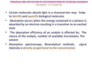
  • 1. PRINCIPLES AND APPLICATION OF SPECTROPHOTOMETRY IN DISEASE DIAGNOSIS Absorption : UV/Visible/IR • Certain molecules absorb light in a characteristic way: helps to identify and quantify biological molecules • Absorption occurs when the energy contained in a photon is absorbed by an electron resulting in a transition to an excited state • The absorption efficiency of an analyte is affected by: The nature of the analyte, number of available microstates, The solvent • Absorption spectroscopy: Bioanalytical methods; signal intensity is directly proportional to the concentration
  • 2. • Pigment Chlorophyll- which absorbs light; in the blue and red region of the visible light spectrum. • For this reason, leaves are- green (because they reflect green). • If Leaf is extracted in an organic solvent, the leaf extract (containing the solute chlorophyll) with a high chlorophyll content will produce: dark green colour • A leaf extract with a low chlorophyll content will yield a pale green extract. Spectrophotometry is • a mean of measuring how densely green the solution is.(concentration)
  • 3. SPECTROSCOPY / SPECTROCHEMICAL ANALYSIS. The study how the chemical compound interacts with different wavelenghts in a given region of electromagnetic radiation Spectrophotometry : Quantitative measurement of the reflection or transmission properties of a material as a function of wavelength.; Involves the use of a spectrophotometer. SPECTROPHOTOMETER : The combination of two devices, a spectrometer and a photometer. •A device that is used to measure intensity of light as a function of the wavelength of light. • An instrument that measures the amount of light of a specified wavelength that passes through (is transmitted through) a sample (medium)
  • 4. BLOCK DIAGRAM OF SPECTROPHOTOMETER
  • 5. The Spectrophotometer Single-beam Double-beam
  • 6. SPECTROPHOTOMETE Spectrometer: for producing light of any selected wavelength or color Photometer: used for measuring the intensity of light. Rx i t s l i t The two devices are placed at either side of a cuvette filled with a liquid E E n tr a n c e s lit D e te c to r Red I0 I R eadout d e v ic e I0= radiant power arriving at the cuvette a = absorptivity of the sample (extinction coefficient) P r is m L = length of the path through the sample C I = radiant power leaving the cuvette V io le tc = concentration vof the absorbing substance C u e tte L ig h t s o u r c e M o n o c h ro m a to r
  • 7. PRINCIPLES : Spectroscopy:  Deals with the production, measurement, and interpretation of spectra arising from the interaction of electromagnetic radiation with matter.  Electromagnetic spectrum of energy: the gamma rays (wavelengths < 0.1 nanometres) to radio waves (wavelengths > 250 millimetres.)  Spectroscopy deals with :  the ultraviolet (180 to 380nm)  the visible (380 to 800nm)  the infrared (0.8 to 50 micrometres).
  • 8.
  • 9. Colors & Wavelengths COLOR WAVELENGTH (λ in nm) Ultraviolet < 380 Violet 380 – 435 Blue 436 – 480 Greenish-blue 481 – 490 Bluish-green 491 – 500 Green 501 – 560 Yellowish-green 561 – 580 Yellow 581 – 595 Orange 596 – 650 si V Red 651 – 780 Near Infrared > 780
  • 10. SPECTROPHOTOMETRY COLORIMETRY • A photometer (a device for • The measurement of color measuring light intensity) • Any technique used to evaluate an • Measure intensity as a unknown color in reference to function of the color, or more known colors specifically, the wavelength of light • It determines color based on the red, • Tungsten or xenon flashlamp blue, and green components of light absorbed by the object or sample, as the source of white light • Tungsten lamp for • Colored light beam through an measurements in visible optical filter, which transmits only region(360-900nm) one particular color / band of • Hydrogen /deuterium lamp wavelengths of light to the for UV region(200-380nm) photodectector
  • 11. Spectroscopy and Spectrophotometry • Light can either be transmitted or absorbed by dissolved substances • Presence & concentration of dissolved substances is analyzed by passing light through the sample • Spectroscopes measure electromagnetic emission • Spectrophotometers measure electromagnetic absorption • Principle: Based on Beer Lambert’s LAW
  • 12.  Spectrometer produces the light of desired wavelength and it passes through the tube and reaches photometer that measures its intensity.  Then the photometer produces a voltage signal to a display device, usually a galvanometer.  As the amount of light absorbed by the liquid changes; the signal also changes.  The concentration of a substance in solution can be measured by calculating the amount of absorption of light at the appropriate wavelength or a particular colour  Reading of Spectrophotometer: (Number)- Absorbance that is directly proportional to the color intensity, and also the concentration of the species responsible for the color.
  • 13. • To use absorbance for analytical purposes, a calibration curve must be generated by measuring the absorbance of several solutions that contain known concentrations of analyte. If development of color is linked to the concentration of a substance in solution then: That concentration can be measured by determining the extent of absorption of light at the appropriate wavelength. For example : Hemoglobin appears red • Hemoglobin absorbs blue and green light rays much more effectively than red.) • Thus, The degree of absorbance of blue or green light is proportional to the concentration of hemoglobin.
  • 14. Terms:/Parameters Transmittance : The passing of light through a sample Absorbance: Amount of light absorbed by a sample (the amount of light that does not pass through or reflect off a sample) %Transmittance: The manner in which a spectrophotometer reports the amount of light that passes through a sample Absorbance units: A unit of light absorbance determined by the decrease in the amount of light in a light beam Absorbance spectrum: A graph of a sample’s absorbance at different wavelengths Lambdamax: The wavelength that gives the highest absorbance value for a sample
  • 15. Absorption: The Beer-Lambert Law August Beer (1825-1863): Added absorption co-efficient and related to conc. in solution. Pierre Bouguer Johan Lambert Astronomer: Light Mathematician, first to is diminished as it prove that π is irrational. passes through the No absorption coefficient atmosphere. A = − log( I1 / I 0 ) = εcl €: Extinction coefficient c: Concentration l : Path length
  • 16. BEER–LAMBERT’S LAW (Beer–Lambert–Bouguer law) • Relates the absorption of light to the properties of the material through which the light is travelling. BEER'S LAW • When monochromatic light (light of a specific wavelength) passes through a solution there is usually a quantitative relationship between the solute concentration and the intensity of the transmitted light • The amount of light absorbed by the a medium ( solution/ sample) is proportional to the concentration of the absorbing material or solute present. • Thus the concentration of a coloured solute in a solution may be determined in the lab by measuring the ABSORBANCY OF LIGHT AT A GIVEN WAVELENGTH
  • 17. BEER–LAMBERT’S LAW (Beer–Lambert–Bouguer law) ….contd LAMBERT'S LAW o Lambert described how intensity changes with distance in an absorbing medium. o The amount of light absorbed by the a medium ( solution/ sample) at a given wavelength is proportional to thickness of the absorbing layer: path length of the light
  • 18. Beer – Lambert Law States that the Absorbance (O.D) of a solution is directly proportional to the concentration of the absorbing species in the solution and the path length. The fraction of the incident light absorbed by a solution at a given wavelength is related to a. thickness of the absorbing layer (path length) and b. concentration of the absorbing species
  • 19. Transmittance Defined as the ratio of the intensity of light emerging from the solution (I) to that of incident light entering (Io) There is a logarithmic dependence between the transmission (or transmissivity), T, of light through a substance and The product of : the absorption coefficient of the substance, α, and the distance the light travels through the material (i.e. the path length), ℓ. • The ABSORPTION COEFFICIENT: (α ) = Molar absorptivity (extinction coefficient) of the absorber, (c) X the concentration (c) of absorbing species in the material I0 : intensity(power) of the incident light I : intensity(power) of the transmitted light ; ℓ : . thickness of the absorbing layer (path length) and cross section of light absorption by a single particle;
  • 20.   T- Transmittance I T= I0 - Original light intensity I0 I- Transmitted light intensity   I % Transmittance (T)= 100 x I0 1 Absorbance (A) = Log T (OPTICAL DENSITY) I0 = Log I = KCL I0 Log I is proportional to : C (concentration of solution) and L (length of light path through the solution).
  • 21. By definition of the Beer - Lambert Law. α = εc A=α ℓ A = ECL   A = Transmission/Transmissivity ; expressed in terms of Absorbance (numerical number only)- (OPTICAL DENSITY) E = Molar Extinction Coefficient of the absorber (ε)- Extinction Coefficient of a solution containing 1g molecule of solute per 1 liter of solution C = concentration of solution ( C; moles per unit vol) L= length of light path through the solution (ℓ ; )
  • 22. IMPLICATIONS OF BEER-LAMBERTS LAW • The absorbance (A) becomes linear with the concentration ( C; number density of absorbers) • Thus, if the path length and the Molar absorptivity ae known; & the absorbance is measured: The concentration of the substance (or the number density of absorbers) can be obtained. • As Concentration (C) increases, light Absorption (A) increases, LINEARLY • As Concentration (C) increases, light Transmission (T) decreases: EXPONENTIALLY (INVERSLY)
  • 23.
  • 24. As Concentration (C) increases, light Absorption (A) increases, LINEARLY . · As Concentration (C) increases, light Transmission (T) decreases, EXPONENTIALLY
  • 25.
  • 26. COMPONENTS OF SPECTROPHOTOMETER 1. Light source(UV and visible) 2. Optical system/Wavelength selector (Monochromator) 3. Sample containers 4. Detector 5. Output: Signal processor and readout
  • 27. SPECTROPHOTOMETER E x it s lit E n tr a n c e s lit D e te c to r Red I0 I R eadout d e v ic e P r is m V io le t C u v e tte I0= radiant power arriving at the cuvette a = absorptivity of the sample (extinction coefficient) L = length of the path through the sample C I = radiant power leaving the cuvette c = concentration of the absorbing substance L ig h t s o u r c e M o n o c h ro m a to r
  • 28. WORKING OF SPECTROPHOTOMETER • White light radiation source that passes through a MONOCHROMATOR ( prism or a diffraction grating that separates the white light into all colors of the visible spectrum) . • After the light is separated, it passes through a FILTER (to block out unwanted light, sometimes light of a different color) and a SLIT (to narrow the beam of light). • Next the beam of light passes through the SAMPLE that is in the sample holder.(cuvette) • The light passes through the sample and the unabsorbed portion (reflected) strikes a PHOTODETECTOR that produces an electrical signal which is proportional to the intensity of the light. • The signal is then converted to A READABLE OUTPUT (absorbance )that is used in the analysis of the sample. • Calibration curve : generated by measuring the absorbance of several solutions that contain known concentrations of analyte. •
  • 29. COMPONENTS OF SPECTROPHOTOMETER 1. LIGHT SOURCE • Deuterium Lamps - Continuous spectrum in the ultraviolet region is produced by electrical excitation of deuterium at low pressure. (160nm- 375nm) • Tungsten Filament Lamps - the most common source of visible and near infrared radiation ( at wavelength 320 to 2500 nm) • Hydrogen Gas Lamp and Mercury Lamp, Xenon (wavelengths from 200 to 800 nm)- in UV Spectrophotometer • Silicon Carbide (SiC) Rod : Radiation at wavelengths:1200 -40000 nm • NiChrome wire (750 nm to 20000 nm); ZrO2 (400 nm to 20000 nm) – for IR Region: • Laser: Used when high intensity line source is required
  • 30. OPTICAL SYSTEM/WAVELENGTH SELECTOR MONOCHROMATOR • Optical device • Disperses a beam of light into its E x it s lit component wavelengths E n t r a n c e s lit D e te c to r • Allows only a narrow band of wavelengths to Red I0 I pass R eadout • Blocks all other d e v ic e wavelengths 1. An entrance slit 2.I =A collimating lens P r is m a = absorptivity of the sample (extinction coefficient) 0 radiant power arriving at the cuvette C u v e tte (concave) L = length of the patht through the sample C V io le I = radiant power leaving the cuvette c = concentration of the absorbing substance 3. A dispersing device (usually a prism or a grating) L ig h t s o u rc e M o n o c h ro m a to r 4. A focusing lens 5. An exit slit
  • 31. MONOCHROMATOR •Czerny-Turner setup • AS A FILTER: It will select a narrow portion of the spectrum (the bandpass) of a given source. • IN ANALYSIS: the monochromator will sequentially select for the detector to record the different components (spectrum) of any source or sample emitting light. • Mirror collimates light (parallel rays) • Gating disperses light ( Prisms were formerly used) • Light coming through entrance slit is polychromatic • Light out of exit slit is monochromatic
  • 32. CUVETTES ( SAMPLE CONTAINERS) • The containers for the sample- usually plastic or quartz: • Reference solution must be transparent to the radiation which will pass through them. • Quartz or fused crystalline silica cuvettes for UV spectroscopy . • Glass cuvettes for Visible Spectrophotometer • NaCl and KBr Crystals for IR wavelengths
  • 33. Cell Types I AND II Open-topped rectangular standard cell (a)
  • 34. • The photomultiplier tube (In UV-Vis spectroscopy) Consists of : Detectors • A photoemissive cathode (a cathode which emits electrons when struck by photons ) • Several dynodes (which emit several electrons for each electron striking them) • An anode. • Produces an electric signal proportional to the radiation intensity • Signal is amplified and made available for direct display • A sensitivity control amplifies the signal • Examples: Phototube (UV); Photomultiplier tube (UV-Vis); Thermocouple (IR); Thermister (IR)
  • 36. 5. OUTPUT: SIGNAL PROCESSOR AND READOUT (DISPLAY DEVICE) DISPLAY DEVICE (Output device) • Consist of a moving–coil meter or a pen recorder displaying % transmission (%T). • At present: Instrument control, operation, standardization and data processing or storage: carried out by a microcomputer or microprocessor built in or interfaced to it.
  • 37. Steps in working with spectrophotomoter  When warming up the spectrophotometer, there should be no cuvettes in the machine  Preparation of samples  A series of standard solutions of known concentration  Set spectrophotometer to wavelength of maximum light absorption • Measure light absorbance of standards  Set the % transmittance of light as 0%  In the sample space, lodge a cuvette, filled with solvent and close the sample space.  Set the transmittance at 100%  For comparing, fill the cuvette with sample and place it in sample space and close the sample space.  Note down the reading on the Photometer for calculations.  Plot standard curve: Absorbance vs. Concentration  Calculating the concentration of sample using Beer Lambert Equation: A = ECL
  • 39. MEASURING THE CONCENTRATION USING STANDARD
  • 40. DIFFERENT TYPES OF SPECTROPHOTOMETERS Classification Based on:  Different measurement techniques Differ with respect to the species to be analysed (such as molecular or atomic spectroscopy)  The sources of intensity variation: Type of radiation-matter interaction to be monitored (such as absorption, emission, or diffraction)  The region of the electromagnetic spectrum (The wavelengths they work with )used in the analysis · Based on the absorption or emission of radiation, in the ultraviolet (UV), visible (Vis), infrared (IR), and radio (nuclear magnetic resonance, NMR) frequency ranges are most commonly encountered
  • 41. TYPES AND APPLICATIONS OF SPECTROPHOTOMETER • Primarily used for QUANTITATIVE Analysis of Known Compounds
  • 42. Tissue absorption Major tissue absorbers include: Hemoglobin, lipids (beta carotene), melanin, water, proteins, blood components, body fluids Oxy and deoxy hemoglobin have distinct spectra. Optical measurements can provide information on tissue oxygenation, oxygen consumption, blood hemodynamics
  • 43. APPLICATIONS OF SPECTROPHOTOMETER  Forensic sciences.  Molecular biology: in measuring the growth of micro organisms like bacteria.  UV-Vis : Most Popular in Pharmaceutical, Foods and Paints Industries, Water Laboratories  In Disease diagnosis/ Pathological states (changes): detected by the analysis of various samples.,taken from the body : are analyzed in three different areas – Chemistry, Hematology and Microbiology section  Blood (the blood plasma, and the formed elements – the blood cells )- The most common substance for analysis
  • 44. TYPES AND APPLICATION OF SPECTROSCOPY…contd Types of Spectroscopy Absorption Spectroscopy :  The power of a beam of light measured before and after interaction with a sample is compared.  Specific absorption techniques tend to be referred to by the wavelength of radiation measured such as ultraviolet, infrared or microwave absorption spectroscopy  Absorption occurs when the energy of the photons matches the energy difference between two states of the material.  The absorption of ultraviolet radiation by molecules is dependent upon the electronic structure of the molecule. So the ultraviolet spectrum is called electronic spectrum
  • 45. Ultraviolet Spectroscopy  All atoms absorb in the Ultraviolet (UV) region because these photons are energetic enough to excite outer electrons.  Used in quantifying protein and DNA concentration, the ratio of protein to DNA concentration in a solution; Amino Acids (aromatic), Pantothenic Acid, Glucose Determination and Enzyme Activity (Hexokinase)  Several amino acids usually found in protein, such as tryptophan, absorb light in the 280 nm range and DNA absorbs light in the 260 nm range. (Ratio of 260/280 nm absorbance- general indicator of the relative purity of a solution)  Used as a detector for high performance liquid chromatography (HPLC). The presence of an analyte gives a response which can be assumed to be proportional to the concentration
  • 46. Visible Spectroscopy  Many atoms emit or absorb visible light.  In order to obtain a fine line spectrum, the atoms must be in a gas phase.  This means that the substance has to be vaporised.  The spectrum is studied in absorption or emission.  Often combined : UV absorption spectroscopy in UV/Vis spectroscopy. spectroscopy  Applications- Estimation of : Niacin, Pyridoxine, Vitamin B12, Metal Determination (Fe), Fat-quality Determination (TBA) and Enzyme Activity (glucose oxidase)
  • 47. Infrared Spectroscopy • The IR spectral region Further subdivided into ; near-infrared (NIR), mid-infrared (MIR), and far-infrared (FIR) based on wavelength. • The MIR region : most familiar to the organic chemist as offers the possibility to measure different types of interatomic bond vibrations at different frequencies. • In organic chemistry the analysis of IR absorption spectra shows types of bonds are present in the sample. • IR-based methods: Most common clinical analytical tests, those involving serum, whole blood, and urine.; fluids that are less commonly assayed (e.g. saliva and amniotic fluid)
  • 48. Near /Mid Infrared Spectroscopy • Near Infrared Spectroscopy : NIRange, immediately beyond the visible wavelength range, -Much greater penetration depth into the sample than in the case of mid IR spectroscopy range. • Allows large samples to be measured in each scan • Practical applications : Medical diagnosis,, pharmaceuticals/medicines, biotechnology, genomics analysis, proteomic analysis, interatomics research, inline textile monitoring, food analysis and chemical imaging/hyperspectral imaging of intact organisms, agricultural: rapid grain analysis; insect detection • Forensic lab application, crime detection and various military applications. • To identify changes in biofluid metabolite concentrations reflecting site and mechanism-specific toxicity, to define novel indices of toxic insult, to evaluate control data, to monitor disease progression and response to therapeutic intervention and to track progression and regression of toxin-induced lesions over a time period
  • 49.
  • 50. X-Ray Spectroscopy • When X-rays of sufficient frequency (energy) interact with a substance, inner shell electrons in the atom are excited to outer empty orbitals, or they may be removed completely, ionizing the atom.  The inner shell "hole" will then be filled by electrons from outer orbitals.  The energy available in this de-excitation process is emitted as radiation (fluorescence) or will remove other less-bound electrons from the atom (Auger effect).  The absorption or emission frequencies (energies) are characteristic of the specific atom.  Used in chemistry and material sciences to determine elemental composition and chemical bonding.
  • 51. Atomic Absorption Spectroscopy -  Uses a pre-burner nebulizer (or nebulizing chamber) to create a sample mist and a slot-shaped burner that gives a longer path length flame.  The nebulizer and flame are used to desolvate and atomize the sample, but the excitation of the analyte atoms is done by the use of lamps shining through the flame at various wavelengths for each type of analyte.  The amount of light absorbed after going through the flame determines the amount of analyte in the sample.  A graphite furnace for heating the sample to desolvate and atomize is commonly used for greater sensitivity.  Good sensitivity and selectivity: Used for trace elements in aqueous (and other liquid) samples.
  • 52. Photo Emission Spectroscopy  Photoelectron spectroscopy  Refers to energy measurement of electrons emitted from solids, gases or liquids by the photoelectric effect, in order to determine the binding energies of electrons in a substance.  Various techniques, depending on whether the ionization energy is provided by an X-ray photon or an ultraviolet photon.
  • 53. Mass Spectroscopy • Unique among the various techniques • Mass spectrometry: Highly sensitive detection and identification technique, allowing determination of molecular structures, and thus of a sample’s composition • Weigh atoms, molecules, cluster, nano-particle, virus, cell and etc. In general, it can only determine mass (mass-to- charge ratio (M/Z) for a particle in gas phase.) • .For most mass spectrometers, Z is equal to 1 so that mass can be determined • Involves the interaction of electromagnetic radiation or some form of energy with molecules. • The molecules absorb the radiation and produce a spectrum : during absorption process or as the excited molecules return to the ground state.
  • 54. Mass Spectrometry The Components of a Mass Spectrometer 1. Ion Source 2. Analyser 3. Detector 4. Data
  • 55. Mass Spectrometry Provides Information on 1. Molecular Mass 2. Molecular Structure (fragmentation) 3. Elemental composition
  • 56. MS Applications Non-biomedical • Pollutant Analysis • Trace Metal Analysis • Explosive Analysis • Illegal Drug Detection • Alcohol Analysis • Organic Chemical Analysis • Inorganic Chemical Analysis Biomedical • Proteomic Analysis • DNA sequencing • DNA fingerprinting for Forensic Applications • Biomolecule structure analysis • Polysaccharide Analysis • Metabolomic Analysis and Pharmacological Applications
  • 57. Mass Spectrometry Hyphenated techniques; GC-MS GC (Gas Chromatograph) Excellent in separation and quantitation Poor in identification MS (Mass Spectrometer) Excellent in identification and quantitation Poor in separation GC-MS Excellent in separation, identification and quantitation!
  • 58. Raman Spectroscopy • Interactions between matter and electromagnetic radiation also give rise to scattering processes, such as elastic scattering, and inelastic scattering • It relies on inelastic scattering, or Raman scattering, of monochromatic light, usually from a laser in the visible, near infrared, or near ultraviolet range. • The laser light interacts with molecular vibrations, phonons or other excitations in the system, resulting in the energy of the laser photons being shifted up or down. • The shift in energy gives information about the phonon modes in the system. • This process,it takes place with no change in frequency • for the radiation forming the beam involved. • To study vibrational, rotational, and other low-frequency
  • 59. Nuclear Magnetic Resonance Spectroscopy • Analyses the magnetic properties of certain atomic nuclei to determine different electronic local environments of hydrogen, carbon, or other atoms in an organic compound or other compound • Used to determine the structure of the compound.
  • 60. APPLICATIONS OF NMR IN MEDICINE BRAIN  Distinguishing gray matter & white matter  Imaging posterior fossae, brain stem, spinal cord  Detect demyelinating lesions, tumors, hemorrhages, infarctions ABDOMEN 1. Metabolic liver disease 2. Measures liver iron over load in hemochromatosis 3. Focal areas of inflammation in chronic active hepatisis KIDNEYS  Distinguishing renal cortex & medulla  To evaluate transplanted kidney PELVIS  Differentiates between BPH & prostatic carcinoma  Detects bladder tumours HEART o Tomographic images of heart muscle, chambers, valvular structures o Discrimination between infarcted, ischemic & normal myocardium o

Editor's Notes

  1. The presence and concentration of various substances dissolved in a water sample is commonly analyzed by passing different types of light (visible, infrared, or UV) through the sample. Light can either be transmitted or absorbed by the dissolved substances.