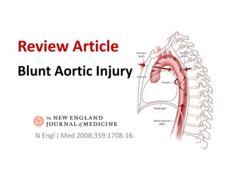
Blunt Aortic Injury
•Download as PPTX, PDF•
7 likes•6,517 views

Blunt Aortic Injury N Engl J Med 2008;359:1708-16.

Recommended
More Related Content
What's hot
What's hot (20)
Viewers also liked
Viewers also liked (20)
Similar to Blunt Aortic Injury
Similar to Blunt Aortic Injury (20)
More from Sun Yai-Cheng
More from Sun Yai-Cheng (20)
Recently uploaded
Recently uploaded (20)
Blunt Aortic Injury
- 1. Review Article Blunt Aortic Injury N Engl J Med 2008;359:1708-16.
- 2. 孫穗芬到院幾無心跳血壓 手術8小時救回【聯合報 2011.01.02】 國父孫女孫穗芬昨天八點十五分發生嚴重車禍,緊急送新光醫院,到院前幾無血壓、心跳,上半身創傷累累、大量失血,經近八小時手術後,轉至外科加護病房觀察,能否安然度過危險期,未來兩天是關鍵。 新光外傷科主任陳慶霖說,孫送醫昏迷指數只有三分,血壓、心跳「幾乎量不到」,兩側瞳孔擴大、對光無反應,「顯示腦部情況相當不好」,胸腹主動脈創傷性破裂出血,造成大量血胸,合併肋骨骨折、橫膈膜破裂,腸、肝及肺撕裂。 經心肺復甦術搶救回血壓、心跳後,立即進開刀房。先剖腹縫補肝臟撕裂傷、斷裂血管;再由心臟外科修補胸主動脈出血,裝上三根血管支架;最後胸腔外科、一般外科修補破裂橫隔膜,取出左側血胸血塊,共輸血約一萬兩千西西,直到三點三十五分結束手術送至加護病房,血壓收縮壓約一百毫米汞柱,心跳恢復約一百一十次,但仍未脫離險境。
- 3. The Clinical Problem Blunt aortic injury occurs in less than 1% of motor vehicle crashes but is responsible for 16% of the deaths. Up to 80% of patients die before their arrival at a hospital. Of those who survive the initial injury, a majority will die without definitive treatment.
- 4. Mechanism of Injury In a prospective study of hospitaladmissions involving blunt aortic injury, the crash impact wasmost often head-on (72%), followed by side impact (24%) andrear impact (4%). In a recentautopsy study, 42% of fatalities involving blunt aortic injurywere due to side-impact crashes.
- 5. Mechanism of Injury Factors that correlation with thoracicaortic injury Change in velocity of 20 mph or more Impacton the patient's side of the car The intrusion of the vehicularwall into the passenger compartment of 15 inches or more
- 6. Pathophysiological Features Aortic rupture during a sudden increase in intra-abdominal pressure may explain the association between blunt aortic injury and diaphragmatic rupture. A "water-hammer" effect, which involves simultaneous occlusion of the aorta and a sudden elevation in blood pressure The "osseous pinch" effect from entrapment of the aorta between the anterior chest wall and the vertebral column.
- 8. Diagnosis Untreated, approximately 30% of surviving patients who are admitted to a hospital with blunt aortic injury will die within the first 24 hours. Aortography was considered the best study to identify blunt aortic injury for more than 40 years.
- 9. Diagnosis The absence of the following signs were valuable to exclude the diagnosis of aortic injury: loss of the aortico-pulmonarywindow abnormality of the aortic arch rightward tracheal shift widening of the left para-spinal line without associatedfracture Between 7.3% and 44% of patients with blunt aortic injury may have a normal mediastinum on CXR.
- 10. Endograft Repair of Blunt Aortic Injury In Panel A, radiography shortly after blunt aortic injury to the chest shows an indistinct mediastinal silhouette (arrows) and loss of normal contour of the aorta (arrowheads). In Panel B, radiography in the same patient 5 months after endograft repair shows the normal contour of the mediastinum and aorta.
- 11. Diagnosis HelicalCT of the thorax is now the diagnostic test of choice. HelicalCT is more sensitive for blunt aortic injury thanangiography and is estimated to have a sensitivity of 100%,as compared with 92% for angiography.
- 13. Diagnosis Other options for the diagnosisof blunt aortic injury include TEE,intravascular ultrasonography, and MRI.
- 14. Minimal Aortic Injury “Minimal aortic injury" is used to describe a lesion of the aorta associated with blunt injury that carry a relatively low risk of rupture. Minimal aortic injury can be present in approximately 10% of patients whose blunt aortic injury is identified by helical CT. It has been reported that up to 50% of minimal aortic injuries that are identified by helical CT are missed on angiography.
- 15. Minimal Aortic Injury Minimal aortic injury was defined as an intimalflap of less than 1 cm with no or minimal peri-aortic hematoma,50% of minimal aortic injuries that were followed up had developedpseudo-aneurysms by 8 weeks after injury. If the injury is associated with significant thrombus,peri-aortic hematoma, lumen encroachment, or pseudo-aneurysm,it is our practice to proceed with endograft coverage, particularlyif the anatomy is favorable.
- 16. Perioperative Decision Making Once the diagnosis is made, immediate operative repair used to be the rule. Using β-blockerswith or without vasodilators to maintain a SBP 100 mm Hg (or 110 to 120 mm Hg in older patients)and a pulse rate of under 100 bpm in selected patientswith blunt aortic injury and a coexisting head injury, pulmonaryinjury, or cardiac insufficiency.
- 18. Surgical Repair Advantages of the veno-arterial bypass include the ability tocool the patient, which potentially enhances spinal-cord protection.
- 19. Two Bypass Procedures for Repair of Blunt Aortic Injury Panel A shows a bypass procedure from the pulmonary vein (left atrium) to the femoral artery with a centrifugal pump. The main drawing depicts open repair with an interposition graft at a typical location in the isthmus. Panel B shows femoral venous–femoral arterial cardiopulmonary bypass, which is performed by inserting a long cannula through the femoral vein into the right atrium.
- 20. Endovascular Repair Endovascular grafting wasfirst described by Parodi et al. in 1991 for the treatment ofabdominal aortic aneurysms.
- 21. Intraoperative Aortography On aortography performed during surgery for blunt aortic injury (Panel A), an aortic pseudoaneurysm (arrow) is visible with a first endograft component in position before deployment (arrowhead). An aortogram obtained after completion of the procedure (Panel B) shows the patent endograft and successful exclusion of the pseudoaneurysm.
- 22. Advantages of Endovascular Repair No physiologicalburden In brain injury, the device can be deployed with the head of thebed elevated Not require single-lung ventilation Minimal or no heparin No need for a bypass of any kind No cause of paraplegia
- 23. Technical Limitations to Endografting Injuries that occur adjacent to a sharp bend in the aorta mayresult in poor apposition of the covered stent to the aorticwall. Lesions adjacent to the left subclavianartery. Coverage ofthe left subclavian artery can result in ischemia of the upperextremity or territory perfused by the left vertebral artery.