Conjunctivitis for Nurses- Easy Explanation
•
40 likes•15,976 views

Conjunctivitis, commonly known as pink eye, is inflammation of the outermost layer of the white part of the eye and inner surface of the eyelid. It can be caused by bacteria, viruses, sexually transmitted infections, allergies, or chemicals. Symptoms include redness, swelling, watering eyes, and thick yellow discharge. Diagnosis is based on distinctive signs seen under a slit lamp and confirmation via smear or culture tests. Treatment depends on the cause, such as antibiotics for bacterial pink eye or corticosteroids for allergic conjunctivitis. Prevention involves proper hand washing and not sharing personal items.

Recommended
More Related Content
What's hot
Similar to Conjunctivitis for Nurses- Easy Explanation
Similar to Conjunctivitis for Nurses- Easy Explanation (20)
More from Swatilekha Das
More from Swatilekha Das (20)
Recently uploaded
Recently uploaded (20)
Conjunctivitis for Nurses- Easy Explanation
- 1. CONJUNCTIVITIS PRESENTED BY SWATILEKHA DAS ASST. PROFESSOR
- 2. EYE ANATOMY & PHYSIOLOGY The human eye functions much like a digital camera. Here's how it works. Light enters the eye through the cornea, the clear front surface of the eye, which acts like a camera lens. The iris works much like the diaphragm of a camera--controlling how much light reaches the back of the eye.
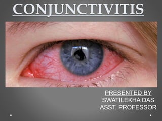
- 3. DEFINITION Conjunctivitis, also known as pink eye, is inflammation of the outermost layer of the white part of the eye and the inner surface of the eyelid.
- 5. TYPES & CAUSES BACTERIAL – due to bacteria VIRAL- caused by adenovirus CLAMYDIA-caused by sexually transmitted infections ALLERGIC-caused by pollens, dust REACTIVE- caused by chemicals such as water of swimming pool
- 7. Clinical Manifestations • Red eye, swelling of the conjunctiva, and watering of the eyes are symptoms common to all forms of conjunctivitis. However, the pupils should be normally reactive, and the visual acuity normal. • Conjunctivitis is identified by irritation and redness of the conjunctiva. • Thick yellow discharge when dries it crusts over eyelashes • Burning sensation • Blurred vision • Increased sensitivity to light
- 8. Assessment and Diagnostic Findings • The four main clinical features important to evaluate are- • the type of discharge (ie, watery, mucoid, purulent, or mucopurulent), • type of conjunctival reaction (ie, follicular or papillary), presence of pseudomembranes or true membranes, • and presence or absence of lymphadenopathy (ie, enlargement of the preauricular and submandibular lymph nodes where the eyelids drain).
- 9. • Conjunctival epithelium in numerous projections that are usually seen as a fine mosaic pattern under slit-lamp examination. • Diagnosis is based on the distinctive characteristics of ocular signs, acute or chronic presentation, and identification of any precipitating events. • Positive results of swab smear preparations and cultures confirm the diagnosis.
- 10. Medical Management Treatment depends on types of conjunctivitis- Viral is self limiting. Cold compression can alleviate some symptoms. Bacterial is treated with antibiotics( Quinolone in form of eye drops, ointment or pills) Wear spectacles to reduce light sensitivity. Discard old pair of contact lenses and use new one when conjunctivitis is gone. Allergic conjunctivitis are treated with corticosteroids in ophthalmic preparations.depending on severity they can be given in oral forms.
- 11. PREVENTION • Don’t touch eyes with hand. • Wash hands often. • Use clean towels & washclothes. • Don’t share towels or washclothes. • Change pillowcases often. • Avoid swimming in a swimming pool. • Don’t share eye cosmetics or personal eye care items. • Use any antibiotics for the complete period prescribed. • Prevention of reinfection is important.