APPROACH TO PINEAL TUMOR
•Download as PPTX, PDF•
28 likes•5,323 views

Pineal gland is essentially an extra axial midline structure lying at the roof of dienchephalon rostral to the quadrigeminal cistern surrounded by important neurovascular structure, occurring in the geometric center of brain with same depth of trajectory had made the surgery in this region a formidable challenge to neurosurgeons, however radical resection must be the goal in selected pathologies, if not pure germ cell tumor.

Recommended
More Related Content
What's hot
What's hot (20)
Similar to APPROACH TO PINEAL TUMOR
Similar to APPROACH TO PINEAL TUMOR (20)
More from suresh Bishokarma
More from suresh Bishokarma (20)
Recently uploaded
Recently uploaded (20)
APPROACH TO PINEAL TUMOR
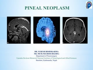
- 1. DR. SURESH BISHOKARMA MS, MCH (NEUROSURGERY) Department of Neurosurgery Upendra Devkota Memorial National Institute of Neurological and Allied Sciences Bansbari, Kathmandu, Nepal PINEAL NEOPLASM
- 2. TIMELINE ON PINEAL GLAND First brain dissection and to describe pineal gland Descartes V. Horsley 1910 Seat of the human soul Pioneered intratentorial supracerebellar approach Dandy 1921 Krause 1913 First attempted resection Herophilus 335-280 B.C Van Wagenen 1931 Trancortical transventricular approach Studnicka 1905 Glandular function Pioneered occipital transcallosal approach Melatonin dicovered Lerne r 1958 Poppen 1960 Stein 1971 Occipital transtentorial approach Revolutionised Krause ITSC approach Pinealis – pine cone Vedas – one of the 7 centers of vital energy “I have never succeeded in exposing pineal region tumor sufficiently well to justify an attempt to remove it” Cushing (1932)
- 3. ~8 mm long and 4 mm, Midline, quadrigeminal cistern Geometric center of brain: same depth of trajectory; Difficult to access Extra-axial structure: readily resectable plane. Diverse pathologies: neoplastic and non neoplastic lesions. Surrounds: Neurovascular structure. Pineal mass originate infratentorially and expand into the 3rd ventricle, thalamus or quadrigeminal plate or midbrain. ANATOMY
- 4. Gland is located in dienchephalic roof at the posterior extremity to 3rd ventricle between the habenular commissure and the posterior commissure, lies between the superior colliculi and basal to the splenium of the corpus callosum. Anteriorly: Posterior commisure and posterior part of 3rd ventricle. Posteriorly: Habernular commisure and cerebral vermis Superiorly: Splenium Inferiorly: Quadrigeminal plate and tectum Intimate with Velum interpositum Boundaries of pineal region
- 5. P1 – Quadrigeminal artery (superior colliculus) P2 – Medial posterior choroidal artery – Pineal body, corpora quadrigemina, tela choroidea, thalamus Lateral posterior choroidal artery – Pineal gland, Choroid plexus lat ventricle, LGB, Thalamus P3, P4 – Medial occipital artery – Calcarine artery – calcarine sulcus – Parieto-occipital artery – parieto-occipital sulcus – Posterior pericallosal artery SCA – Inferior colliculus ARTERIAL SUPPLY
- 6. ICV Rosenthal Pre centralcerebellar Internal occipital Posterior mesenchephalic Posterior ventricular GALEN INF SAG. SINUS STRAIGHT SINUS The great vein of Galen and its tributaries form a roof-like, dense venous network above the pineal body and the quadrigeminal plate. VENOUS DRAINAGE
- 7. Reptiles: Photoreceptor Hormone secretion for circadian rhythms Neurotransmitter secretory function Inhibits gonadal development and regulates menstruation, adrenal function, and thyroid function. It is innervated by sympathetic nerves from the superior cervical ganglion that release norepinephrine to increase the pineal gland’s melatonin secretion. Light stimuli thereby reach the pineal gland from the retina via a polysynaptic pathway and affect the production of the neuroendocrine substance, melatonin. The production of melatonin is inhibited by light. Apparently, in many animals the major function of the pineal gland is to regulate the reproductive cycle; in fall and winter when days are short, more melatonin is produced, causing a gonad suppressing effect. The exact function of the pineal gland in humans remains unclear. Recent studies could prove a diagnostic value of the melatonin profile in case of tumors of the pineal region. These studies showed a dramatically reduced melatonin rhythm in cases of undifferentiated or invasive tumors as well as the absence of melatonin variation as a consequence of pineal distortion after surgery. Also, the evidence for melatonin deficiency is recognized as a predictive factor to prevent post- pinealectomy syndrome. The absence of production of melatonin, as after pinealectomy, causes a “Jet-lag- like ” syndrome. Function of pineal gland
- 8. • Rare: 0.4 to 1.0 % (adults) and 3-8 % in children. • Most common type: • Germ cell tumors. • Pineal parenchymal cell tumors and glial cell tumor. • Other : Meningiomas, PNET, Neurocytomas, Hemangioblastomas, Cavernomas and Mets. PINEAL TUMORS
- 9. TYPES OF PINEAL TUMORS: 2007 WHO Tumor Origin Frequency 1. Germ Cell Tumors: A. Germinoma: 50-70% of GCT B. Non-Germinomatous GCT: 30-35% of GCT. i. Embryonal carcinoma ii. Yolk sac tumor (endodermal sinus tumor) iii. Choriocarcinoma iv. Teratoma: Mature, Immature, Teratoma with Malignant Transformation v. Mixed Rest of germ cells ~60% 2. Pineal Parenchymal Tumors i. Pineocytoma (WHO grade I): 45% ii. Pineal parenchymal tumor of intermediate differentiation (WHO grade II or III): 10% iii. Pinealoblastoma (WHO grade IV) :45% iv. Papillary tumor of pineal region: Ependymal cell origin: 10% Pineal glandular tissue 14-30% 3. Tumors of Supportive cells (Glial cell, mesenchymal, ependymal, arachnoid) and Adjacent Structures ~10% • Astrocytoma: Glial cell or pleuripotent pineal parenchymal cells • Glioma (glioblastoma or oligodendroglioma) • Medulloepithelioma Glial cells • Ependymoma • Choroid plexus papilloma Ependymal lining • Meningioma: Velum interpositum, Falx, tentorium Arachnoid cells • Hemangioma • Hemangiopericytoma or blastoma • Chemodectoma: from Sympathetic plexus • Craniopharyngioma Vascular cells • Malignant solitary fibrous tumor Mesenchymal tissue Very rare 4. Non-neoplastic Tumor-like Conditions <1% • Arachnoid cyst Arachnoid cells • Degenerative cysts (pineal cysts) Glial cells • Cysticercosis Parasites • Arteriovenous malformations Vascularization • Cavernomas Aneurysms of the vein of Galen 5. Metastases Absence of blood-brain barrier <0.1% • Lung (most common), breast, stomach, kidney, melanoma
- 10. Superior colliculus: Perinaud (50-75%): Paralysis of upgaze: Medial rectus and inferior oblique. Convergence or retraction nystagmus (60-70%): Co-contraction of all the horizontal extraocular muscles occurs, and the eyes are pulled into the orbit. Light near pupillary dissociation (Argyll Robertson's pupil): Accommodate but react. Midbrain: Sylvian aqueduct syndrome: paralysis of downward or horizontal gaze. Dorsal midbrain: Settling sun sign, lid retraction (Collier’s) (10%) or ptosis. Sup. Cerebellar peduncle: Cerebellar efferent fibres: Ataxia and dysmetria. Inferior colliculus: rare: hearing dysfunction Rarity: Seeding: Seizure, Radiculomyelopathy, thalamic pain, Extrapyramidal (ganglia): rare synchronous lesion. Compressive symptoms
- 11. A midline lesion of the posterior commissure, interrupts most projections from both Pretectal olivary nucleus (PON), causing bilateral symmetric loss of the PLR with preservation of the near response (bilateral light-near dissociation [LND]). It is suspected that the near-reflex fibers approach the Edinger-Westphal nucleus through the ventral region, unlike the afferent retinal light-reflex fibers that follow a dorsal route. Retinal light-reflex fibers that enter the Edinger-Westphal nucleus dorsally in the rostral midbrain are is interfered in the periaqueductal region, which causes semi dilated fixed pupil. Coupled with loss of upward gaze, it manifest as Perinaud’s syndrome. Note Argyll Roberson pupil has damage in peri aqueductal area and manifest similarly. Mechanism of light near pupillary dissociation
- 12. Endocrine: 10% Secondary effect of HCP. Hypothalamic involvement: DI. Precocious puberty: only boys: Gonadotropin independent Precocious Puberty: Choriocarcinoma or germinoma containing syncytiotrophoblastic cells: B-HCG subunit is similar to LH: LH stimulate leydig cells of testes to produce testosterone. Endocrine symptoms GIPP: gonadotrophin-independent precocious puberty
- 13. Germ cell tumors derive from pluripotential germ cells and span a wide range of differentiation and malignant characteristics. Most germ cell tumors of the CNS occur in the first three decades (98%) with a peak in the second decade (65%) between 11 and 20 years. Intracranial germ cell tumors (GCTs) usually arise in midline structures, including the pineal or suprasellar regions of children and young adults. Only germinoma and teratoma are likely to be encountered as pure tumor types. Intracranial germinomas have been reportedly associated with genetic diseases, including Klinefelter syndrome, Down syndrome , Retinoblastoma (RB1 gene) and neurofibromatosis type 1 (Merlin). GERM CELL TUMOR
- 14. The multiple histologic subtypes of GCTs are presumed to share a common cell of origin. Teilum’s “germ cell theory” proposes that extragonadal GCTs arise from primordial germ cells that have migrated aberrantly during embryonic development and then undergo malignant transformation. An alternative theory is the “embryonic cell theory,” which suggests that a mis-migrational pluripotent embryonic cell gives rise to GCTs. Sano and colleagues suggested that pure germinomas are the only tumors that truly arise from germ cells, and other GCTs develop secondary to misfolding and misplacement of embryonic cells into the lateral mesoderm causing these cells to become entrapped in different brain regions. Gonadotropins play a role in the development or progression of CNS germ cell tumors. Klinefelter syndrome,: elevated gonadotropinsGCT. Teilum regarded embryonal carcinomas as tumors of totipotential cells that may give rise to either embryonal neoplasms, with the potential to differentiate into the derivatives of all three germ layers (teratoma), or extraembryonal neoplasms (yolk sac tumor and choriocarcinoma). Theory of GCT development Maria E et a. Pediatric Central Nervous System Germ Cell Tumors: A Review. The oncologist. 2008
- 15. Teilum’s “germ cell theory Common cell of origin. Teilum’s “germ cell theory” proposes that extragonadal gcts arise from primordial germ cells that have migrated aberrantly during embryonic development and then undergo malignant transformation.
- 16. Studies: Gonadotropins play a role in the development or progression of CNS germ cell tumors. Klinefelter syndrome,: elevated gonadotropinsGCT. Gonadotropin in GCT
- 17. Germinomas are the most common GCTs in the neuraxis: Pineal and suprasellar (neurohypophyseal) regions. Germinoma are the most radiosensitive malignant tumor in the CNS. Typical germinomas contain two cell populations (two-cell pattern). One cell population contains large neoplastic tumor cells with prominent nucleoli, and the other consists of lymphocytes. Necrosis and hemorrhage are usually absent. Germinoma
- 18. Cell membrane labeling for the proto-oncogene c-kit is also characteristic of germinomas and not shared by other GCTs. 90% long term control rate for these tumors. Germinoma with elevated B-HCG level have a less favourable outcome: Benefited with aggressive treatment of chemotherapy+Radiation. 5 year survival: 75%; 10 year survival: 69% with 5000cGy. Adverse outcome of XRT: Cognitive deficit, hypothalamic and endocrine dysfunction, cerebral necrosis, secondary glioma. Intraventriculuar plaques are histopathologically identical to the primary germinomas. Treatment of germinomas radiation volume should involve the whole ventricular system including obex of the fourth ventricle and pituitary fossa. With complete diagnostic craniospinal evaluation, spinal irradiation is not necessary and the cure rates for localized germinomas are excellent with extended focal irradiation including the third and lateral ventricles as well as the sella and the pineal region (whole ventricular field)
- 19. Previously, this cytological pattern was considered to be almost pathognomonic, but later, it was realized that other diseases can also show similar cytological patterns. Thus, CNS germinoma cannot be diagnosed by the CSF cytological findings alone, although it may be helpful in establishing the clinical diagnosis.
- 20. Teratomas differentiate along ectodermal, endodermal and mesodermal lines. These tumors in the pineal region most often affect males. They are usually identified within the first two decades of life but occur more often in much younger children than other germ cell tumors 3 subtypes Mature teratomas contain 3 well-differentiated germ cell layers: ectoderm, endoderm and mesoderm layer: prognosis good. aggregates of keratin debris, fat, cartilage and bone Immature teratomas are composed of incompletely differentiated tissues resembling fatal tissue, most frequently associated with malignant course. Immature teratomas are the most common type of teratomas arising from the CNS. Teratomas that, like carcinomas and sarcomas, contain elements exhibiting unequivocal malignant transformation are referred to as teratomas with malignant transformation. Teratoma
- 21. Yolk sac tumors are highly malignant GCTs mimicking normal yolk sac structures. About half of primary intracranial yolk sac tumors are mixed GCTs containing various proportions of other germ cell elements. Marker: An elevated level of AFP alone in blood and/or CSF is strongly suggestive of yolk sac tumor. Macroscopy The tumors are firm in consistency and contain gelatinous or myxomatous areas. Hemorrhagic and necrotic areas are frequently seen. Microscopy: Schiller-Duval bodies and Eosinophilic hyaline globules are diagnostic. Yolk sac tumor
- 22. Intracranial embryonal carcinomas are rare variants of GCTs. Tumor cells may exceptionally replicate the structure of an early embryo, forming ‘embryoid bodies’, which include germ discs and miniature amniotic cavities. Embryonal carcinomas often differentiate into yolk sac tumor and choriocarcinoma, accompanied by elevation of serum AFP and β-HCG levels. Embryonal carcinomas contain primitive epithelial cells growing in solid sheets or poorly formed glands with necrosis. DIsseminate via the CSF spontaneously, or iatrogenically (shunting) Spinal axis evaluation and CSF analysis. Prophylactic craniospinal XRT is indicated following surgical removal, but cranial XRT is avoided if at all possible before 3 years of age to avoid intellectual impairment and growth retardation. Collin’s law: AKA period of risk of recurrence (PRR) PRR = the age at diagnosis plus 9 months. Embryonal carcinoma
- 23. Choriocarcinomas are extremely rare forms of intracranial GCTs. Pure: rare; Mixed Two characteristic cell types: Cytotrophoblastic cells and syncytiotrophoblastic. Cytotrophoblastic cells are round to polygonal with clear cytoplasm, round nuclei and prominent nucleoli. syncytiotrophoblastic typically contain pleomorphic densely hyperchromatic multiple nuclei, and frequently show mitotic figures. Extensive hemorrhage and necrosis are diagnostic. These tumors are highly malignant and usually invade adjacent structures. Metastasis, especially in the lungs. Choriocarcinoma
- 25. Pineocytomas are well-differentiated bening neoplasm composed of relatively small, uniform, mature cells resembling mature pineocytes. Young adult. Degenerative changes including cyst formation and focal hemorrhage can be present. They may disseminate widely along the CSF pathways. There is no evidence of local infiltration into adjacent brain parenchyma and leptomeninges. IHC: synaptophysin, neurofilaments, class III β-tubulin, and chromogranin A , Interstitial cells are usually positive for GFAP and S-100 protein. PINEALOCYTOMA
- 26. Highly malignant (WHO IV) primitive embryonal tumor of the pineal gland and represents a true primitive neuroectodermal tumor (PNET). Young age. Hemorrhage and/or necrosis are common, and craniospinal dissemination through the CSF occurs frequently. Extracranial metastatic spread of the tumor may occur to the peritoneal cavity via the ventriculoperitoneal shunt. Extremely rare extracranial metastases to the bone or lung. Pineal anlage tumors. Hypercellular and closely packed patternless sheets of small cells, mimicking primitive neuroectodermal tumor or medulloblastoma. Numerous Homer Wright rosettes and occasional Flexner-Wintersteiner rosettes are observed. IHC: synaptophysin, neurofilaments, class III β-tubulin, chromogranin A, and retinal S-antigen Pinealoblastoma
- 27. This rather newly defined group consists of rare neuroepithelial tumors in adults characterized by a typical papillary structure and epithelial structure. Their biological behavior is variable and shows a similar course to the one of grade II or III tumors. The origin of that group of tumors is specialized ependymal cells from the subcommissural region. The incidence seems extremely rare; no more than 100 cases. Tumor progression occurred in more than 70%. Papillary tumor
- 28. The absence of a blood-brain barrier Lung, Breast, stomach, kidney, and melanomas. METASTASES TO THE PINEAL REGION
- 29. TUMOR OF PINEAL REGION Tumor features Germinoma Teratoma P.blastoma P.cytoma Glioma Meningioma Age Child Child Child Adult Child Adult Sex predilection Male Male Male None None None Pineal vs. parapineal Pineal Pineal Pineal Pineal Parapineal (usually) Parapineal (usually) Signal intensity Homo Hetero Hetero Homo Homo Homo T1W Iso-Hyper Hyper Iso Hypo-Iso Hypo Iso-Hypo T2W Iso-Hyper Iso-Hyper Hyper Hyper Hyper Hyper Gd-C_MRI Avid Peripheral rim of © Variably Prominent diffuse Variable Avid Hemorrhage Common Typical Common Common Rare Rare Calcification Rare, central Typical, Rim Rare, Blasted calcification Common Uncommon Common Cystic Variably Brain edema or invasion Common Variable Common Uncommon Primarily midbrain neoplasm Occasional Metastasize Yes Variable Yes Disseminate via CSF Variable No Enhancement Dense Variable Dense Dense Variable Dense Prognosis with 10-year survival rate, if available Good with additional therapy: 85% to >35% Variable Poor Variable Variable Excellent
- 30. Age group Most common Less common Infant Pinealoblastoma Arachnoid cyst V of Galen Malf Childhood Germinoma Glioma Tuberculoma Pinealoblastoma Pineal cyst Young adult NGGCT Glioma Pinealoblastoma Pineal cyst Older adult Pinealocytoma Glioma Meningioma Epidermoid Mets Pineal mass with Age
- 32. Hydrocephalus Size Synchronous lesion: Sellar and pineal: 10%, Occurrence : Basal ganglia. Trilateral retinoblastoma syndrome: Pinealoblastoma in RB1gene abnormality patients. Pineal calcification: 60% above 20yrs, rare below 6yrs, Size >1cm high suspicion. Extent: Lateral or supratentorial extension. Anatomical relationship Involvement of 3rd ventricle Involvement of brainstem Vascularity: Location of deep venous system and its relation to the tumor. Germinoma: Displaces it superiorly along the dorsal periphery of tumor: SCITapproach (Krause) Meningioma (velum interpositum), Epidermoid, C. Callosum tumor: displaces it ventrally and inferiorly. Supratentorial approach: OTT (Poppen) Spinal seeding IMAGING
- 33. IMAGING Germinoma Teratoma Embryonal ca Pinealocytoma Pinealoblastoma
- 34. IMAGING Lipoma Epidermoid cyst Arachnoid cystPapillary tumor
- 35. ICH PROFILE (TUMOR MARKERS) OF TUMOR OF PINEAL REGION Type (Normal value) Alpha Fetoprotein (<5ng/mL) Human Chorionic Gonadotropin (<5mIU/mL) Human Placental Lactogen HPL Placental Alkaline Phosphatase PLAP Cytokeratins (CAM 5.2, AE 1/3) c-kit (CD 117) OCT 4 Melatonin Germinoma – + <770ng/mL – ++ – + + – Teratoma + (<1000ng/m L) – – – + +/- – – Yolk sac tumor +++ – – +/- + – – – Embryonal carcinoma ++ (<1000ng/m L) ++ (<770ng/mL) – + + – + – Choriocarci- noma – +++ (>2000ng/mL) ++ +/- + – – – Pinealocytoma – – – – – – – + Pinealo- blastoma – – – – – – – ++ Papillary tumor – – – – ++ – – – CSF tends to be more sensitive than serum levels. PLAP is detected in cytoplasmic staining in Germinoma. A-FP: Fetal yolk sac element: B-HCG: Trophoblastic product; Although the presence of germ cell markers indicate a malignant germ cell tumor, the converse is not necessarily true. Absence of marker should be interpreted cautiously. If A-FP is present in Germinoma; think of mixed tumor. (Youman) A TYE; Before ECG; HPL: Cardio, PG Ellenbogen 3rd ed TUMOR MARKERS
- 36. 1. Tissue diagnosis and treatment: 2. Pinealocytoma: surgery may be curative 3. Pinealoblastoma: No evidence surgery improves the outcome. 4. Choice of post op adjuvant therapy: i. Radiation: Malignant germ cell tumor, Germinoma ii. Chemotherapy: NG-GCT, Germinoma with synctiotrophoblastic with elevated B-HCG. iii. Teratoma: Doesnot responds to Chemo or radiotherapy: Surgery is indicated. 5. Metastatic work up: High: Germinoma, Choriocarcinoma, pinealoblastoma; None: Pinealocytoma, Variable: Glioma, Teratoma. 6. Estimation of Prognosis: i. Good prognosis: Germinoma, Mature teratoma, Pinealocytoma ii. Intermediate prognosis: Germinoma with elevated levels of B-HCG, Immature teratoma, Extensive/multifocal/germinoma, Teratoma with malignant transformation, Mixed tumors composed mainly of germinoma or teratoma. iii. Poor prognosis: Choriocarcinoma, Yolk sac tumor , Embryonal carcinoma, Pinealoblastoma, Mixed tumors composed mainly of choriocarcinoma, yolk sac tumor, or embryonal carcinoma. 7. Planning of long term follow up: Histology is must
- 37. Surgical aspects
- 38. 1. Location of tumor (Tentorial incisura) 2. Tumor morphology (lateral extent) 3. Displacement of great veins 4. Probable diagnosis on imaging 5. Angle of tentorium/ posterior fossa size 6. Surgeons preference CHOICE OF APPROACH
- 39. 1. The posterior transcallosal approach : Dandy. 2. The trans-ventricular approach : VanWagenen. 3. The occipital transtentorial approach : Foerster and Poppen. 4. The infratentorial-supracerebellar approach : Krause (Stein). 5. Three-quarter prone, operated side down, OTT : Ausman. Common surgical approach
- 40. Preoperative evaluation of leptomeningeal dissemination is essential for selecting appropriate postoperative adjuvant therapies and for predicting the prognosis of the patients. Whole neuraxis magnetic resonance imaging and cytological examination of the cerebrospinal fluid should be undertaken depending on whether the patient’s condition permits it. Tumor markers including human chorionic gonadotropin β-subunit, α- fetoprotein, and placental alkaline phosphatase in the serum or cerebrospinal fluid are assayed Neuroophthalmological evaluation is useful for detecting Parinaud’s syndrome and papilloedema Obstructive hydrocephalus with increased intracranial pressure Administration of corticosteroids. Ventricular drainage is instituted before or during the craniotomy. Shunt operations should be avoided: Hydrocephalus generally improves after tumor resection Peritoneal dissemination of the tumor cells via the shunting system. Pre-operative management
- 41. • Hydrocephalus • EVD • Shunt • ETV • Tissue diagnosis • ETV + biopsy • Stereotacatic biopsy • Open surgery • Tumor control • Surgery • Radiotherapy • Chemotherapy Management • Microsurgical excision is still nowadays the mainstay of management for most pineal region tumors. • An exception is germ cell tumors in which treatment policies include radiotherapy and chemotherapy after obtaining a biopsy, radiotherapy and chemotherapy without biopsy, • Endoscopic tumor biopsy with simultaneous endoscopic third ventriculostomy (ETV) has emerged as a minimally invasive and highly effective strategy for initial management since it addresses the issue of tissue diagnosis and offers a solution for the associated hydrocephalus.
- 42. • Present in almost all cases • Must be addressed prior to tumor surgery • Stable patient, complete resection likely, temporary EVD at time of surgery • Symptomatic raised ICP • ETV +/-‐ biopsy » Gradual reduction of ICP » Avoids peritoneal seeding » Avoids shunt related complications • VP shunt: Risk of metastasis. Hydrocephalus
- 43. SURGICALANATOMY • Most arise from or attached to undersurface of velum interpositum, rarely extends above • Blood supply comes from within velum mainly from M P.ch & L P.ch with anastomoses to pericallosal & quadrigeminal artery • Most tumors are centered at pineal gland, some extend to Foramen of Monroe
- 44. SURGICALANATOMY • Mostly ICV, Galen , Rosenthal & precentral cerebellar veins surround or cap the periphery of these tumors. • Rarely, ICV are ventral to tumor. • Highly vascular tumors – Pineoblastomas – Hemangioblastomas – Hemangiopericytomas
- 45. Infratentorial approaches Infratentorial supracerebellar (Krause and stein) Paramedian infratentorial Supratentorial Occipital transtentorial (Foerester and Poppen) Interhemispheric posterior transcallosal (Dandy) Transventricular (van Waganen) Combined supra-infratentorial. Surgical approaches
- 46. • Infratentorial supracerebellar: • Approach to center of tumor • Minimizes risk to veins • Good exposure • No violation of normal tissue • Occipital transtentorial / Transcallosal interhemispheric • Tumors extending superiorly • Extending laterally • Displaces veins ventrally • Large tumors • Greater exposure Surgery common approaches
- 47. Infratentorial supracerebellar approach • Position • Sitting preferred – Can also be done in Concorde position • Large ventricle/ <3 years – 3 quarter prone • Table should be able to go low • Head flexed to keep tentorium parallel to floor • Patient tilted forward
- 49. INFRATENTORIAL SUPRACEREBELLAR APPROACH • Exposure • Incision – inion to C4, spinous process of C2 exposed • Burrhole – above torcula, lateral aspect of transverse sinus • Craniotomy – exposing transverse sinus and torcula • If dura tense release CSF (ventricular tap)
- 50. Dural incision – curved between lateral most aspect of transverse sinus Dura retracted Avoid excess retraction – sinus occlusion SUPRACEREBELLAR INFRATENTORIAL APPROACH
- 52. INFRATENTORIAL SUPRACEREBELLAR APPROACH • Surgical technique – Cauterize and divide adhesions and veins between cerebellum and tentorium. – Retract vermis poster inferiorly. – Open arachnoid over the tumor (opaque white), – The precentral cerebellar vein and the superior vermian vein are identified and in most cases cut before the tumor is dissected. – Small branches of choroidal and SCA over tumor divided. – Trajectory of dissection changed towards the tumor. Precentral cerebellar vein in the center of the field. This vein can be sacrificed to gain direct access the pineal gland and lesion
- 53. Internal debulking of tumor Lateral walls dissected, vessels on it are choroidal and may be sacrificed Dissection of inferior tumor from brainstem Most dangerous parts Assistant retracts capsule upwards Final dissection – superior along velum interpositum, great veins at risk
- 54. INFRATENTORIAL SUPRACEREBELLAR APPROACH • Complications • Transient ocular movement dysfunction • Ataxia • Cognitive impairment, akinetic mutism – brainstem handling • Bleed in incompletely resected tumor
- 55. Advantage • Dissection proceed without transgressing the galenic system. • Gravity aided drainage of blood/ CSF: less pooling of blood in field. • Gravity aided cerebellar retraction • Midline – orientation easy • Strictly midline trajectory through which disorientation is less likely. (C.F OTT) • No neural structures en route INFRATENTORIAL SUPRACEREBELLAR Disadvantage • Air embolism • Surgeon fatigue • A narrow angle to the lateral and caudal side of the surgical field. • Difficult in very young and old. • No ideal in steep inclination of tentorium. • Quadriplegia from excessive flexion in elderly • Hypotension
- 56. • POSITION – On the side: usually right side down – Upper part of trunk raised 30` – Head flexed with neck stretched – & rotated 45` face down • SURGICAL TECHNIQUE – S-shaped incision behind mastoid – Oval craniectomy close to sigmoid sinus laterally & transverse sinus superiorly – Durotomy : cruciate – Bridging veins divided, petrosal & precentral cerebellar veins preserved. – Tentorial incisura reached, preserving SCA. LATERAL PARAMEDIAN INFRATENTORIAL
- 57. • INDICATIONS • Biopsy • Small quadrigeminal area tumor • ADVANTAGE • Minimal damage to neural tissues • Useful in steep tentorium • Reduced risk of air embolism (lateral position) • DISADVANTAGES • Narrow space • Difficult to reach tumor portion extending to infero posterior part of 3rd ventricle LATERAL PARAMEDIAN INFRATENTORIAL
- 58. • Commonest supratentorial approach • Indications – Predominantly supratentorial – Corpus callosum extension – Lateral extension into cerebral hemisphere – Thalamic extension – Predominantly third ventricular mass. • Advantage – Extensive tumor view. – Managing bleeding is easier. – Working distance is smaller – Access to pineal, third ventricle, midbrain, superior vermis • Disadvantages – View obstructed by Galenic venous system – Restricted view of opposite side – Oblique trajectory, which may at times be disorienting for surgeons not familiar with the approach OCCIPITALTRANSTENTORIALAPPROACH
- 59. • Position – Lateral decubitus with • Rt side down. • Midsagittal plane 30’ above horizontal – Three quarter prone – Prone – Sitting Occipital transtentorial approach
- 60. • Craniotomy – Incision: U SHAPE (6-10cm flap) – Craniotomy : 6 burr holes – 2 on left, 2 on right of sagittal Sinus – 1 just rostral to transverse sinus – 1 parietal. – Durotomy: Reflected along sagittal sinus with “T” incision. – Retractor on inferior surface of occipital lobe. Exposure
- 61. 1. Occipital retraction to be kept minimum. 2. CSF release (from posterior callosal/ dorsal mesencephalic cisterns) 3. Opening of arachnoid (venous system lies in it) 4. Yasargil – positively identify vein of Rosenthal- Galen junction (Vein of Rosenthal may be mistaken for darkly colored dorsal mesencephalic cistern) 5. Tentorium incised 5 – 10 mm from the midline to avoid the superior cerebellar veins, which join the straight sinus from a paramedian position. SURGICAL STEPS 6. Medial flap sutured to falx. 7. Identify and preserve IV nerve when manipulating tent. 8. Precentral cerebellar vein may be sacrificed. 9. Tentorial splitting*: Inferior sagital sinus and falx may be divided to facilitate further falcial retraction. 10. At this point, arachnoid overlying the tumor and quadrigeminal plate may be seen. 11. Resection: • Cleavage plane found in small tumor. • Debulking in large tumor. • For hypervascular tumor: feeding arteries identified & coagulated prior to debulking . • To avoid venous injury, total resection is not necessary & should not be attempted. 12. Immaculate hemostasis, water-‐tight dura closure. Occipital lobe retraction was greatly reduced as the occipital lobe sufficiently fell away from the operative field and required minimal retraction. Significantly decrease the incidence of postoperative visual field defects Splitting the tentorium offers a panoramic supra‐ and infratentorial view of the tumor, the surrounding deep venous structures and the collicular plate of the midbrain.
- 62. INDICATIONS • Predominantly supratentorial tumor • Position • Sitting/ prone • Lateral / 3 quarter prone. TRANSCALLOSAL INTERHEMISPHERIC SURGERY – U shapes skin flap across the midline – Bone flap across the midline – Position of bone flap depending on centering of the tumor – Wide craniotomy for alternate corridors to avoid bridging veins – Avoid sacrifice of more than 1 bridging vein – Pericallosal retracted – Callosotomy <2 cm centered over the tumor bulge. – Identify deep veins early.
- 64. • Indication – Tumor extending into lateral ventricle • Disadvantage • Limited exposure • Cortical incision required • Stereotactic guidance may be required TRANSCORTICAL TRANSVETRICULAR
- 65. COMBINED SUPRA – INFRATENTORIAL TRANS SINUS
- 66. COMBINED SUPRA – INFRATENTORIAL TRANS SINUS
- 69. Hemiparesis – Brain retraction – Sacrifice of bridging veins Sensory stereognostic deficits – Parietal lobe retraction injury Visual field defects – Occipital lobe retraction injury Disconnection syndrome – Corpus callosum section Memory defecits – Fornix injury Bleed in residual tumor Thrombosis of the basal veins of Rosenthal and internal cerebral veins: Fatal hemorrhagic infarction: Vein should be coagulated as far as possible from the confluence of the basal veins of Rosenthal. COMPLICATIONS OF SUPRATENTORIAL APPROACH
- 70. Biopsy failure of a mixed GCT is not rare. If a mixed GCT is mistakenly interpreted as ‘pure germinoma’ after an initial biopsy, at least the germinoma component can be eradicated by adjuvant therapy, and other components may remain. After that, a second-look surgery is feasible to resect the residual tumor that was resistant to the adjuvant therapy. When the level of HCG is normalized, the residual mass is almost always a teratoma Emphasis must be placed on performing an extremely careful histological examination in which the entire part of the resected specimen is searched. Biopsy failure of a mixed GCT
- 71. Indications: Non germinomatous malignant germ cell tumors Germinoma with syncytiotrophoblastic giant cells Recurrent /disseminated pineal cell tumors Regimens: Testicular cancer protocal: Einhorn regimen: Cisplatin, Vinblastine, bleomycin. Cisplatin/carboplatin + Etoposide – Widely used. Others: Vincristine/lomustine/cyclophosphamide. Success with various regimen been limited, so no clear cut recommendation. Platinum-based chemotherapy yielded a complete response (CR) rate of 85–100% in patients with germinomas. This has led to the speculation that chemotherapy may eventually replace radiotherapy Germinomas tend to be treated with a lower dose of irradiation than those used with conventional radiotherapy of 40–55 Gy. Pre-irradiation chemotherapy has been advocated as an adjuvant therapy to further decrease the total volume of radiation therapy. The combined chemoradiotherapy approach has afforded the ability to decrease the dose and volume of radiation therapy without affecting survival rates and with minimal endocrinopathy and minimal neurocognitive dysfunction ADJUVANT CHEMOTHERAPY Maria E et a. Pediatric Central Nervous System Germ Cell Tumors: A Review. The oncologist. 2008
- 72. It has been a matter of controversy whether the partially removed mature teratoma should be treated with adjuvant therapy or simply observed until relapse. Even if histological diagnosis of the resected specimen is mature teratoma, the residual part may contain a small portion of immature or other malignant tissue. A true mature teratoma is resistant to chemoradiation therapy and eventually grows during the adjuvant therapy. Immature teratomas require adjuvant therapy because they often recur even after a gross total resection.
- 73. Non-germinomas into two groups, intermediate prognosis (moderate malignancy), and poor prognosis (high malignancy) groups
- 74. Primary: Germinoma Radiation therapy (RT) plays a primary role in the treatment of germinomas and a 90–100% curative rate can be expected after RT alone. Patients with these B-HCG– secreting GCT appear to have inferior outcomes with the use of irradiation alone. The recommended radiation dose is 55 Gy given in 1.8 Gy daily fractions, with 40 cGy to the ventricular system and an additional 15 cGy to the tumor bed. Pinealoblastoma (55 Gy to bed, 35 Gy to spinal axis) Pinealocytoma (No effect on survival in incompletely excised tumors) All malignant germ cell/ pineal cell neoplasm – can be with held for histologically benign completely resected pinealocytoma, ependymoma. CSI is not necessary for localized germinomas. Dose reductions to the primary site have maintained cure rates >90%. Patterns of relapse following CSI versus reduced volume radiation, either with whole-brain or whole- ventricular radiation therapy are not significantly different. Treatment with whole-ventricular irradiation does not result in a greater number of relapses than with CSI. Radiation therapy remains a vital accepted treatment strategy in CNS NGGCTs; however, controversy still exists over the necessity of CSI versus focal or whole- ventricular radiotherapy, especially in patients with localized disease. On the other hand, nongerminomatous GCTs (NGGCTs) are generally aggressive and most patients with non- germinomatous tumors treated conventionally with surgery and RT fail to survive longer than 3 years RADIOTHERAPY
- 75. Pineal tumor Germinoma Residual NGCGT/G*BHG Residual Teratoma MANAGEMENT FLOW XRT: 55G in 1.8Gy F+ 40Gy Ventricula+15G bed Einhorn regimen: Cisplatin, Vinblastine, Belomycin Mature Teratoma is chemoradioresistant G*BHG :B-HCG secreting GCT: Germinoma with Synctiotrophoblastic component
- 76. Indications Invasive disseminated tumor at diagnosis Multiple medical problems Selected cases with very large tumors Neonate with large tumor (highly malignant, poor prognosis) Presentation suggestive of infectious/ metastatic disease with diffuse systemic disease Target selection – Avidly enhancing tumor, preferably from the centre – Multiple sites STEREOTACTIC BIOPSY
- 77. Orthogonal lateral Traverses the temporalis muscle Technically difficult using a stereotactic frame Oblique anterolateral: Most preferred Low frontal trajectory below the plane of the internal cerebral veins. Anterior to the coronal suture and lateral to the midpupillary line is usually preferred. Often a point just behind the hairline, at the superior temporal line. Trajectory traverses the frontal lobe and internal capsule, staying lateral to the lateral ventricle Posterolateral superior approach: Entry point at the parietooccipital junction Lesions with significant lateral extension TRAJECTORIES Oblique anterolateral:
- 78. Histologically confirmed Maximum experience with pinealocytoma. Used as an adjuvant therapy. Possibly primary therapy for pinealocytoma Indications still evolving – Current possible indications 1. Pineal parenchymal tumors 2. Germinoma 3. NGGCT 4. Astrocytoma GKRS
- 79. High-dose chemotherapy with autologous hematopoietic cell rescue (AuHCR) has traditionally been reserved for patients with refractory or recurrent GCTs; however, this approach has also shown promise as consolidation therapy for some high-risk gonadal GCTs at diagnosis AUTOLOGOUS HEMATOPOIETIC CELL RESCUE (AUHCR)
- 80. Overall Survival Five-year survival rates >90% Germinoma : using irradiation alone >70% Mixed germinoma, teratoma, immature teratoma with malignant elements or mixed tumors that predominantly consisted of germinomas or teratomas 67% NGGCT patients Treated with two cycles of chemotherapy followed by surgical resection, 36 Gy of CSI, and 14 Gy to the tumor bed followed by two further courses of chemotherapy. 9.3% Choriocarcinoma, endodermal sinus tumor, or embryonal carcinoma
- 81. Pineal region surgery is certainly not trivial, but within the last 25 years major morbidity and mortality (50%) from published surgical series have improved dramatically and have dropped to 0 to 2% in published surgical series. Significant improvement in outcome Mortality: 50% 0-2%* Improved neuroimaging, Microneurosurgery, Neurointensive and anaesthesia care. Mortality • Regis J et al. Retrospective study: Pineal region tumors and the role of stereotactic biopsy: review of the mortality, morbidity, and diagnostic rates in 370 cases. Neurosurgery. 1996. • Bruce J. Youman’s Neurological Surgery: Pineal Tumors. Philadelphia: WB Saunders, 2011:1011–1029
- 82. 1. Gross total resections, however, are not currently recommended because of the risk for postsurgical morbidity. 2. The value of partial or gross total resections in germinomas has not been proven, and the role of radical resection in NGGCTs is still unclear. 3. For patients with mature teratomas, however, gross total resection of the tumor is usually curative. BOTTOM LINE
- 83. References: Ellenbogen Principle of Neurological surgery 4th edition Greenberg Handbook of Neurosurgery; 8th edition Youman’s Winn Neurological surgery; 7th edition Schmidek and Sweet operative Neurosugical technique 5th edition Pineal region tumor editor. Lunsford, Vol 23, 2009 Maria E et a. Pediatric Central Nervous System Germ Cell Tumors: A Review. The oncologist. 2008 PINEAL NEOPLASM TAKE HOME MESSAGE 1. Pineal gland lies in Geometric center of brain in dienchephalic roof at the posterior extremity to 3rd ventricle 2. Pineal tumor are rare: 0.4 to 1.0 % (adults) and 3-8 % in children. 3. Teilum’s germ cell theory proposes that extragonadal GCTs arise from primordial germ cells that have migrated aberrantly during embryonic development and then undergo malignant transformation. 4. Association of GCT with Klienfelter syndrome has unfolded the role of gonadotropin. 5. Pineal calcification of size >1cm: high suspicion. 6. Center calcification is rare presentation of germinoma while blasted calcification of Pinealoblastoma. 7. Pure tumor are only Germinoma and Mature teratoma. Rest are mostly mixed. 8. AFP and B-HCG help to differentiate germinomas from NGGCTs. If AFP and B-HCG is present in Germinoma; think of mixed tumor. 9. Elevated serum or CSF concentrations of AFP and B-HCG are strong predictors of NGGCTs. 10. Although the presence of germ cell markers indicate a malignant germ cell tumor, the converse is not necessarily true. 11. Invasive nature is seen with Germinoma and pinealoblastoma.