Narrow qrs tachy i.tammi raju
•Download as PPTX, PDF•
23 likes•2,977 views

arrhythmias

Recommended
More Related Content
What's hot
What's hot (20)
Similar to Narrow qrs tachy i.tammi raju
Similar to Narrow qrs tachy i.tammi raju (20)
More from Tammiraju Iragavarapu
More from Tammiraju Iragavarapu (13)
Recently uploaded
Recently uploaded (20)
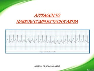
Narrow qrs tachy i.tammi raju
- 2. • DEFINITION • Tachycardia with a QRS complex width of < 120msec. • A narrow QRS complex (<120 msec) reflects rapid activation of the ventricles via the normal His-Purkinje system. • The site of origin may be in the – sinus node, – the atria, – the atrioventricular node, – the His bundle, or – some combination of these.
- 3. Classification of SVT by site of origin and regularityNARROW QRS TACHYCARDIA
- 4. • Sinus Tachycardia • frequency between 100 and 180 beats/min, but it can be higher with extreme exertion and in young individuals. • Sinus tachycardia generally has a gradual onset and termination. • The P-P interval can vary slightly from cycle to cycle, especially at slower rates. • P waves have a normal contour, a larger amplitude can develop, and the wave can become peaked. • They appear before each QRS complex with a stable PR interval unless concomitant AV block ensues REGULAR SVT
- 5. With very fast heart rates the P waves may be hidden in the preceding T wave, producing a ‘camel hump’ appearance. Sinus Tachycardia REGULAR SVT NARROW QRS TACHYCARDIA
- 6. • Inappropriate Sinus Tachycardia • Typically seen in young healthy female adults. • Sinus rate persistently elevated above 100 bpm in absence of physiological stressor. • Exaggerated rate response to minimal exercise. • ECG indistinguishable from sinus tachycardia. REGULAR SVT
- 7. • Sinus Node Reentrant Tachycardia (SNRT) • Caused by reentry circuit close to or within the sinus node. • Abrupt onset and termination. • P wave morphology is normal. • Rate usually 100 – 150 bpm. • May terminate with vagal manoeuvres. REGULAR SVT
- 8. Atrial Tachycardia • Tachycardia resultant from one ectopic foci within the atria, distinguished by a consistent p-wave of abnormal morphology that fall before a narrow, regular QRS complex.
- 9. Atrial Tachycardia • Includes various conditions such as – automatic AT, – macroreentrant AT, – scar-related AT, – atrial flutters. • Automatic AT usually presents with long RP tachycardia. • A single site located anywhere in atria exhibits inherent automaticity at a cycle length shorter than that of sinus node. • P wave morphology pattern depends on exact site of origin. • AV nodal blocking agents do not terminate the tachycardia.
- 11. • Typical Atrial Flutter (Common, or Type I Atrial Flutter) • Involves the inferior vena cava – tricuspid isthmus in the reentry circuit. Can be further classified based on the direction of the circuit: • Anticlockwise Reentry – Commonest form of typical atrial flutter 90% cases – Positive flutter waves in V1 – Negative flutter waves in leads II,III, aVF • Clockwise Reentry (Reversed Typical Atrial Flutter) – Wide negative flutter waves in V1 – Positive flutter waves in leads II, III, aVF REGULAR SVT
- 12. • Atypical atrial flutter (Uncommon, or Type II Atrial Flutter) • Does not fulfil criteria for either type of typical atrial flutter. • Often associated with higher atrial rates and rhythm instability. • Less amenable to treatment with ablation. REGULAR SVT
- 13. • ECG Features • Regular rhythm in presence of fixed AV block. • Ventricular rate ~150 bpm in presence of 2:1 AV block. • Flutter waves / ‘saw-tooth pattern’ best seen in leads II, III, aVF and V1. • Flutter wave morphology depending on type of atrial flutter (see above). • QRS complexes usually < 120 ms unless pre-existing bundle branch block, accessory pathway, or rate related aberrant conduction. • Variable AV block will result in an irregular rhythm. • Absence of an isoelectric baseline REGULAR SVT
- 14. • Atrial flutter should be considered in all regular narrow complex tachycardia with a ventricular rate of ~150 bpm. • Vagal manoeuvres may help differentiate sinus tachycardia from atrial flutter. In atrial flutter vagal manoeuvres may be ineffective or result in a rapid decrease in rate allowing flutter waves to be more easily seen. • In atrial flutter with variable block the R-R distances will be multiples of each other unlike atrial fibrillation in which no relationship exists e.g. assuming atrial rate of 300bpm the R-R distance in 2:1 block is 400ms, in 3:1 block 600ms, in 4:1 block 800ms. REGULAR SVT
- 15. Atrial flutter with 2:1 block:
- 17. • ECG Features Of Atrial Fibrillation • Irregularly irregular rhythm. • No P waves. • Absence of an isoelectric baseline. • Variable ventricular rate. • QRS complexes usually < 120 ms unless pre-existing bundle branch block, accessory pathway, or rate related aberrant conduction. • Fibrillatory waves may be present and can be either fine (amplitude < 0.5mm) or coarse (amplitude >0.5mm). • Fibrillatory waves may mimic P waves leading to misdiagnosis. Atrial Fibrillation most common sustained arrhythmia. IRREGULAR SVT
- 18. • First episode – initial detection of AF regardless of symptoms or duration • Recurrent AF – More than 2 episodes of AF • Paroxysmal AF – Self terminating episode < 7 days • Persistent AF – Not self terminating, duration > 7 days • Long-standing persistent AF – > 1 year • Permanent (Accepted) AF – Duration > 1 yr in which rhythm control interventions are not pursued or are unsuccessful Classification Of Atrial Fibrillation
- 21. • A rapid, irregular atrial rhythm arising from multiple ectopic foci within the atria. • Most commonly seen in patients with severe COPD or congestive heart failure. • It is typically a transitional rhythm between frequent premature atrial complexes (PACs) and atrial flutter / fibrillation. Multifocal Atrial Tachycardia IRREGULAR SVT
- 23. • Commonest cause of palpitations in patients with structurally normal hearts. • AVNRT is typically paroxysmal and may occur spontaneously or provocation). • It is more common in women than men (~ 75% of cases occurring in women) • complain of the sudden onset of rapid, regular palpitations, presyncope. angina. • The patient may complain of shortness of breath, anxiety and occasionally polyuria. • The tachycardia typically ranges between 140-280 bpm and is regular in nature. It may cease spontaneously (and abruptly) or continue indefinitely until medical treatment is sought. • The condition is generally well tolerated and is rarely life threatening in patients with pre-existing heart disease. AVNRT REGULAR SVT
- 24. • The slow pathway (alpha): a slowly-conducting pathway with a short refractory period. • The fast pathway (beta): a rapidly-conducting pathway with a long refractory period. REGULAR SVT AVNRT
- 25. AVNRT - ECG • Presence of a narrow complex tachycardia with regular R-R intervals and no visible p waves. • P waves are retrograde and are inverted in leads II,III,AVF. • P waves are buried in the QRS complexes –simultaneous activation of atria and ventricles – most common presentation of AVNRT –66%. • If not synchronous –pseudo s wave in inferior leads ,pseudo r’ wave in lead V1---30% cases . • P wave may be farther away from QRS complex distorting the ST segment ---AVNRT ,mostly AVRT. REGULAR SVT NARROW QRS TACHYCARDIA
- 26. • Slow-Fast AVNRT (common type) REGULAR SVT
- 27. REGULAR SVT NARROW QRS TACHYCARDIA
- 28. REGULAR SVT
- 29. • Accounts for 10% of AVNRT • Associated with Fast AV nodal pathway for anterograde conduction and Slow AV nodal pathway for retrograde conduction. • Due to the relatively long ventriculo-atrial interval, the retrograde P wave is more likely to be visible after the corresponding QRS. Fast-Slow AVNRT (Uncommon AVNRT) REGULAR SVT
- 30. • Slow-Slow AVNRT (Atypical AVNRT) • 1-5% AVNRT • Associated with Slow AV nodal pathway for anterograde conduction and Slow left atrial fibres as the pathway for retrograde conduction. • ECG features: • Tachycardia with a P-wave seen in mid-diastole… effectively appearing “before” the QRS complex. • Confusing as a P wave appearing before the QRS complex in the face of a tachycardia might be read as a sinus tachycardia. REGULAR SVT
- 31. AVNRT(theJaeggialgorithm), • Pseudo S/R waves, • RP interval, • Lack of significant ST depression in multiple leads • A correct diagnosis of typical AVNRT can be made by ECG analysis 76% of the time NARROW QRS TACHYCARDIA
- 32. Slow-Fast (Typical) AVNRT: •Narrow complex tachycardia at ~ 150 bpm. •No visible P waves. •There are pseudo R’ waves in V1-2. NARROW QRS TACHYCARDIA
- 36. Summary of AVNRT subtypes
- 37. • PR interval <120ms • Delta wave – slurring slow rise of initial portion of the QRS • QRS prolongation >110ms • ST Segment and T wave discordant changes – i.e. in the opposite direction to the major component of the QRS complex • Pseudo-infarction pattern can be seen in up to 70% of patients – due to negatively deflected delta waves in the inferior / anterior leads (“pseudo-Q waves”), or as a prominent R wave in V1-3 (mimicking posterior infarction). WPW in sinus rhythm REGULAR SVT-AVRT
- 38. Atrioventricular Reentry Tachycardias .AVRT • AVRT is a form of paroxysmal supraventricular tachycardia. • A reentry circuit is formed by the normal conduction system and the accessory pathway resulting in circus movement. • During tachyarrythmias the features of pre-excitation are lost as the accessory pathway forms part of the reentry circuit. • AVRT often triggered by premature atrial or premature ventricular beats. • AVRT are further divided in to orthodromic or antidromic con duction based on direction of reentry conduction and ECG morphology. REGULAR SVT NARROW QRS TACHYCARDIA
- 39. • AVRT With Orthodromic Conduction • In orthodromic AVRT antegrade conduction occurs via the AV node with retrograde conduction occurring via the accessory pathway. This can occur in patients with a concealed pathway. REGULAR SVT
- 40. Orthodromic AVRT using a rapidly conducting accessory pathway: Most common type of AVRT Initiated by either an APB or VPB AV conduction is over the AV node & VA conduction over accessory pathway Activation of ventricle & atrium follow sequentially P waves are separated from the QRS complex. Retrograde conduction is rapid P wave closer to the preceding QRS RP < PR. The QRS may be narrow or if aberrant conduction occurs, a typical BBB will be present. NARROW QRS TACHYCARDIA
- 41. • AVRT With Antidromic Conduction • In antidromic AVRT antegrade conduction occurs via the accessory pathway with retrograde conduction via the AV node. Much less common than orthodromic AVRT occuring in ~5% of patients with WPW. • ECG features of AVRT with antidromic conduction are: – Rate usually 200 – 300 bpm. – Wide QRS complexes due to abnormal ventricular depolarisation via accessory pathway. REGULAR SVT
- 42. REGULAR SVTNARROW QRS TACHYCARDIA
- 44. AVNRT AVRT Incidence Most common Less than AVNRT sex female males Pathway Slow-fast, Ventricles not required for activation Accesory Ventricles required for activation Activation Simultaneous activation Sequential activation Rate <200 >200 P-wave Burried in QRS Will be seen after QRS Pseudo-r,pseudo-s,pseudo-q present absent RP-interval <70msec >70msec ST-T changes Less common more ST elevation in aVR lesss more Notch in aVL more less QRS alternans Rare common Abberancy Rare common BBB Doesnot alter rate Alters rate(coumel’s law) AV block Possible Not possible in its presence
- 45. WAYSTODIFFERENTIATE • Physical examination • Carotid sinus massage • Adenosine • ECG
- 46. Physical examination during SVT • Pulse, BP, S1 : they are regular & constant in regular tachycardias. In AF & A.flutter with variable AV block, pulse, BP & loudness of S1 varies. • Neck veins : SVT – rapid, regular pulsations (frog sign) A.Flutter – flutter waves AT & sinus tachycardia – no abnormal pulsations The Frog sign: in AVNRT or AVRT, the atria contract against closed AV valves rapid, regular, expansive venous pulsations in the neck (that resemble the rhythmic puffing motion of a frog). It is due to simultaneous activation of atria & ventricles.
- 47. CAROTIDSINUSMASSAGE • Site- • At the level of cricoid cartilage and at the angle of mandible. • Procedure- • Gentle pressure is applied over the carotid sinus for 5 -10 seconds. • In case of no response – try on the other side. • Precaution- – Check for carotid bruit before massage. – Ecg monitor must – Simultaneous pressure not to be applied both sides. • Alternative manuevres are valsalva, gag reflex, ice water pouring over the face. NARROW QRS TACHYCARDIA
- 48. • Carotid sinus massage Contraindications • A carotid bruit. • Prior stroke or transient ischemic attack, unless imaging has shown no significant carotid disease. • A history of serious cardiac arrhythmias (ventricular tachycardia or fibrillation). NARROW QRS TACHYCARDIA
- 49. • RESPONSE • The slowing of SA nodal activity can cause a temporary decrease in the atrial rate (in patients with sinus tachycardia). • The slowing of AV nodal conduction can lead to AV nodal block, which may "unmask" atrial electrical activity (ie, reveal P waves or flutter waves) by decreasing the number of QRS complexes that obscure the electrical baseline. • Carotid sinus massage NARROW QRS TACHYCARDIA
- 50. • With some narrow QRS complex tachycardias that require AV nodal conduction (especially AVNRT and AVRT), the transient slowing of AV nodal conduction can terminate the arrhythmia by interrupting the reentry circuit. • Less commonly, CSM can cause some atrial tachycardias to slow and terminate. • In some cases, no response is obtained. Carotid sinus massage NARROW QRS TACHYCARDIA
- 51. Carotid sinus massage NARROW QRS TACHYCARDIA
- 54. • Termination of the arrhythmia • Termination with a P wave after the last QRS complex is – most common in AVRT or AVNRT and is rarely seen with AT. • Termination with a QRS complex can be seen with – AVRT, AVNRT, or AT. • If the tachycardia continues despite successful induction of at least some degree of AV nodal blockade, the rhythm is almost certainly AT or atrial flutter; AVRT is excluded and AVNRT is very unlikely Carotid sinus massage NARROW QRS TACHYCARDIA
- 55. ADENOSINE • MECHANISM OF ACTION • Adenosine interacts with A1 receptors present on the extracellular surface of cardiac cells, activating K+ channels. • The increase in K+ conductance shortens atrial APD, hyperpolarizes the membrane potential, and decreases atrial contractility. • Similar changes occur in the sinus and AV nodes. • Adenosine antagonizes catecholamine-stimulated adenylate cyclase to decrease cyclic adenosine monophosphate accumulation and to decrease ICa.L and the pacemaker current If in sinus node cells .
- 56. • Adenosine slows the sinus rate in humans, which is followed by a reflex increase in sinus rate. • In the region of the AV node, conduction is depressed, along with decreases in action potential amplitude, duration, . • Adenosine does not affect conduction in normal accessory pathways. Conduction may be blocked in accessory pathways that have long conduction times. • A 6-6-12 mg dose of adenosine, administered intravenously in rapid fashion, terminates the nodal-dependent SVT (i.e., AVRT and AVNRT) over 90% of the time
- 57. ECG • The atrial rate • Rhythm • The P wave morphology (ie, identical to normal sinus rhythm, retrograde, or abnormal). • The position of the P wave in relation to the preceding and following QRS complexes (ie, the RP relationship). • The relationship between atrial and ventricular rates (1:1 or otherwise). • QRS alternance • Lead Avr, and aVL • ST – T changes NARROW QRS TACHYCARDIA
- 58. Narrow qrs tachycardia rate Sinus tachy 100-180 SVT Reentrant- AVNRT,AVRT,sinoatrial Automatic Atrial tachy Junctional tachy 200±50 Atrial flutter 300 ± 50 Atrial fibrillation 400±50 ATRIAL RATE
- 59. • IRREGULAR • AF, • Atrial tachycardia with variable AV conduction, • MAT • REGULAR • Atrial flutter • Atrial atchycardia • AVNRT • AVRT • PJRT RHYTHM NARROW QRS TACHYCARDIA
- 60. P wave morphology • Present or not -Absent in AF,AVNRT • If present – Similar to sinus rhythm – Retrograde – Abnormal NARROW QRS TACHYCARDIA
- 61. • SIMILAR TO SINUS RHYTHM • Inappropriate sinus tachycardia • SNRT • Atrial tachycardia originating near the sinus node
- 62. Retrograde P waves • AVNRT, • AVRT, and, • Abnormal P waves • Atrial tachycardia NARROW QRS TACHYCARDIA
- 63. Pwaves no Irregular R-R interval ATRIAL FIBRILLATION Regular R-R interval AVNRT yes NORMAL MORPHOLOGY SINUS TACHYCARDIA SINUS NODE REENTRY INAPPROPRIATE SINUS TACHYCARDIA NARROW QRS TACHYCARDIA
- 64. P wave present but not of same morphology as sinus rhythm Pseudo r’ wave in V1 AVNRT Pseudo S wave on lead II AVNRT Pwave ST-T changes Positive in lead I AVRT Right posteroseptal Accessory pathway Negative in lead I AVRT Left sided accessory pathway NARROW QRS TACHYCARDIA
- 69. NARROW COMPLEX QRS TACHYCARDIA SHORT RP INTERVAL TYPICAL AVNRT AVRT LONG RP INTERVAL ATYPICAL AVNRT AVRT slow retrograde conduction Permanent Form junctional tachycardia ATRIAL TACHYCARDIA SANRT INAPPROPRIATE ST NARROW QRS TACHYCARDIA
- 71. STsegmentdepression • Represent either repolarization changes or a retrograde atrial activation • More commonly seen in those with an AV reentrant tachycardia associated with an accessory pathway NARROW QRS TACHYCARDIA
- 74. • aVL notch: any positive deflection at the end of the QRS during tachycardia and its absence during sinus rhythm. • The aVL notch was found during AVNRT in 51.3 vs. 7.4% in AVRT NARROW QRS TACHYCARDIA
- 77. • ST-segment elevation in aVR lead. • The percentage of patients with aVR ST-segment elevation was significantly greater in AVRT than in AVNRT • The ST segment elevation in this lead is thought to be due to deformation of the ST segment by retrograde atrial activation rather than from a true repolarization abnormalit
- 81. • QRS Alternans • QRS alternans describes a beat-to-beat variation in the morphology of the QRS complex, most easily seen as a difference in amplitude from one beat to the next. • This finding is most often associated with accessory pathway- mediated SVTs, such as orthodromic AVRT, • solely a rate-related phenomenon and is independent of tachycardia mechanism. • In summary, its use as a discriminating factor is limited
- 82. The mechanism of QRS alternans during narrow QRS tachycardia is not known. It has been attributed to non-specific intraventricular conduction abnormalities. QRS alternans has been considered to be strongly suggestive of AV re-entrant tachycardia. However, it may also occur during AV nodal re- entrant tachycardia and can also be induced by abrupt rapid atrial pacing.
- 85. SUMMARY
- 86. AV ACCESSORY PATHWAYS • 46 to 60 percent of accessory pathways are found within the left free wall space • 25 percent are within the posteroseptal space • 13 to 21 percent of pathways are within the right free wall space • 2 percent are within the anteroseptal space
- 87. • Pathways located between annulus fibrosus and epicardial reflection of atrium • Variable depth from epicardium/endocardium • LCX,CS-in left free wall space • CS, MCV,PDA- in posterolat space • RCA-anteroseptal and right free wall spaces
- 94. A, Patient with a right anterior pathway: The retrograde P wave is negative in lead V1 and positive in leads II, III and aVF.
- 95. Patient with a right posterior pathway: The retrograde P wave is negative in leads V1, II, III and aVF.
- 96. Patient with a right midseptal pathway: The retrograde P wave is biphasic in lead V1 and negative in leads II, III and aVF.
- 97. Patient with a right posteroseptal pathway: The retrograde P wave is positive in lead V1, negative in leads II, III aVF and biphasic in lead I.
- 101. • The software used for T-wave subtraction is a customdesigned software incorporated in the Bard EP recording system (Labsystem Pro). • the rapid identification of the localization of an accessory pathway during an EP study just by analyzing P-wave polarity during AVRT,
- 102. • However, the presented algorithm may virtually also be applied to the analysis of retrograde P-wave polarity without the use of this specific software.
- 105. • Lead V1 was used to differentiate right (negative or isoelectric) from left (solely positive) APs. • Retrograde P-wave in lead I was negative in left posterior APs exclusively and became more positive with an AP location shifting towards right anterior.
Editor's Notes
- 3