
Recommended
More Related Content
What's hot
What's hot (20)
Viewers also liked
Similar to Beam modification devices
Similar to Beam modification devices (20)
More from Vibhay Pareek
More from Vibhay Pareek (11)
Recently uploaded
Recently uploaded (20)
Beam modification devices
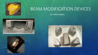
- 1. BEAM MODIFICATION DEVICES DR. VIBHAY PAREEK
- 2. WHY THE NEED FOR BEAM MODIFICATION: • Irregularity in patient exterior contour • Tissue deficit • Dose inhomogeneity • Acute normal tissue reactions. • Detraction from overall effective treatment.
- 3. DEFINITION: • Beam Modification Defined as desirable modification in the spatial distribution of radiation - within the patient - by insertion of any material in the beam path.
- 4. HURDLES: • Radiation reaching any point is made up of primary and scattered photons • Introduction of modification devices results in alteration of dose distribution • The phenomenon of scattering results in an “blurring” of the effect of beam modification
- 5. TYPES OF BEAM MODIFICATION: • Shielding: To eliminate radiation dose to some special parts of the zone at which the beam is directed. • Compensation: To allow normal dose distribution data to be applied to the treated zone, when the beam enters a or obliquely through the body or where different types of tissues are present • Wedge filtration: Where a special tilt in isodose curves is obtained. • Flattening: Where the spatial distribution of the natural beam is altered by reducing the central exposure rate relative to the peripheral.
- 6. TYPES OF BEAM MODIFICATION DEVICES: • Field blocking and shaping devices: • Shielding blocks. • Custom blocks. • Asymmetrical jaws. • Multi leaf collimators. • Compensators. • Beam spoilers • Wedge filters. • Beam flattening filters. • Bolus • Breast Cone • Penumbra trimmers • Electron Beam modification
- 8. FIELD BLOCKING AND SHAPING DEVICE:
- 9. • SHEILDING BLOCKS • Aims of Shielding: • Protect critical organs • Avoid unnecessary radiation to surrounding normal tissue • Matching adjacent fields • An ideal shielding material should have the following characteristics: • High atomic number. • High-density. • Easily available. • Inexpensive. The most commonly used shielding material for photons is Lead (Pb).
- 10. CHOICE OF SHIELDING ALSO DEPENDS ON TYPE OF BEAM USED:
- 11. • The thickness of shielding block used depends upon the energy of the radiation. • The shielding material which reduces beam transmission to 5% of its original is considered acceptable. • The term half value-layer is an expression for the attenuation produced by any material. • Half-value layer is defined as the thickness of material, which will reduce the intensity of the primary beam by 50%. • Practically thickness of lead between 4.5 - 5 half-value layers results in 5% or less of primary beam transmission.
- 14. CUSTOM BLOCKS • Material used for custom locking is known as the Lipowitz metal or Cerrobend. • Melting point 70°C. • Density 9.4 g /cm3 at 20°C (83% of lead). • 1.21 times thicker blocks necessary to produce the same attenuation. • Most commonly thickness of 7.5 cms used.
- 17. ADVANTAGES: • Low melting point than lead and hence can be cast in any shape • At room temperature it is harder than lead
- 18. STYROFOAM CUTTER Caution -avoid bubbles while pouring Spray silicon inside for easy removal
- 19. Outline of the treatment field being traced on radiograph using a Styrofoam cutting device. Electrically heated wire pivoting around a point (simulating the source) cutting the styrofoam block Cavities in the styrofoam block being used to cast the Cerrobend blocks.
- 24. DISADVANTAGES: • Production is time consuming • Cumbersome to use • Increase in treatment time • Production of toxic fumes while fabricating (exhaust reqire) • Increase setup time • Space requirement • Inaccurate daily variation (manual placement using template)
- 25. ASYMMETRICAL JAWS: • Used when we want to block of the part of the field without changing the position of the isocenter. • Independently movable jaws, allows us to shield a part of the field, and this can be used for “beam splitting”. • Here beam is blocked off at the central axis to remove the divergence. • Use of independent jaws and other beam blocking devices results in the shift of the isodose curves. • This is due to the elimination of photon and electrons scatter from the blocked part of the field.
- 27. MULTILEAF COLLIMATOR: • Multileaf collimators are a bank of large number of collimating blocks or leaves • Can be moved automatically independent of each other to generate a field of any shape. • 40 pairs of leaves or more having a width of 1 cm on less (projected at the isocenter). • Thickness = 6 – 7.5 cm • Made of a tungsten alloy. • Density of 17 - 18.5 g/cm3. • Primary x-ray transmission: • Through the leaves < 2%. • Interleaf transmission < 3%. • For jaws 1% • Cerrobend blocks 3.5% .
- 28. RATIONALE: * To improve efficiency of treatment delivery over Conventional Radiotherapy by the use of Conformal radiotherapy * To make delivery of 3DCRT or IMRT possible * Dynamic wedge * ETC
- 29. MLC TERMINOLOGY • Leaf Design - “Truncated pie” shape (thickness at lower end of leaf > at upper end) to account for beam divergence - Altered sides to overlap adjacent leaf (Staggered sides, Sinusoidal pattern) - “Focused” Leaves : Leaf ends follow beam divergence as field opens and closes; keeping each leaf aligned to primary fluence at all times for all field sizes; reduces width of penumbra, allows better conformality - “Double Focused” Leaves : Both Leaf Ends and Leaf Sides match Beam Divergence
- 34. MLC CONFIGURATIONS • Upper Jaw Replacement (Total / Partial) • Lower Jaw Replacement (Total / Partial) • Tertiary Collimation ELEKTA SIEMENS VARIAN
- 37. MLCS TYPE A ● The Y jaws are replaced by bank of MLCs. ● Small traveling range of leaves ● Shorter leaf length & compact head diameter ● Require tight tolerances on leaf travel & leaf dimension. ● Average radiation leakage increase. ● Elekta
- 38. MLCS TYPE B The lower jaws i.e. X jaws are replaced with bank of MLCs. Lengthy downtime if mechanical problem arise Used by Siemens
- 39. MLC TYPE C ● MLC placed just below standard upper & lower jaws. ● Tolerance on leaf positioning & leaf dimension is relaxed. ● Useful to avoid lengthy downtime. ● But collimator will have over bulk. ● Treatment Head & patient clearance is considerably reduced. ● Must attenuate primary beam to same extent as customised block (5 HVL)
- 41. ADVANTAGES • Time for shaping and inserting of custom blocks is not required. • The hardening of beam, scattered radiation, and increase in skin doses and doses outside the field, as seen with physical compensators is avoided. • Automation of reshaping and modulation of beam intensity in IMRT. • MLCs can also be used to as dynamic wedges and electronic compensators (2D).
- 42. DISADVANTAGES • Island blocking is not possible. • As the physical penumbra is larger than that produced by Cerrobend blocks, treatment of smaller fields is difficult, as is the shielding of critical structures, near the field. • The jagged boundary of the field makes matching difficult.
- 43. COMPENSATORS: • A beam modifying device which evens out the skin surface contours, while retaining the skin-sparing advantage. • It allows normal depth dose data to be used for such irregular surfaces. • Compensators can also be used for • To compensate for tissue heterogeneity. This was first used by Ellis, and is primarily used in total body irradiation. • To compensate for dose irregularities arising due to reduced scatter near the field edges (example mantle fields), and horns in the beam profile
- 45. • The dimension and shape of a compensator must be adjusted to account for : • Beam divergence. • Linear attenuation coefficients of the filter material and soft tissue. • Reduction in scatter at various depths due to the compensating filters, when it is placed at the distance away from the skin. • To compensate for these factors a tissue compensator is always has an attenuation less than that required for primary radiation. • As the distance between the skin and compensator increases the thickness ratio (h’/h) decreases.
- 51. 2D COMPENSATORS: • Used when proper mould room facilities are not available. • Thickness varies, along a single dimension only. • Can be constructed using thin sheets of lead, Lucite or aluminum. This results in production of a laminated filter. • Constructed by gluing together sheets of lead or other material in a stepwise fashion to form a laminated filter. • The total thickness of the filter at any point is calculated to compensate for the air gap at the point below it.
- 53. 3D COMPENSATORS: • 3-D compensators are designed to measure tissue deficits in both transverse and longitudinal cross sections. • Various devices are used to drive a pantographic cutting unit. • Cavity produced in the Styrofoam block is used to cast compensator filters. • Medium density materials are preferred to reduce errors. • Various systems in use for design of these compensators are: • Moiré Camera. • Magnetic Digitizers. • CT based compensator designing systems.
- 55. Compensating wedges: • Compensating wedges are useful where the contour can be approximated with a straight line for an oblique beam. • Three important differences between compensating wedges and wedge filters are: • Standard isodose curves, can be used • No wedge transmission factors are required. • Partial field compensation can be done. Set up • At the filter-surface distance calculated ≥ 20 cm. • Nominal SSD measured from a plane perpendicular to beam axis touching the highest point in the contour. • In SAD technique the depth of the isocentre is measured from the same elevated point only.
- 56. BEAM SPOILERS: • Special beam modification device where shadow trays made from Lucite are kept at a certain distance from the skin. • Based on the principle that relative surface dose increases when the surface to tray distance is reduced. • First used by Doppke to increase dose to superficial neck nodes in head and neck cancers using 10 MV photon beams. • Also used in TBI to bring the surface dose to at least 90% of the prescribed TBI dose.
- 57. BEAM SPOILER
- 58. BEAM SPOILER
- 59. WEDGE FILTERS: • A beam modifying device, which causes a progressive decrease in intensity across the beam, resulting in tilting the isodose curves from their normal positions. • Degree of the tilt depends upon the slope of the wedge filter. • Material: tungsten, brass. Lead or steel. • Usually wedges are mounted at a distance of 15 centimeters from the skin surface. • The sloping surface is made either straight or sigmoid in shape. • A sigmoid shape produces a straighter isodose curve. • Mounted on trays which are mounted on to the head of the gantry.
- 62. • Types of wedge systems: • Individualized wedge . • Universal wedge. • Dynamic wedges • Virtual wedges • Pseudo wedges • The two dimensions of wedges are important – “X” or width and “Y” or length. • All wedges are aligned so that the central axis of the beam is at the central axis of the wedge. • If the X dimension of field is longer then we can’t use the wedge without risking a hot spot!!
- 63. • Photograph of a 45* wedge for a 4MV linear accelerator • Wedge angles used are: 60°, 45°, 30° & 15°.
- 68. • Definition of wedge angle Wedge angle : Line drawn through two points a quarter of a field size on either side of central axis which lie on the isodose contour that intersects that central axis at a 10 cm depth.
- 69. WEDGES CAN BE OF TWO TYPES : - INDIVIDUALIZED WEDGE - UNIVERSAL WEDGE
- 70. • Individual wedges are useful in Cobalt beams • Using bigger wedges than necessary will reduce output of the machine → increased treatment time. • The width (W) of the wedge is fixed and important. • The same wedge can however be used for fields with lesser lengths or breadths. • The wedge systems available are: • 6W ( x 15) • 8W ( x 15) • 10W ( x 15) • All systems have the following four angles 15°, 30°, 45°, 60°.
- 71. • Universal wedges are designed so that the same wedge can be used with all field sizes. • This is useful as it saves time. • However not suitable for cobalt beams because of excessive reduction of beam output with smaller fields. • Come in one size of 20 x 30 cms (except 60°). • Wedge angles used are: 60°, 45°, 30° & 15°.
- 72. Individualized wedge Universal wedge
- 73. • Wedged fields are generally used for relatively superficial tumors. • Beams are usually directed from the same side of the patient. • The broad edges of the wedges should be aligned together. • The wedge angle chosen depends on the angle between the central rays of the two beams also called the “hinge angle”(φ). • Wedges: • Reduce the hot spots at the surface • Rapid dose falloff beyond the region of overlap. • The overlap region is also called the “plateau region”.
- 74. Thus the 2 factors on which the wedge angle is chosen are: The hinge angle. The wedge separation The wedge angle that will make the isodose curves parallel to each other and the hinge angle bisector is obtained using the equation. Hinge angle = 90-Wedge angle/2
- 76. WEDGE FILTER •Primarily tilts standard isodose curves through specific angle •Alters the isodose distribution in the entire treatment field COMPENSATING WEDGE •Mainly used as missing tissue compensator so that standard isodose charts can be used without modification. No wedge transmission factors required •Can be used to compensate for part of a contour that’s irregular in shape
- 77. Problem Solution A different value of wedge angle is required for different beam angles A small range of hinge angles can be covered by a given wedge angle without producing significant variation(±5%). The body contour can be more curved as a result, the isodose curves are not obtained in the manner desired. Compensators may be used to overcome the deficit or a different wedge angle can be used, so that part acts as a compensator.
- 78. MOTORIZED WEDGE • MW is a single 60° physical wedge which generates desired wedge angle from 0 to 60° by the combination of open and wedged beam. • It is mounted above the asymmetrical collimator and it can be moved in and out of the radiation field to create the different wedged profiles.
- 79. MOTORIZED WEDGE • Dose calculation : * The dose distribution of fields containing a MW is calculated based on weighted dose percentages for wedged and open parts of the field. * Weight Factor – Parameter indicating the % dose contributions of wedged and open part of the field containing the MW WF 0 Fully open field WF 1 Fully wedged field WF 0 - 1 Partly wedged field • Total Treatment time is set as the sum of Open Beam time and MW Beam time, as determined by the TPS software Wedge Angle Wedged Beam weight factor Open Beam weight factor 15 0.16 0.84 30 0.38 0.62 45 0.63 0.37 60 1 -
- 80. DYNAMIC WEDGE: • Treatment delivery technique in which wedged dose profile is created electronically, by the sweeping action of an independent collimator jaw from open to closed position while the beam is on. • Because of the collimator motion, different parts of the field are exposed to the primary beam for different lengths of time. This creates wedged dose gradient across the field. • Dose rate is modified according to jaw position • Dose is delivered throughout the treatment. • In VARIAN dynamic wedge, one Y jaw moves, while other Y jaw and X jaws stand still during treatment.
- 82. FLATTENING FILTERS • A beam flattening filter reduces the central exposure rate relative to that near the edge of the beam. • Used for Linear accelerators. • Due to the lower scatter the isodose curves are exhibit “forward peaking”. • The filter is designed so that the thickest part is in the centre. • Material: copper or brass.
- 83. • Penetrating power should not increase as this will alter the PDD as well as reduce the flattening. • Not required in cobalt beams, as : • The beam is almost monoenergetic. • Source emits uniform radiation all around Creates beam of sufficient area & uniformity for clinical use • Compensates for rapid variation of central beam & the periphery • Found that Al is material of choice
- 84. • The beam flatness is specified at 10 centimeters. • The extent of flatness should be ± 3% along the central axis of the beam at 10 centimeters. • Should cover 80% or more of the field, or reach closer than one centimeter from the edge. • No point parallel to the surface should receive a dose > 107% of the central axis dose. • There is usually over flattening of isodoses, near the surface. This results in production of “horns” or hot spots. • Because of the thinner outer rim, the average beam energy is lower at the periphery as compared to the centre
- 86. BOLUS: • A tissue equivalent material used to reduce the depth of the maximum dose (Dmax). • Better called a “build-up bolus”. • A bolus can be used in place of a compensator for kilovoltage radiation to even out the skin surface contours. • In megavoltage radiation bolus is primarily used to bring up the buildup zone near the skin in treating superficial lesions.
- 87. • The thickness of the bolus used varies according to the energy of the radiation. • In megavoltage radiation: • Co60 : 2 - 3 mm • 6 MV : 7- 8 mm • 10 MV : 12 - 14 mm • 25 MV: 18 - 20 mm
- 88. • Properties of an ideal bolus: • Same electron density and atomic number • Pliable to conform to surface. • Usual specific gravity is 1.02 -1.03
- 89. • Commonly used materials are: • Cotton soaked with water. • Paraffin wax. • Other materials that have been used: • Mix- D (wax, polyethylene, magnesium oxide) • Temex rubber (rubber) • Lincolnshire bolus (sugar and magnesium carbonate in form of spheres) • Spiers Bolus (rice flour and soda bicarbonate) • Commercial materials: • Superflab: Thick and doesn't undergo elastic deformation. Made of synthetic oil gel. • Superstuff: Add water to powder to get a pliable gelatin like material. • Bolx Sheets: Gel enclosed in plastic sheet.
- 91. BREAST CONE: • A beam modifying and directing device used for a tangential fields therapy. • Advantages: • Directs beam to the central axis of the area of interest, where a tangential beam is applied to a curved surface. • Helps position, the patient with an accurate SSD. • Endplate provides compensation, enhances surface dose and presses down the tissue. • Effective shielding of lungs.
- 92. PENUMBRA TRIMMERS: • Refers to the region at the edge of the beam where the dose-rate changes rapidly as a function of distance from the beam axis. • Types: • Transmission penumbra: Transmission through the edge of the collimator block. • Geometrical penumbra : Finite size of the source. • Physical penumbra: Lateral distance between to specified isodose curves at a specific depth (90% & 20% at Dmax). • Takes scattered radiation into account.
- 93. • Penumbra width depends upon: • Source diameter. • SSD. • Depth below skin. • Source to diaphragm distance (inversely) • Consists of extensible, heavy metal bars to attenuate the beam in the penumbra region. • Increase the source to diaphragm distance, reducing the geometric penumbra. • Another method is to use secondary blocks placed close to the patient ( 15 – 20 cms).
- 94. ELECTRON BEAMS: • Electron field shaping is done using lead cutouts. • For a low-energy electrons (<10 MeV), sheets of lead, less than 6 mm thickness are used. • The lead sheet can be placed directly on the skin surface. • Shields can also be supported at the end of the treatment cone if too heavy at the cost of greater inaccuracies. • Design is easier, because the size is same as that of the field on the patients skin. • To avoid variation in output and electron scatter, jaws cannot be used to collimate electron beams. • An electron beam cone is therefore used to provide the collimation. • A primary collimator is provided close to source – defines the maximum field size. • A secondary collimator, near the patient defines the treatment field.
- 96. DIRECT/INDIRECT SHIELDING: • Used for electron beam shielding. • A lead shield can be placed where shielding of structures against backscatter electrons is required. • A tissue equivalent material is coated over the lead shield like wax/ dental acrylic/ aluminum. • Example of areas requiring these techniques are the buccal mucosa and eye lids.
- 97. SCATTERING FOIL: • A device to widen the thin pencil beam (3 mm) of electrons. • Metallic plates of tin, lead or aluminium are used. • Disadvantages: • Beam attenuation. • Generation of bremsstrahlung radiation. • Advantages: • Less prone to mechanical errors. • Less expensive. • Requires less instrumentation. • Nowadays dual foil systems are used, which compare well with scanning beam systems.
- 98. CONCLUSION: • Beam modification increases conformity allowing a higher dose delivery to the target, while sparing more of normal tissue simultaneously. • Megavoltage radiotherapy is better suited for most forms of beam modification due to it’s favorable scatter profile.