SlideShare a Scribd company logo
1 of 100
Animal physiology & Biochemistry, Animal
Biotechnology
Lab Manual
NAME:……………………………………
Reg.No:……………………………….
COURSE CODE……………………..
ACADEMIC YEAR…………………………..
Content
S.No Title of the Experiment Page
No
1. Rate of oxygen consumption in a fish
2. Effect of temperature on operculum
movement of fish
2
3. Qualitative analysis of carbohydrate
4. Qualitative analysis of protein
5. Qualitative analysis of lipid
6. Demonstration of blood pressure using
sphygmomanometer
7. Estimation of haemoglobin
8. Action of salivary amylase in relation to
enzyme concentration
9. Action of salivary amylase in relation to
temperature
10. Counting of different kinds of blood cells
using haemocytometer
11. Isolation of genomic DNA from E. coli
12. Estimation of protein by Lowry’s Method
Dos and Don’ts in the Lab
Important Precautions to be taken
 Do plan and prepare for every laboratory exercise before
coming to lab. Always do some homework before carrying
out experiments in the lab.
3
 Keep work bench neat and clean before leaving the
laboratory. Do wipe with detergents if necessary.
 Dispose of biological materials such as microbes by
sterilizing and in special containers for biological hazards. Do
not throw other materials into these bags.
 Tie back long hair to avoid contamination and fire hazard.
 Be very careful with Bunsen burners. Always keep the burner
at distance from the organic solvents. The burner must be
turned off soon after the use.
 Wash your hands before leaving the laboratory, even if you
wore gloves, wash with disinfectant in running tap water
before and after the work.
 Be familiar with the chemicals in the laboratory and take
maximum care in handling each chemical.
 Keep chemicals back in position after use.
 You must cut your nails regularly.
 Tightly close all bottles and caps
Don’ts
 Don’t mishandle the chemical solutions, spirit lamp, UV
light, instruments/apparatus or electricity.
 Don‘t roam here and there in the laboratory without work or
any aim.
 Never misplace lab wares and other equipment.
 Don‘t eat, drink, chew gum or apply cosmetics in the lab.
Never use a lab microwave, laboratory fridge or freezer for
food.
4
LAB EXPERIMENT-
Animal physiology
and Biochemistry
5
Ex. No: 1
Date:
RATE OF OXYGEN CONSUMPTION IN A FISH
BACKGROUND:
Respiration by fish includes the uptake of oxygen from the environment
at sites of gas exchange (typically the gills), the use of oxygen at
mitochondria within individual cells, and the excretion of waste gases to the
environment. Respiration provides oxygen for aerobic conversion of the
energy contained in food to high-energy chemical bonds, such as those
formed when adenosine diphosphate (ADP) is changed to adenosine
triphosphate (ATP). The energy thus stored is then used to keep fish alive
(maintenance) and to allow fish to move, digest food, grow, reproduce, and
carry out all other energy-requiring functions. Measurements of respiration
often can tell us how a fish is responding to environmental conditions and
what its physiological state may be. Respirometry gives quantitative
measures of how rapidly energy and oxygen are used. Respiratory data are
important in the construction of bioenergetics models that can be used, for
example, to calculate capacities for growth and reproduction. Respiration
rates can be sensitive indicators of altered environmental conditions or
physiological states, and thus can reveal much about a fish's recent and
current activity, acclimation, and stress.
AIM:
To determine the rate of oxygen consumption by the given fish.
PRINCIPLE OF TEST
When manganous sulphate is added to the sample of water followed
by alkaline iodide, manganous hydroxide is formed. This combines with
6
dissolved oxygen present in the water forming manganic hydroxide. This on
acidification in the presence of concentrated sulphuric acid release certain
amount of iodine which is equivalent to the amount of oxygen dissolved in
the water. The amount of iodine released is determined by titrating against
0.01 N sodium thio sulphate solution using 1% starch as an indicator. The
reactions are shown below:
MnSO4 + 2KOH -------àMn(OH)2 + K2SO4
2 Mn(OH)2 +O2 ----à 2 MnO(OH)2 (Basic manganic oxide)
MnO(OH)2 + H2SO4 ----àMnSO4 + 2H2O + [O]
2KI + H2SO4 + [O] -----à K2SO4 + H2O + I2
I2 + 2Na2S2O3 ------à 2NaI + Na2S4O6
MATERIALS REQUIRED:
 100 ml beaker with siphon arrangement burette
 Burette stand
 Pipette
 Conical flask
 Reagent bottles
 Thermometer
 Balance
REAGENT REQUIRED:
 Manganese Sulphate
 Alkaline iodide
 Concentrated H2SO4
 0.01 N Sodium thiosulphate
 1% Starch
METHOD:
7
A living tilapia fish is kept in container containing one litre of tap
water. The same tap water is closed in a glass stopper control of bottle
known volume (250 ml). A fine layer of kerosene is added to the surface of
the experimental medium to prevent mixing with atmospheric air. The
starting time of the experiment is noted. In this condition the fish is allowed
to respire for half an hour in room temperature. The tap water in the control
bottle is kept under control during the time of experiment, so that no change
in the medium affect the control, after half an hour the water in the control
bottle is siphoned out into the analyzing bottle. The volume of analyzing
bottle is determined by Winkler’s method. The difference between the oxygen
in control water and the experimental water gives the volume of oxygen
consumed by the fish in half an hour.
ESTIMATION OF OXYGEN IN THE WATER SAMPLE:
A reagent bottle is taken and filled with water. The stopper is replaced
without enclosing any air bubble. After removing the stopper, 1 ml of
Manganous Sulphate and 1 ml of alkaline mixture were added. The
stopper is replaced and the bottle is shaken well to ensure maximum
chemical reaction and hence maximum absorption of oxygen. This is done
for the uniform distribution by precipitation. Then the precipitate is allowed
by settle down for few minutes. After this 1 ml of con H2SO4 is added to the
solution and is shaken well till the precipitate is not seen in the bottle. The
burette is rinsed with water and 0.1 N Sodium thiosulphate, filled with 0.1
N Sodium thiosulphate solution. Initial reading of the burette is noted.
About 50 ml of solution from the bottle is transferred to a conical flask. To
this 1 ml of 1% starch is added as an indicator. Now the color of the solution
turns to dark blue. This solution is titrated against 0.01 Sodium
thiosulphate and the titration is continued till the blue color disappeared.
This is the achromic point and the final reading is noted. The volume of
Sodium thiosulphate required for titrated is noted. The titration is repeated
for concordant values. The difference between oxygen content of the control
and of the experimental bottle gives the volume of oxygen consumed by the
8
fish in half an hour. Then the fish is removed from the experimental
medium. Weight of the fish is taken. Oxygen consumed by the fish in one
hour is calculated. The rate of oxygen consumption by the fish is calculated
by using the formula.
TABULATION:
RATE OF OXYGEN CONSUMPTION IN A FISH
S.No
Volume of
sample(ml)
Burette
reading
Volume
of
Na2S2O3
Concordant
value(ml)
Indicator
Weight
of fishInitial
(ml)
Final
(ml)
CALCULATION:
O2/ml/litre= CD* M*E*0.698*1000*Vt
vs
CD= volume of reagent bottle
Volume of reagent bottle-volume of reagent used
M= Molarity of sodium thiosulphate(0.01)
E= Equivalent weight of oxygen
1000= To express litre
Vt= Volume of sodium thiosulphate used in titration
vs= Volume of sample titrated
O2 in control water=………………………parts/ml/hr.
9
RATE OF OXYGEN CONSUMPTION IN THE EXPERI MENTAL WATER
S.No
Volume of
sample(ml)
Burette
reading
Volume
of
Na2S2O3
Concordant
value(ml)
Indicator
Weight
of fishInitial
(ml)
Final
(ml)
CALCULATION:
O2/ml/litre= CD* M*E*0.698*1000*Vt
vs
CD= volume of reagent bottle
Volume of reagent bottle-volume of reagent used
M= Molarity of sodium thiosulphate(0.01)
E= Equivalent weight of oxygen
1000= to express litre
Vt= Volume of sodium thiosulphate used in titration
vs= Volume of sample titrated
O2 in experimental water=………………………parts/ml/hr.
10
Oxygen consumed by the fish in half an hour= 02 content in the tap water-
O2 content in the experimental water
Oxygen consumed by the fish in one hour=……………..*2
Rate of oxygen consumed by the fish=…..…………../weight of the fish
=….…………..parts/ml/hr
RESULT:
Rate of oxygen consumption by the fish= …………………….parts/ml/hr.
DISCUSSION:
RVIEW QUESTIONS:
1. What is dissolved oxygen (DO)?
2. What are the approved methods for determination of DO?
3. What reagents are required for D.O. determinations using the
modified Winkler titration method?
4. What interferences affect DO determinations by the modified Winkler
titration procedure?
11
Ex. No: 2
Date:
EFFECT OF TEMPERATURE ON OPERCULUM MOVEMENT OF FISH
BACKGROUND
Fish obtain their oxygen from water by ventilating their gills.
These breathing apparatus are highly vascularized out pocketing of the body
surface. The operculum, a protective flap, covers and protects the gills,
which are soft and delicate. The gills are ventilated by pumping water
across them in one direction. Both the mouth and operculum work together
to pump the water through the mouth, into the gills, and then out to the
sides of the body. Counting gill cover (operculum) movements is a way to
calculate respiration rates in fish. An oxygen-carbon dioxide exchange
occurs. Then the operculum opens to allow the carbon dioxide-rich water to
exit. Fish are exothermic; their body temperature is the same as water
temperature and changes directly with it. Many physiological processes,
such as metabolic rate, heart rate, and enzyme activities occur faster as
body temperature increases and slow down at colder body temperature. By
counting gill cover movements, students are getting an idea of a fishes'
response to an ecological change.
12
AIM:
To determine the rate of operculum movement of a given fish at
different temperature.
MATERIALS
 Fish
 Crushed ice
 Large beaker
 Hot water
 Glass stirring rod
 Thermometer
 Stop clock
METHOD
A living fish is taken gently without rubbing on the mucous
membrane and placed into a plastic jar containing water at room
temperature. The temperature of the water sample is recorded with the help
of thermometer. After 5 minutes a fish is exposed to the sample water, the
number of operculum movement per minute at this temperature is recorded
with the help of stop watch. The observation is repeated 3 times until two
13
concordant values are obtained. After that hot is added to the water sample
until the temperature of the medium is increased by 5˚c. Again the
experiment is repeated and the number of gill movement per minute are
calculated. This same experiment is repeated with different temperature like
30˚c, 35˚c, 40˚c. The temperature of sample water is decreased by the
addition of ice water and the time taken at 25˚c 20˚c and 15˚c is noted. The
rate of operculum movement (1/t) is calculated.
TABULATION
S.No Temperature Time taken for
seconds
Concordant
value
1/t rate of
activity
FORMULA:
Q10 is the factor by which the reaction rate increases when the temperature
is raised by ten degrees.
Q10=K2 (10)/K1 (t2-t1)
Speed of rate in reciprocal=1/t
Where k1, K2 represent the velocity constant of particular temperature t2& t1
respectively
t1= Lower temperature
t2= Higher temperature
14
K1= Rate of activity at low temperature.
K2= Rate of activity at high temperature.
RESULTS:
Effects of temperature Q10 on operculum movement of fish Q10=
Q102=
DISCUSSION:
REVIEW QUESTIONS:
1. Are fish warm-blooded or cold-blooded animals?
2. Do you think that all animals would be affected in this manner by
temperature changes? Explain.
15
Ex. No: 3
Date:
QUALITATIVE ANALYSIS OF CARBOHYDRATE
BACKGROUND:
Carbohydrates are polyhydroxy aldehydes and ketones or substances
that hydrolyze to yield polyhydroxy aldehydes and ketones. Aldehydes (–
CHO) and ketones (= CO) constitute the major groups in carbohydrates.
Carbohydrates are mainly divided into monosaccharides, disaccharides and
polysaccharides. The commonly occurring monosaccharides includes
glucose, fructose, galactose, ribose, etc. The two monosaccharides combine
together to form disaccharides which include sucrose, lactose and maltose.
Starch and cellulose fall into the category of polysaccharides, which consist
of many monosaccharide residues.
AIM:
To detect the presence of different group carbohydrate in the given solution.
GENERAL MATERIALS:
0.1% of glucose, Test solution
I. ANTHRONE TEST:
PRINCIPLE:
16
Carbohydrates are dehydrated with concentrated H2SO4 to form “Furfural”,
which condenses with anthrone to form a green color complex which can
be measured by using colorimetrically at 620nm (or) by using a red filter.
Anthrone react with dextrins, monosaccharides, disaccharides,
polysaccharides, starch, gums and glycosides. But they yields of color where
is to form carbohydrate to carbohydrate.
REAGENTS:
 0.2% anthrone in con H2SO4
 Test tubes
 Spirit lamp
 2ml pipette.
PROCEDURE:
 Take 2 ml of sample solution in a test tube.
 Add 1 ml of anthrone reagent and mix thoroughly
OBSERVATION:
A bluish green color develops in the test tube indicates the presence of
sugar.
II. FEHLING’S TEST:
This is an important test to detect the presence of reducing sugars. Fehling’s
solution A is copper sulphate solution and Fehling’s solution B is potassium
sodium tartrate. On heating, carbohydrate reduces deep blue solution of
copper (II) ions to red precipitate of insoluble copper oxide.
MATERIALS:
 Fehling’s reagent
17
 Test tube
 Holder
 Spit lamp
PROCEDURE:
 Take 5 ml of fehling’s solution in a test tube.
 Add 1ml of test solution to it.
 Mix well and boil in water bath.
OBSERVATION:
The production of brownish red cuprous oxide precipitate indicates the
presence of reducing sugar.
III. BENDICT’S TEST:
PRINCIPLE:
The copper sulfate (CuSO4) present in Benedict's solution reacts with
electrons from the aldehyde or ketone group of the reducing sugar. Reducing
sugars are oxidized by the copper ion in solution to form a carboxylic acid
and a reddish precipitate of copper (I) oxide
MATERIALS:
 Benedict’s reagent
 Test tube
 Test tube holder
 Sprit lamb
 Measuring cylinder
PROCEDURE:
18
 Take 5 ml of sucrose solution in a test tube
 Add 2 ml of Benedict’s reagent
 Boil for two minutes.
OBSERVATION:
A yellowish- red precipitate occurs within 2 to 5 minutes indicates the
presence of monosaccharide.
IV. IODINE TEST
Iodine test is used to detect the presence of starch. Iodine is not much
soluble in water so iodine solution is prepared by dissolving iodine in water
in presence of potassium iodide. Iodine dissolved in an aqueous solution of
potassium iodide reacts with starch to form a starch/iodine complex which
gives characteristics blue black color to the reaction mixture.
PROCEDURE:
 Take 2 ml of sample solution in a test tube.
 Add 0.5 ml of iodine solution.
 Dilute the solution with distilled water.
OBSERVATION
Appearance of blue coloration indicates the presence of starch
RESULT:
Sample…………… indicates the presence of carbohydrates in the test
solution.
DISCUSSION:
REVIEW QUESTIONS:
1. Write a test which helps to differentiate between a disaccharide and a
monosaccharide?
2. If a sample being tested gives a positive result for Benedict’s test and
Fehling's test, then what will the sample be?
19
Ex. No: 4
Date:
QUALITATIVE ANALYSIS OF PROTEIN
BACKGROUND:
Proteins are large biological molecules composed of α-amino acids (Amino
acid in which amino group is attached to α-carbon, which exist as zwitter
ions and are crystalline in nature). They contain carbon, hydrogen, oxygen,
nitrogen and sometimes phosphorous and sulphur. Protein is an important
macronutrient essential for survival. They are constituent of calls and hence
are present in all living bodies. 10-35% of calories should come from protein.
Protein is found in meats, poultry, fish, meat substitutes, cheeses, milk etc.
AIM:
To detect the presence of protein in the given sample.
BIURET TEST:
PRINCIPLE:
This test is used to detect the presence of peptide bond. When treated
with copper sulphate solution in presence of alkali (NaOH or KOH), protein
reacts with copper (II) ions to form a violet colored complex called biuret
20
MATERIALS
Biuret reagent
PROCEDURE:
 Take 2 ml of sample solution in a test tube
 Add 2 ml of Biuret reagent solution to it.
 Mix well and observe the color.
OBSERVATION:
Development of violet color in 15 minutes indicates the presence of protein.
XANTHO PROTEIN TEST:
PRINCIPLE:
Aromatic amino acids, such as Phenyl alanine, tyrosine and
tryptophan, respond to this test. In the presence of concentrated nitric acid,
the aromatic phenyl ring is nitrated to give yellow colored nitro-derivatives.
At alkaline pH, the color changes to orange due to the ionization of the
phenolic group.
MATERIALS
• Conc.HNO3
• Blank solution
PROCEDURE:
 Take 1 ml of sample solution in a test tube.
 Add 0.5 ml of concentrated HNO3.
21
 Observe the yellow color
 Boil the tube in a water bath, cool and add few drops of NaOH
solution.
OBSERVATION:
The yellow colored solution is turn into orange.
RESULT:
Sample………….. Indicates the presence of protein in the solution.
DISCUSSION:
REVIEW QUESTIONS:
1. All the amino acids with aromatic side chains give positive test to
Xanthoprotein test- justify.
2. Define “activated benzene ring”.
3. Write the reaction(s) involved in Biuret’s Test.
22
Ex. No: 5
Date:
QUALITATIVE ANALYSIS OF LIPID
BACKGROUND:
Lipids are one of the major constituents of foods, and are important in
our diet for a number of reasons. They are a major source of energy and
provide essential lipid nutrients. Nevertheless, over-consumption of certain
lipid components can be detrimental to our health, e.g. cholesterol and
saturated fats. In many foods the lipid component plays a major role in
determining the overall physical characteristics, such as flavor, texture,
mouth feel and appearance. For this reason, it is difficult to develop low-fat
alternatives of many foods, because once the fat is removed some of the
most important physical characteristics are lost. Finally, many fats are
prone to lipid oxidation, which leads to the formation of off-flavors and
potentially harmful products. Some of the most important properties of
concern to the food analyst are:
• Total lipid concentration
• Type of lipids present
• Physicochemical properties of lipids, e.g., crystallization, melting
point, smoke point, rheology, density and color
• Structural organization of lipids within a food.
23
AIM:
To determine the presence of lipids in the given sample.
GREASE SPOT TEST:
PRINCIPLE:
The working principle is that most grease or fat have a high boiling
point. So, they are non-volatile. In room temperature, the spot of water can
absorb enough heat from the air and evaporated. But the spot of grease can
never absorb enough heat to evaporate. When the liquid is inside the sheet
of paper, it diffracts light. So, light can pass from one side of the paper to
another side. This gives the phenomenon of "translucent". When there is no
liquid in the paper, there is no diffraction. So, light cannot pass it through.
MATERIALS:
 Coconut oil
 Paper
PROCEDURE
A drop of oil is put over a piece of ordinary paper.
OBSERVATION:
Oil spreads and leaves on oily impression on the paper. The translucent spot
indicates the presence of fat.
TEST FOR SOLUBILITY:
PRINCIPLE:
The oil will float on water because of lesser specific gravity.
MATERIALS:
 Take 5 ml of water and chloroform in a separate test tube.
 Add to it a few drops of sample solution and shake well.
OBSERVATION:
24
The oil drops are completely dissolved chloroform. This indicates the
presence of lipids in the given solution.
EMULSIFICATION TEST:
PRINCIPLE:
When oil and water, which are immiscible, are shaken together, the oil
is broken up into very tiny droplets which are dispersed in water. This is
known as oil in water emulsion. The water molecule due to the high surface
tensions has a tendency to come together and form a separate layer. This is
why the oil and water emulsion is unstable in the presence of substances
that lower the surface tension of water. Eg: Sodium carbonate, soap, bile
salts etc.
MATERIALS:
 Coconut oil
PROCEDURE:
 Take 2 drops of sample solution in a test tube
 Add 2 ml of water to it and shake vigorously.
OBSERVATION:
A milky white emulsion indicates the presence of lipid.
RESULTS:
Sample…………indicates the presence of lipids in the test solution.
DISCUSSION:
REVIEW QUESTIONS:
1. Give the classification of Lipids and write the biological importance.
2. Describe the structure of the triglycerides.
3. Define obesity. Name three diseases associated with obesity.
25
Ex. No:6
Date:
DEMONSTRATION OF BLOOD PRESSURE USING
SPHYGMOMANOMETER
BACKGROUND
Blood Pressure (BP) is the force or pressure which the blood exerts on the
wall of blood vessels. When the left ventricle contracts and pushes blood
into the aorta the pressure produced within the arterial system is called
systolic blood pressure and in the complete cardiac diastole stage, the heart
is resting following the ejection of blood, the pressure within the arteries is
called diastolic blood pressure. In adult normal systolic BP ranges about
110-130 mm Hg and the diastolic BP ranges about 70-85 mm Hg in adult.
A sphygmomanometer is a device that measures blood pressure. It is
composes of an inflatable rubber cuff, which is wrapped around the arm. A
measuring device indicates the cuff's pressure. A bulb inflates the cuff and a
valve releases pressure. A stethoscope is used to listen to arterial blood flow
sounds.
Types
There are two types of sphygmomanometers:
 Manometric
26
 Digital.
A manometric sphygmomanometer is the old-fashioned blood pressure-
measuring device. It is the more accurate of the two types of
sphygmomanometers. A digital sphygmomanometer uses digital readouts.
With this type of sphygmomanometer, the blood pressure reading appears
on a small screen or is signaled in beeps.
Parts of the Device
A sphygmomanometer usually consists of a:
 A rubber bag surrounded by a cuff.
 A manometer (usually a mechanical gauge, sometimes
electronic, rarely a mercury column).
 An inflating bulb to elevate the pressure.
 A deflating valve.
WORKING PRINCIPLE:
A sphygmomanometer consists of a pump, dial, cuff and a valve. To
measure blood pressure, the cuff is wrapped around the arm and then
inflated using the pump. The pressure applied by the cuff closes off the
brachial artery so that no blood flows through. The pressure is then slowly
released and sounds are detected using a stethoscope placed on the brachial
artery. Blood pressure readings are given as a fraction of two numbers,
120/80 mm Hg for example. The first number is the systolic pressure; this
27
is the blood pressure when the ventricles of the heart are contracting. The
number is recorded when a sound is heard for the first time as pressure in
the cuff is slowly relaxed. The second number is the diastolic pressure,
which is the blood pressure in between contractions when the heart is
relaxed. This number is recorded when the thumping of blood squeezing
through the narrowed brachial artery is no longer heard. It is important to
note that multiple measurements of a person’s blood pressure must be
made in order to gain an accurate reading.
APPARATUS:
 Sphygmomanometer
 Stethoscope.
METHODS OF USING SPHYGMOMANOMETER
Open the adjusting screw at the proximal end of the pumping bulb. So
that air is removed from the cuff and tubes. Then wrap the cuff around the
upper arm just above the elbow joint and fix it with the help of attached
hook in a manner that neither it is too tight nor too loose. While wrapping
the cuff care should be taken that the tubes attached to it must face
towards the lower end. Then tighten the adjusting screw and pump the air
be pumping rubber bulb rapidly so that mercury is raised up to 110-120
mm Hg mask in the manometer. Observe the sound of arterial blood
branchial artery with the help of a stethoscope. Again pump the air slowly
and keep on observing the sound of arterial blood in the branchial artery
with stethoscope to disappear. As and when sound is not felt, note the
mercury level in the manometer.Pump a litter more air and again note the
proper arterial sound to reappear. This would be the systolic pressure, now
loose the screw, remove all air and open the cuff and keep the apparatus in
a safe place. The difference in a pressure between systole and diastole is
called the pulse pressure which is normally 30-35 mm hg. The lower limit
systole pressure in a normal adult is estimated about 105 mm Hg and the
upper limit about 150 mm Hg. In women the blood pressure is from 5 to 10
mm Hg less than in man.
28
REVIEW QUESTIONS:
1. What are the benefits of monitoring blood pressure?
2. What are the home remedies for controlling high blood pressure?
3. Why do we use mercury in a sphygmomanometer even though it is
toxic to our body?
Ex. No:7
Date:
ESTIMATION OF HAEMOGLOBIN
BACKGROUND:
The apparatus consists of haemeter having haemoglobin pipette, glass
stirrers and a dropper.
Haemometer Housing:
This is a chamber with 3 compartments and is open on oneside and
closed behind with an opaque glass plate. The central compartment holds a
Hn flux which can be inserted and removed on eitherside of this
compartment 2 vertical tubes are present that are fixed. The Hb tube is
graduated on one side with gm % one side and on the other side % alone.
29
Pipette and Stirrer:
The pipette is known as sucking pipette. It is having a capillary pore
and marker on the top as 20 micro litre. The blood sample is sucked upto
this mark. The glass stirrer has a flattened end and is used for mixing blood
and acid in the Hb-tube.
AIM:
To determine the percentage of haemoglobin in human blood sample.
PRINCIPLE:
To estimate the percentage of haemoglobin a define quality of
Haemoglobin (Hb) is converted to acid haematin by addition of 0.1 N
hydrochloric acid and resulting brown color is compared with standard
brown glass reference blocks of a haemometer.
MATERIAL REQUIRED:
 Haldon’s haemometer
 haemoglobin pipette
 blood sample
 Glass rod
REAGENT:
 N/10 Hcl
METHOD:
Haemoglobinometer has a graduated tube or dilution tube which may
be round or square shaped and standard light brown glass plate rod.
Usually haemometer with square meter or glass rod is used. The
haemoglobin pipette has uniform diameter and 20 cubic meter marked on it.
The graduated haemoglobin tube was sterilized and picked with sterilized
needle up to 20mm3. Blood was poured into the haemoglobin tube already
containing N/10 Hcl. They prevent the production air bubbles. The mixture
is stirred well with the glass rod and the preparation is left for 10 minutes.
The haemoglobin of the sample blood is converted into acid haematin. The
mixture is diluted by N/10 Hcl is added drop into the haemometer till the
30
color matches with that by drop of standard brown color glass rod. The
reading of haemoglobin tube showing percentage of haemoglobin is noted.
DISCUSSION:
REVIEW QUESTIONS:
1. Write the composition of blood and functions of blood.
2. Can a healthy diet and nutrition help keep optimal hemoglobin levels?
Ex. No:8
Date:
ACTION OF SALIVARY AMYLASE IN RELATION TO ENZYME
CONCENTRATION
BACKGROUND:
Saliva mixes up with the food and helps its digestion. That is, the
enzyme ptyalin or amylase present in human saliva hydrolyzes the big
molecules of food into many molecules. For example starch into mono-
saccnaricies maltose and glucose; proteins into amino acids and fats into
fatty acids and glycerol. Thus saliva not only helps in digestion of food but
convert it into energy generating substances. Further, enzymes and their
activity are very sensitive to temperature and pH. Even a slight variation in
these two factors, can disturb the action of enzymes. In other words,
digestion of food by salivary amylase is also effected by pH and temperature
and can be verified experimentally. For example, hydrolysis of starch can be
verified by testing it with iodine solution. Starch forms blue colored complex
with iodine. If no starch is present in a system it will not give blue color with
iodine.
AIM:
To determine amylase activity of human saliva in relation to enzyme
concentration.
MATERIALS:
31
 Test tubes
 Stop watch
 Thermometer
 Pipette
 Porcelain tile
 Glass rod
 Iodine
PRINCIPLE:
The enzyme salivary amylase is present in saliva and is responsible for the
partial digestion by facilitating the formation of easy absorbable amylase
also known as ptyalin. Amylase activity can be studied by time taken for the
digestion of standard starch solution. When digestion is proceeding starch is
hydrolyzed to dextrin and the color produced by iodine is blue. It slowly
changes to reddish violet, brownish violet brown and finally the iodine color.
This point is called acromic point and time taken for complete hydrolysis of
known amount of starch by a known strength of enzyme is the measure of
amylase activity. First product of the action of the enzyme amylase in the
substrate. Starch is the formation of an intensive blue color in combination
with summarized as follows.
32
PROCEDUR
E:
Mouth is washed thoroughly with water and about 5 ml of saliva is
collected in a clean dry test tube. 1 ml of starch is taken in a test tube and a
drop of iodine is added. The starch gives blue color. To this mixture 1ml of
25% saliva is added and the stop clock is switched on. The test tube is
continuously shaken till the blue color is disappeared. At the time of
disappearance of blue color (achromic point) the stop clock is switched off
and the time taken is noted. The same procedure is repeated for various
enzyme concentrations like 50%, 75% and 100%. Tabulate the results and
draw a graph marking the enzyme concentration in “X” axis and the rate of
activity in “Y” axis. Calculate the Q10 value.
TABULATION:
S. No
Enzyme
concentration
Time taken
in seconds
Rate of
activity(1/t)
33
RESULT:
The rate of digestion of different concentration of enzymes
 25%=
 50%=
 75%=
 100%=
DISCUSSION:
REVIEW QUESTIONS:
1. What is an achromic point?
2. What are the functions of the salivary enzyme amylase during
digestion?
Ex. No:9
Date:
ACTION OF SALIVARY AMYLASE IN RELATION TO
TEMPERATURE
BACKGROUND:
All enzymes are proteinaceous in nature. At a lower temperature, the
enzyme salivary amylase is deactivated and at the higher temperature, the
enzyme is denaturated. Therefore, more time will be taken by an enzyme to
digest the starch at lower and higher temperatures. Optimum temperature
for the enzymatic activity of salivary amylase ranges from 32 °C to 37 °C.
The optimum temperature means that the temperature at which the enzyme
34
shows the maximum activity. At this optimum temperature, the enzyme is
most active and hence, takes less time to digest the starch.
AIM:
To determine amylase activity of human saliva in relation to
temperature.
MATERIALS:
 Test tubes
 Stop watch
 Thermometer
 Pipette
 Porcelain tile
 Glass rod
 Iodine and starch
PROCEDURE:
5ml of saliva is collected in a clean beaker. In a test tube 3 ml of 1%
starch solution is taken. The test is immersed in water bath in a beaker. The
temperature of water bath is noted. A drop of iodine is added in the test tube
after adding 1ml of saliva. The color change is noted. The stop clock is
35
switched on at the time of introducing enzyme (saliva). At the achromic
point, the stop watch is stopped and the time taken for the complete
digestion is recorded. This indicates the stage in which all the starch is
completely
hydrolyzed
by amylase
at room
temperature
of 30˚c. Thus
the
experiment
is repeated
with different temperature (25 ˚c, 30 ˚c, and 35˚c). Tabulate the results and
draw a graph marking temperature in “X” axis and the rate of activity in “Y”
axis. Calculate the Q10 value.
S. No Temperature
Time taken
in seconds
Rate of
activity(1/t)
36
Tabulation:
FORMULA:
Q10=K2/K1* 10/t2-t1
T= Time taken in seconds
t2= Higher temperature
t1= Lower temperature
K2= Rate of activity at higher temperature
K1= Rate of activity at lower temperature
RESULTS:
The rate of digestion at
25 ˚c=
30 ˚c=
35˚c=
37
Q10=
DISCUSSION
REVIEW QUESTIONS:
1. The experiment set up to study the effect of temperature on the
activity of salivary amylase on starch is carried out at 10 °C. The
solution mixture that contains amylase and starch keeps on giving
blue color for iodine test about half an hour. What is the reason for
this?
2. What is the optimum temperature for the enzymatic activity of salivary
amylase?
38
Ex. No:10
Date:
COUNTING OF DIFFERENT KINDS OF BLOOD CELLS USING
HAEMOCYTOMETER
AIM:
To count the RBC and WBC of human blood sample.
PRINCIPLE:
Whole blood is diluted with a 3% acetic acid solution, which
hemolyzes mature erythrocytes and facilitates leukocyte counting. The
standard dilution for leukocyte counts is 1:20. This dilution is prepared
using the leukocyte Unopette system. The dilution is mixed well and
incubated to permit lysis of the erythrocytes. Following the incubation
period, the dilution is mounted on a hemacytometer. The cells are allowed to
settle and then are counted in specific areas of the hemacytometer chamber
under the microscope. The number of leukocytes are calculated per µL (x
109/L) of blood.
MATERIALS:
 Haemocytometer
 Pipette
 Counting chamber
 Compound microscope
REAGENTS:
Dilution fluid (Haem’s solution) and 15% acetic acid.
PROCEDURE:
39
Finger tips and pins were sterilized carefully by 70% alcohol. When it
becomes dry, finger is punctured to allow a free flow of blood the tip of the
pipette is place over the blood and sucking up to 0.5 mark in the RBC
pipette. The tip of the pipette is wiped out with a piece of clean cloth and is
immediately placed in the dilution fluid and it is sucked until the mark
shake the pipette well to ensure uniform suspension of cells mean while a
coverslip is placed on the counting chamber.After discarding 3 to 4 drops of
diluted blood. The blood from the pipette is placed on the raised center of
the counting chamber at the edge of the coverslip. So that a drop is drawn
under the coverslip by capillary action.
After 5 minutes the counting chamber is placed under high power
microscope and adjusted in such a way that squares can be seen clearly.
Red blood corpuscles were counted for WBC another pipette having the
white blood is taken. Similar to RBC counting of the blood cells the blood is
drown up to 5 marks and diluted to 1.1 using 1.5% acetic acid as a dilution
40
fluid. The diluted blood is placed in the counting chamber focused in low
power microscope. The WBC corpuscles were seen as blank spots and they
were seen as black spots and they were counted in the four corners of the
square.
CALCULATION:
RBC COUNTING
Total RBC/mm3 = Number of RBC counted × Dilution factor × Depth factor
No. of chambers counted
WBC COUNTING
Total WBC/mm3 = Number of WBC counted × dilution factor × depth factor
No. of chambers counted
RESULT:
No. of RBC in one cubic mm………………………../mm3
No. of WBC in one cubic mm…………………/mm3
DISCUSSION:
REVIEW QUESTIONS:
1. Which are the different dyes used to differentiate between viable and
non-viable cells?
2. Explain the different cell counting techniques available other than
Haemocytometer.
41
LAB EXPERIMENT-
ANIMAL
BIOTECHNOLOGY
42
Ex. No: 11
Date:
ISOLATION OF GENOMIC DNA FROM E. coli
AIM:
To isolate the genomic DNA from E .coli cells.
PRINCIPLE:
The isolation and purification of DNA from cells is one of the most
common procedures in contemporary molecular biology and embodies a
transition from cell biology to the molecular biology (from in vivo to in vitro).
The isolation of DNA from bacteria is a relatively simple process. The
organism to be used should be grown in a favorable medium at anoptimal
temperature, and should be harvested in late log to early stationary phase
for maximum yield. The genomic DNA isolation needs to separate total DNA
from RNA, protein, lipid, etc. Initially the cell membranes must be disrupted
in order to release the DNA in the extraction buffer. SDS (sodium dodecyl
sulphate) is used to disrupt the cell membrane. Once cell is disrupted, the
endogenous nucleases tend to cause extensive hydrolysis. Nucleases
apparently present on human fingertips are notorious for causing spurious
degradation of nucleic acids during purification. DNA can be protected from
endogenous nucleases by chelating Mg2++ ions using EDTA. Mg2++ ion is
considered as a necessary cofactor for action of most of the nucleases.
Nucleoprotein interactions are disrupted with SDS, phenol or proteinase K.
Proteinase enzyme is used to degrade the proteins in the disrupted cell
soup. Phenol and chloroform are used to denature and separate proteins
from DNA. Chloroform is also a protein denaturant, which stabilizes the
rather unstable boundary between an aqueous phase and pure phenol layer.
The denatured proteins form a layer at the interface between the aqueous
and the organic phases which are removed by centrifugation. DNA released
from disrupted cells is precipitated by cold absolute ethanol or isopropanol.
43
MATERIALS REQUIRED:
 LB Broth, E. coli cells
REAGENTS
 TE buffer (pH 8.0)
 10% SDS
 Proteinase K
 Phenol-chloroform mixture
 5M Sodium Acetate (pH 5.2)
 Isopropanol
 70% ethanol
 Autoclaved Distilled Water
 Eppendorf tubes 2 ml
 Micropipette & Microtips
 Microfuge
PROCEDURE:
1. 2 ml overnight culture is taken and the cells are harvested
bycentrifugation for 10 minutes
2. About 500 μl of TE buffer is added to the cell pellet and the cells
areresuspended in the buffer by gentle mixing.
3. 100 μl of 10% SDS and 5 μl of Proteinase K are added to the cells.
4. The above mixture is mixed well and incubated at 37º C for an hour inan
incubator.
5. 1 ml of phenol-chloroform mixture is added to the contents, mixedwell by
inverting and incubated at room temperature for 5 minutes.
6. The contents are centrifuged at 10,000 rpm for 10 minutes at 4º C.
7. The highly viscous jelly like supernatant is collected using cut tipsand is
transferred to a fresh tube.
8. The process is repeated once again with phenol-chloroform mixtureand
the supernatant is collected in a fresh tube.
9. 100 μl of 5M sodium acetate is added to the contents and is mixedgently.
44
10. 2 ml of isopropanol is added and mixed gently by inversion till white
strands of DNA precipitates out.
11. The contents are centrifuged at 5,000 rpm for 10 minutes.
12. The supernatant is removed and 1ml 70% ethanol is added.
13. The above contents are centrifuged at 5,000 rpm for 10 minutes.
14. After air drying for 5 minutes 200 μl of TE buffer or distilled water
isadded.
15. 10 μl of DNA sample is taken and is diluted to 1 or 2 ml with
distilledwater.
16. The concentration of DNA is determined using a spectrophotometer at
260/280 nm.
17. The remaining samples are stored for further experiments.
RESULTS:
DISCUSSION:
REVIEW QUESTIONS:
1. Can I use a microscope to see the DNA that I extract?
2. Why must we add the proteinase K enzymes to the mixture in order to
extract the DNA?
3. Why does the DNA clump together?
45
Ex. No: 12
Date:
ESTIMATION OF PROTEIN BY LOWRY’S METHOD
AIM:
To estimate the amount of protein in the given sample by Lowry’s method.
PRINCIPLE:
The method is based on both the Biuret reaction, where the peptide
bonds of proteins react with copper under alkaline conditions producing
Cu+, which reacts with the Folin reagent, and the Folin-Ciocalteau reaction,
which is poorly understood but in essence phosphomolybdotungstate is
reduced to heteropolymolybdenum blue by the copper-catalyzed oxidation of
aromatic amino acids. The reactions result in a strong blue color, which
depends partly on the tyrosine and tryptophan content. The Lowry method
is sensitive to pH changes and therefore the pH of assay solution should be
maintained at 10 - 10.5. . The Lowry method is sensitive to low
concentrations of protein.
REAGENTS:
A. 2% Na2CO3 in 0.1 N NaOH
B. 1% NaK Tartrate in H2O
C. 0.5% CuSO4.5 H2O in H2O
D. Reagent I: 48 ml of A, 1 ml of B, 1 ml C
E. Reagent II- 1 part Folin-Phenol [2 N]: 1 part water.
PROCEDURE:
1. Different dilutions of BSA solutions are prepared by mixing stock BSA
solution (1 mg/ ml) and water in the test tube as given in the table. The
46
final volume in each of the test tubes is 5 ml. The BSA range is 0.1 to 0.5
mg/ ml.
2. From these different dilutions, pipette out 5.5 ml reagent I to different
test tubes .Mix the solutions well.
3. This solution is incubated at room temperature for 10 mins.
4. Then add 0.5 ml of reagent II to each tube and incubate for 30 min. Zero
the colorimeter with blank and take the optical density (measure the
absorbance) at 660 nm.
5. Plot the absorbance against protein concentration to get a standard
calibration curve.
6. Check the absorbance of unknown sample and determine the
concentration of the unknown sample using the standard curve plotted
above.
Tabulation:
S.No Vol. of
BSA (ml)
Conc of
BSA
(mg/ml)
Vol. of
Distilled
water
(ml)
Vol. of
reagent
I (ml)
Incubationfor10min
Vol. of
Reagent
II (ml)
Incubationfor30min
OD At
660nm
1. 0.1 100 3.9 5.5 0.5
2. 0.2 200 3.8 5.5 0.5
3. 0.3 300 3.7 5.5 0.5
4. 0.4 400 3.6 5.5 0.5
5. 0.5 500 3.5 5.5 0.5
6. Unknown 100 3.9 5.5 0.5
7. Blank 0 4 5.5 0.5
47
Calculations:
Result:
The amount of protein present in the given sample was found to be …………
Review Questions:
1. Explain the principle behind lowry’s method?
2. Beer-Lamberts law.
3. Compare Lowry’s method with other methods of protein estimation.
48
SPOTTERS
49
Content
1. Glucose
2. Fructose
3. Glycogen
4. Sucrose
5. Amino acids
6. Cholesterol
7. Intestinal villi
8. Haemoglobin
9. ECG
10. Sphygmomanometer
11. Haemometer
12. Haemocytometer
13. Kymograph
14. Cardiac Muscle
15. Striated & Non striated muscle
16. Simple muscle Twitch
17. PBR
322
18. Plasmid
19. Lambda phage
20. Recombinant DNA
21. Gene cloning
22. Southern blotting
23. Stem cells
50
24. RFLP
25. Dolly
26. Transgenesis
27. Animal cloning
51
Glucose
Glucose
1. The most common carbohydrate is glucose (C6H12O6).
2. Glucose is a monosaccharide, an aldohexose and a reducing sugar.
3. Glucose contains six carbon atoms and an aldehyde group.
4. Glucose exists as D-(+)-glucose and L-(-)-glucose.
5. The D-(+)-glucose form is used as fuel by your body and is also
called dextrose.
6. L-(-)-glucose cannot be used by your body as a source of energy.
7. Glucose is a white, sweet-tasting powder, so it can be used as a
sweetener.
8. Glucose is one of the products of photosynthesis in plants and
some prokaryotes.
9. In animals and fungi, glucose is the result of the breakdown of
glycogen but in plants the breakdown substrate is starch.
52
10.Glucose is found in the human bloodstream where it is referred to
as "blood sugar".
11.The first step in the breakdown of glucose in all cells is glycolysis,
producing pyruvate which is the starting point for all other
processes in cellular respiration.
12.A major part of the use of the energy from glucose oxidation is the
conversion of ADP to ATP, with the energy-rich molecule ATP
being subsequently used as the energy currency of the cell.
Fructose
Fructose
1. Fructose (or levulose) is a simple sugar (monosaccharide) with the
same chemical formula as glucose (C6H12O6) but a different atomic
arrangement.
2. Sources of fructoseinclude honey, fruits, and some root vegetables.
53
3. Fructose is also known as Fruit sugar, levulose, D-fructofuranose,
D-fructose, D-arabino-hexulose.
4. The D-fructose is the most predominant isomer in nature
compared to L-fructose.
5. It is the most reactive and most water soluble sugar compared to
other natural sugars.
6. Fructose is a 6-carbon keto sugar and also known as D-
fructopyranose.
7. Fructose is not a product of photosynthesis.
8. Fructose is produced commercially from sugarcane, sugar beets,
and corn.
9. Fructose is difficult to crystallize from an aqueous solution
compared to glucose.
10.Fructose is mainly important for beverage industries and bakery
products because it contributes to enhancing palatability and taste,
and for browning or color development.
Sucrose
54
Sucrose
1. Sucrose is a disaccharide (glucose + fructose) with the molecular
formula C12H22O11.
2. Sucroseis a non- reducing sugar in which alpha glucose is linked
to fructo furanoseby a glycosidic link.
3. Sucroseis fine and colorless. It is odor-free and is a crystalline
powder with a sweet taste
4. It is best known for its role in human nutrition and is formed by
plants but not by higher animals.
5. Sucroseis soluble in water with melting point 180° C.
6. Sucroseis heated above its melting point, it forms a brown
substance known as caramel.
7. Hydrolysis of sucroseyields D-glucose and D-fructose
Sucrose + water =hydrolysis=> Glucose + Fructose
C12H22O11 + H2O =hydrolysis=> C6H12O6 + C6H12O6
8. Sucrose has the ability to dissolve in water and form crystals when
heated properly.
9. Sucrose is too large to cross cell membranes.
10.So our body break down the large molecule in to glucose and
fructose with an assistance of enzyme sucrose and the process
called hydrolysis.
55
11. The bacterial metabolism of sucrose can cause abdominal
cramping, bloating, constipation and diarrhea.
Amino Acids
Amino Acids
1. Amino acids are the building blocks of proteins, each one
corresponding to one or more DNA codon.
2. The physical and chemical properties of a protein are determined by
its constituent amino acids.
3. They consist of an amine group, a carboxylic acid group, a hydrogen
and a group unique to each residue around a central carbon atom.
4. Amino acids are forming a peptide bond between the amine group of
one amino acid and the carboxyl group of the next.
5. Amino acids are more soluble in water than they are in ether,
dichloromethane, and other common organic solvents.
6. In a polypeptide chain, the end with the free amine group is called the
N-terminus and the end with the free carboxylic acid the C-terminus.
56
7. They can be classified according to the core structural functional
groups' locations as alpha- (α-), beta- (β-), gamma- (γ-) or delta- (δ-)
amino acids.
8. Humans can synthesize about half of the amino acids needed to make
proteins. Other amino acids, called the essential amino acids, must be
provided in the diet.
9. Amino acids have high melting points, generally over 200 °C.
10.All natural amino acids are L‐amino acids.
11.Essential Amino Acids: Histidine, Isoleucine, Leucine, Lysine,
Methionine, Phenylalanine, Threonine, Tryptophan, and Valine.
12.Nonessential Amino Acids: Alanine, Asparagine, Aspartic Acid,
Glutamic Acid.
13.Conditional Amino Acids: Arginine (essential in children, not in
adults), Cysteine, Glutamine, Glycine, Proline, Serine, and Tyrosine.
14.classification depends on the side chain structure, and experts
recognize 5 types in this classification:
Containing sulfur (Cysteine and Methionine)
Neutral (Asparagine, Serine, Threonine, and Glutamine)
Acidic (Glutamic acid and Aspartic acid) and basic (Arginine and
Lysine)
Alphatic (these include Leucine, Isoleucine, Glycine, Valine, and
Alanine)
Aromatic (these include Phenylalanine, Tryptophan, and Tyrosine)
Cholesterol
57
Cholesterol
1. Cholesterol is a sterol and a lipid found in the cell membranes of all
body tissues.
2. It is transported in the blood plasma of all animals.
3. Its molecular formula is C27H46O.
4. Its pure state it is a white, crystalline substance that is odourless and
tasteless.
5. Cholesterol is present in higher concentrations in tissues or densely
packed membranes, for example, the liver, spinal cord and brain.
6. Cholesterol is required to build and maintain cell membranes and it
makes the membrane's fluidity, degree of viscosity and stable over
wider temperature.
7. Cholesterol also serves as a precursor for the biosynthesis of steroid
hormones and bile acids.
8. High levels of cholesterol in the bloodstream will cause
atherosclerosis.
58
9. Cholesterol is insoluble in the blood; it must be attached to certain
protein complexes called lipoproteins in order to be transported
through the bloodstream.
Glycogen
1. Glycogen is a homo polysaccharide.
2. Glycogen is a polymer of glucose residues in the main chains are
connected by α (1→4) glycosidic bonds.
3. The branches are attached by α (1→6) glycosidic linkages.
4. It is a major reserve carbohydrates in animals. So it is called as
animal starch.
5. It is a white powder, readily dissolves in water.
6. Glycogen is a large molecule produced in the liver and stored
mainly in the liver and muscle cells.
7. Glycogen is the analogue of starch, a glucose polymer that
functions as energy storage in plants.
59
8. It is a non-reducing sugar. It gives red color with iodine. The red
color disappears on boiling and reappears on cooling.
9. The blood always contains 1% glucose. When it exceeds 1% the
excess glucose is transported to the liver and is converted into
glycogen. This process is called glycogenesis.
10.When blood sugar level is below 1% the liver glycogen is changed
into blood glucose. This process is called glycogenolysis.
11.Muscle glycogen is utilized as the energy source during muscle
contraction.
Intestinal Villi
Intestinal Villi
60
1. The wall of the small intestine contains millions of fingerlike
projections called villi.
2. Their fingerlike shape greatly increases the surface area of the
intestine and increase the overall rate of absorption.
3. The villi and intestinal walls are richly supplied with blood vessels
and lymphatic vessels to carry away absorbed food substances.
4. Each villus transports nutrients to a network of capillaries and fine
lymphatic vessels called lacteals close to its surface.
5. They have very thin walls, only one cell thick, this enables molecules
to pass through easily.
6. The villi also have digestive enzymes on their surface.
7. Digested food passes into the villi through diffusion.
8. The villi are connected to blood vessels to allow nutrients to be
carried away by the blood.
9. Villi also help the food to move along the digestive tract.
10.. Villus capillaries collect the simple sugars and amino acids into the
bloodstream.
Hemoglobin
61
Hemoglobin
1. Hemoglobin is the oxygen binding protein of red blood cells
2. It is a globular protein with quaternary structure.
3. A hemoglobin molecule is composed of a globin protein group and
four heme groups.
4. Each RBC contains over 600 million hemoglobin molecules.
5. Each heam group associated with one Fe2+ atom.
6. Each hemoglobin molecule can bind 4 oxygen molecules.
7. The heme molecules give hemoglobin its red color.
8. Hemoglobin consists of four polypeptide subunits are 2 alpha chains
and two beta chains.
9. Hemoglobin transports oxygen in the blood from the lungs to the
rest of the body.
10.Hemoglobin develops in cells in the bone marrow that become red
blood cells.
62
11.During phagocytosis globin protein is broken down to amino acids,
which are recycled into new proteins and the heme is broken down
into Iron, Biliverdin and Carbon Monoxide.
12.Hemoglobin plays an important role in the modulation of
erythrocyte metabolism.
Electrocardiogram
Electrocardiogram
1. An electrocardiogram is abbreviated as EKG or ECG.
2. The electrocardiogram can measure the rate and rhythm of the
heartbeat, as well as provide indirect evidence of blood flow to the
heart muscle.
3. The ECG as a graph, plotting electrical activity on the vertical axis
against time on the horizontal axis.
4. Standard ECG paper moves at 25 mm per second during real-time
recording.
63
5. ECG paper is marked with a grid of small and large squares. Each
small square represents 40 milliseconds (ms) in time along the
horizontal axis and each larger square contains 5 small squares, thus
representing 200 ms.
6. Standard paper speeds and square markings allow easy measurement
of cardiac timing intervals. This enables calculation of heart rates and
identification of abnormal electrical conduction within the heart
7. The first electrical signal on a normal ECG originates from the atria
and is known as the P wave.
8. Depolarisation of the ventricles results in usually the largest part of
the ECG signal and this is known as the QRS complex.
9. The Q wave is the first initial downward deflection
10.The R wave is then the next upward deflection.
11.The S wave is then the next downward deflection.
12.ST segment and the T wave electrical signal reflecting repolarisation
of the myocardium.
13.Doctors use ECGs to detect and study many heart problems, such as
heart attacks, arrhythmias and heart failure.
64
Sphygmomanometer
Sphygmomanometer
1. A sphygmomanometer is a device that measures blood pressure.
There are two types of sphygmomanometers:
 Manometric
 Digital.
2. A manometric sphygmomanometer is the old-fashioned blood
pressure-measuring device. It is the more accurate of the two types of
sphygmomanometers.
3. A digital sphygmomanometer uses digital readouts. With this type of
sphygmomanometer, the blood pressure reading appears on a small
screen or is signaled in beeps.
65
4. A sphygmomanometer usually consists of a:
 A rubber bag surrounded by a cuff.
 A manometer (usually a mechanical gauge, sometimes
electronic, rarely a mercury column).
 An inflating bulb to elevate the pressure.
 A deflating valve.
5. To measure blood pressure, the cuff is wrapped around the arm and
then inflated using the pump. The pressure applied by the cuff closes
off the brachial artery so that no blood flows through.
6. The pressure is then slowly released and sounds are detected using a
stethoscope placed on the brachial artery.
7. Sphygmomanometer is kept at the level of heart of the subject and
cuff is wrapped around the upper arm just above the elbow.
8. The chest piece of the stethoscope is placed upon the brachial artery.
9. The other ends of the stethoscope are connected with two ears.
10.The bag of the cuff is filled by the pump up to 240 mm Hg. pressure.
11.The pressure inside the cuff is released slowly by losing the air with
the air adjustment screw.
12.As the pressure is released sudden appearance and disappearance of a
sound is heard and recorded.
13.Sudden onset of trapping sound is systolic blood pressure and the
sudden disappearance of sound is diastolic pressure.
14.Blood pressure readings are given as a fraction of two numbers,
120/80 mm Hg.
66
Haemometer
1. Haemometer is an apparatus used for the estimation of blood
haemoglobin. It is otherwise called as haemoglobinometer.
2. It consists of a chamber and a capillary pipette. The chamber contains
three test tubes held against a black background.
3. The two lateral tubes are filled with standard acid haematin and are
sealed. They are called comparison tubes.
4. The central tube is graduated and is called experimental tube.
5. The experimental tube is graduated and is called experimental tube.
6. The experimental tube is graduated from 2 to 22gm% on one side and
form 10 to 140gm% on the other side.
7. This tube has a glass rod to stir the blood when it is mixed with Hcl.
8. The capillary pipette is called Pasteur pipette. It is marked with 20
cu.mm. It is connected to a sucking rubber tube.
9. There is a small bottle to take N/10 Hcl and cleaning brush.
67
10.To estimate haemoglobin the experimental tube is filled up to the
level of 2 gm. with N/10 Hcl.
11.The finger is picked with the needle and blood is sucked into the
capillary pipette up to the level of 20 cubic mm. the blood is poured
into the experimental tube.
12.The blood is mixed with Hcl by sucking and releasing with the pipette
several times.
13.Allow 5 minutes to complete the reaction.
14.The red color of blood changes to brown indicating the formation of
acid haematin.
15.Now start diluting acid haematin by adding distilled water drop by
drop with the help of a dropper till the brown color matches the
standard solution color.
16.The result was observed in bright sunshine.
17.The amount of haemoglobin is represented in g%. It is the measure of
the height of the solution in the experimental tube.
18.The normal level of haemoglobin is 12g%.
Haemocytometer
68
Haemocytometer
1. The haemocytometer was originally invented by Louis-Charles
Malassez to count blood cells.
2. A haemocytometer has two chambers areas separated by an H-shaped
moat and each chamber has a microscopic grid etched on the glass
surface.
3. The chambers are overlaid with a glass coverslip that rests on pillars
exactly 0.1 mm above the chamber floor.
4. These two squares are identical and each has a total area of 9 mm2
(1
mm on each side).
5. These squares are divided into nine primary squares with an area of
1mm2
.
6. The four corner primary squares are used when counting leukocytes.
7. These 4 large corner squares contain 16 smaller secondary squares,
each with an area of 0.04 mm2
.
69
8. All 25 secondary squares of the center primary square are used to
count platelets.
9. The raised edges of the haemocytometer hold the coverslip 0.1 mm
off the marked grid, giving each square a defined volume.
10.The distance between the bottom of the coverslip and the surface of
the counting area of the chamber is 0.1 mm
Kymograph
1. A kymograph is a device that graphically records changes in position
over time.
2. This instrument was invented in the 1840s by German physiologist
Carl Ludwig.
3. The kymograph has been used most commonly in the field of
medicine to study various physiological and muscular processes, for
example blood pressure, respiration and muscle contractions.
70
4. This instrument consists of a motor, a central rod, a rotating drum, a
stand, a battery, a switch and an induction coil.
5. The muscle is suspended from the clamp of a stand.
6. The free end of the muscle is tied with a lever. The lever has a pointer
which is directed to the rotating drum.
7. A battery supplies the electric current which acts as a stimulus.
8. The current passes through an induction coil.
9. A smoked paper is pasted on the rotating drum.
10.A clockwork or electric motor drives the drum, rotating it slowly.
11.The stylus traces a graphic record on a piece of paper wrapped around
the drum.
12.The two terminals of the wires coming out from the induction coil are
connected to the two ends of the muscle (ex. Frog thigh muscle).
13.When an electric shock is given, the muscle contracts and hence
shortens.
14.As a result, the pointer moves up and down makes a lines on the
smoked paper of the moving drum.
15.The graphic record generated by the kymograph instrument is
commonly translated into a graph, showing changes in pressure or
motion on the horizontal x-axis, and time elapsed on the vertical y-
axis.
Cardiac muscle
71
Cardiac muscle
1. Cardiac muscle (heart muscle) is involuntary striated muscle.
2. It is found in the walls and histological foundation of the heart.
3. Cardiac muscle fibres are essentially long, cylindrical cells with one
nuclei.
4. The arrangement of actin and myosin is similar to skeletal
striated muscle.
5. Intercalated disks are located between cardiac muscles cells.
6. The intercalated disks form tight junctions between the cells so that
they cannot separate under the strain of pumping blood.
7. The cardiac muscles are autorhythmic because they contract
independently of the nervous system and has an intrinsic rhythm.
8. The muscle fibres are uninucleate and do not have a sarcolemma.
9. The cytoplasm consists of a large number of mitochondria and
numerous glycogen granules.
10.They have a stable resting membrane potential of -90 mV. It has a
prolonged action potential.
72
11.Millions of cardiac muscle cells working together are easily able to
pump all of the blood in the body through the heart in less than a
minute.
Striated Muscle
Striated Muscle
1. Skeletal muscle is called "striated" because of its appearance
consisting of light and dark bands visible using a light microscope.
2. Skeletal muscle, also called voluntary muscle in vertebrates.
3. A single skeletal muscle cell is long and approximately cylindrical in
shape, with many nuclei located at the edges of the cell.
4. A skeletal muscle fiber is around 20-100 µm thick and up to 20 cm
long.
5. It is a form of striated muscle tissue which is under the voluntary
control of the somatic nervous system.
73
6. Most skeletal muscles are attached to bones by bundles of collagen
fibres known as tendons.
7. They have number of glycogen granules and mitochondria.
8. The most striking feature of the striated muscle fibre is the presence
of al-ternating dark and light transverse bands called stripes or
striations.
9. The dark bands are called anisotropic or A bands.
10. Each A band has at its middle a light zone called the H zone or
Henson's line.
11.Skeletal muscle fibres are bound together by connective tissue and
communicate with nerves and blood vessels.
12.The lighter band is iso-tropic and is called the I band.
13.Each I band has at its centre a dark line called the membrane of
Krause or Z line or Z band.
14. Myofilaments are composed of thick (myosin) and thin (actin)
filaments, which interact to cause muscle contractions.
Non-Striated muscle
74
Non-Striated muscle
1. Smooth muscle are also called non-striated or non-straight muscles.
2. The muscle fibres are made up of long narrow spindle shaped cells
that are generally shorter than striated muscle cells.
3. They average a length of about 0.2 mm.
4. Each muscle fibre has a single nucleus in the central thick portion.
5. The sarcoplasm consists of a number of parallely arranged myofibrils.
6. The walls of blood vessels, the tubes of the digestive system, and the
tubes of the reproductive systems are composed primarily of smooth
muscle.
7. Contractions of smooth muscle move food through the digestive
tracts and push blood through the blood vessels.
8. The muscle cells are not bound by a true sarcolemma.
9. Smooth muscle fibres may occur individually or in the form of
bundles of sheets.
75
10.The muscle fibres of viscera are usually arranged in the form of
sheets made up of many layers of fibres.
11.In villi of the small intestine the muscle fibres occur singly.
12.Smooth muscles undergo slow and rhythmic contraction regulated by
the autonomous nervous system. So they are called involuntary
muscles.
Simple Muscle Twitch
1. A muscle twitch is contraction of a muscle in response to a stimulus
that causes an action potential in one or more muscle fibres.
2. A twitch can last for a few milliseconds or 100 milliseconds,
depending on the muscle type.
76
3. The tension produced by a single twitch can be measured by a
myogram, an instrument that measures the amount of tension
produced over time.
4. A simple twitch can be divided into a latent period, a contraction
phase, and a relaxation phase.
5. The latent period begins at stimulation and typically lasts about two
milliseconds.
6. The time it takes the muscle to react to the stimulus is called the
latent period.
7. The contraction period is the time where the muscle is actually
contracting and tension rises to a peak.
8. The last period is when the muscle length returns back to normal and
it continues for about another twenty-five milliseconds.
Plasmid pBR322
Plasmid pBR322
1. Plasmid pBR322 is one of the first multipurpose cloning vectors
constructed for use in Escherichia coli.
77
2. It is cloning vector used for reproducing the DNA fragment.
3. pBR322 was the first artificial plasmid created by Mexicans Bolivar
and Rodriguez in 1977.
4. The name pBR denotes the p stands for "plasmid," and BR for "Bolivar"
and "Rodriguez."
5. pBR322 is 4363 base pairs in length and contains a replicon region
(source plasmid pMB1).
6. It has two antibiotic resistance genes like ampicillin (ampR) and
tetracycline (tetR).
7. The gene for ampicillin resistance are Pst I, Pvu I, and Sca I.
8. It have the gene for tetracycline resistance are BamH I, BspM I, EcoR
V, Nhe I, Nru I, Sal I, Sph I, and Xma III.
9. It contains the origin of replication of pMB1, and the rop gene, which
encodes a restrictor of plasmid copy number.
10.It also contains the unique EcoRI restriction site GAATTC and splices
the DNA between G and A from 5' to 3' position.
Ti plasmid
78
Ti plasmid
1. Tumor inducing plasmid or Ti plasmid are double stranded circular
DNA molecule present in Agrobacterium tumefaciens.
2. Ti plasmids are classified based on the type of opines
(octopine and nopaline. ) they produce in the host cell during
infection.
3. Almost all Ti plasmids have identical structure except in their
sequence for opine metabolism.
4. The plasmid has 196 genes that code for 195 proteins.
5. Size of the plasmid is ~250 kbp and 206,479 nucleotides long, the GC
content is 56% and 81% of the material is coding genes.
6. It has T DNA which is of 20kb and in addition it has several genes
such as vir genes for virulence, ori gene for origin of
replication, tra genes for transfer and genes for opine synthesis.
7. Vir region contains eight operons they are Vir A, Vir B, Vir C, Vir D,
Vir E, Vir F, Vir G and Vir H which together span about 40kb of DNA.
8. Virulence genes are responsible for the transfer of T DNA into the
host cell and integration of T DNA with host genome.
79
9. T DNA is region of Ti plasmids common to both octopine and
nopaline plasmids. But generally T DNA segment of Octopine
plasmids is shorter than in Nopaline plasmids.
10.Ti plasmids have a single repeated sequence at both ends of T DNA.
This sequence is called as the bordered sequence.
11. T DNA encode for the production of growth hormones
like Cytokinin and Auxin which is necessary for tumor induction.
12.Vir region is activated by the phenolic compounds, namely
Acetosyringone and alpha hydroxy acetosyringone produces by plant
wounds.
13.These bind to Vir A proteins activates Vir G gene by phosphorylation
which in turn activates other genes.
14.Vir D proteins together with Vir D proteins participate in the
formation of conjugal tube formation between the bacterial cell and
plant tissue during the infection process.
15.In transferring gene of interest to Ti plasmid, an intermediate vector
is used such as pBR 322. T DNA portion of the Ti plasmid is separated.
T DNA is inserted into the pBR 322 vector which results in formation
of ashuttle vector. This shuttle vector can replicate in E. coli and
in Agrobacterium.
16.Ti plasmids serve as ideal vectors for plant genetic engineering as it
has the capability of transferring gene of interest into the target site
with a high efficiency. It is easy to screen recombinant cells as marker
genes are present.
80
Lambda phage
Lambda phage
1. Easter Lederberg discovered lambda phage in E.coli K12 strain in
1951.
2. Enterobacteria phage λ (lambda phage, coliphage λ) is a temperate
bacteriophage that infects Escherichia coli.
3. It belongs to a Group I, dsDNA phages, Order-Caudovirales, Family-
Siphoviridae, Genus- lambda like viruses and Species lambda phage.
4. A lambda phage has a head measuring around 50-60 nanometers in
diameter and a flexible tail that is around 150 nanometers long.
81
5. The head functions as a capsid that contains its genome, which
contains 48,490 base pairs of double-stranded linear DNA.
6. The λ genome contains of
 an origin of replication
 genes for head and tail proteins
 enzymes for DNA replication
 gene for lysis and lysogeny cycle
 single-stranded protruding cohesive ends of 12 bases
(5’GGGCGGCGACCT; the other end is complementary to it,
i.e., CCCGCCGCTGGA5′).
7. The single-stranded parts are known as sticky sites and are also called
the cos site, which encircles the DNA in the host cytoplasm.
8. They can have either lytic or lysogenic cycle, depending on the
environment.
9. The lysogenic cycle to the lytic cycle transformation is mainly
controlled by the proteins cI (also known as λ repressor) and Cro.
10. λ phages are commonly used in DNA cloning and it can be used to
clone up to 20-25 kb DNA.
Recombinant PBR
322 (rDNA)
82
1. One of the most notable plasmids, termed pBR322 after its
developers Bolivar and Rodriguez (pBR) pBR322.
2. pBR322 has sequences derived originally from three different
plasmids as follows:
 The blaR
gene from plasmid Rl, a typical antibiotic resistance
plasmid found in natural populations of E. coli.
 The tet gene is derived from plasmid R6-5, another antibiotic
resistance plasmid.
 The origin of replication from plasmid pMBl, which is a close
relative of plasmid ColEl.
3. The useful features of this vector are as follows:
 Plasmid pBR322 has been completely sequenced.
83
 There are over 40 enzymes with unique cleavage sites on the
pBR322 genome.
 Small size (-4.4 kb) enables easy purification and manipulation.
 Two selectable markers (bla and tet) permit easy selection of
recombination DNA,
 High copy number of 15 copies per cell, which
 Can be amplified to up to 1,000 to 3,000 when protein synthesis is
blocked, e.g., by applying chloramphenicol.
Gene Cloning
84
Gene Cloning
1. It is technique used in genetic engineering that involves the
identification, isolation and insertion of gene of interest into a vector
such as a plasmid or bacteriophage to form a recombinant DNA
molecule.
2. This technique is also used to produce large quantities of that desire
gene fragment or product encoded by that gene.
3. The major steps involved in cloning a gene are:
 Step1: Identification and isolation of gene of interest (from
Genomic library, cDNA library, Chemical synthesis of gene).
 Step II: joining of this gene into a suitable vector (construction
of recombinant DNA Eg: pBR 322)
I. In the process, restriction enzymes functions as scissors
for cutting DNA molecules.
II. Ligase enzyme is the joining enzyme that joins the
vector DNA with gene of interest.
III. The resulting DNA is called the recombinant DNA,
chimera or recombinant vector.
 Step III: Introduction of this vector into a suitable organism
Introduction of recombinant vector into host cell is achieved by
different gene transfer methods like Electroporation,
Microinjection, Liposome mediated gene transfer etc.
 Step VI: Selection of transformed recombinant cells with gene of
interest
85
Test like antibiotic resistance, visible characters, assay for
biological activity are used to isolate the gene of interest.
 Step V: Multiplication or expression of the gene of interest.
Southern Blotting
Southern Blotting
1. A Southern blot is a method used in molecular biology for
detection of a specific DNA sequence in DNA samples.
2. Southern blotting combines transfer of electrophoresis-separated
DNA fragments to a filter membrane and subsequent fragment
detection by probe hybridization.
3. The method is named after its inventor, the British biologist Edwin
Southern.
4. Southern blot are used in gene discovery and mapping, evolution
and development studies, diagnostics and forensics.
86
Method:
 Restriction endonucleases are used to cut high-molecular-weight
DNA strands into smaller fragments.
 The DNA fragments are then electrophoresedon an agarose gel to
separate them by size.
 Once separated, the fragments are denatured from double-stranded
to single-stranded form by treatment with sodium hydroxide
(NaOH).
 After the denaturation step a positively charged nylon filter and a
stack of paper towels are placed atop the gel and fragments are
pulled towards the positively charged nylon by capillary action.
 The membrane is then baked in a vacuum or regular oven at 80 °C
for 2 hours or exposed to ultraviolet radiation (nylon membrane)
to permanently attach the transferredDNA to the membrane.
 The membrane is then exposed to a hybridization probe solution.
 Hybridization of the probe to a specific DNA fragment on the filter
membrane indicates that this fragment contains DNA sequence
that is complementary to the probe.
 Excess probe is washed off by reducing NaCl concentration.
 Finally, the radio-labelled fragments are exposed to X rays film by
autoradiography.
 The position of the fragments on the nylon sheet corresponds to
black bands where X-rays have been absorbed.
87
STEM CELL
1. Stem cells are cells which can generate new copies of themselves,
and can turn into the more specialized cells (e.g. red blood cells)
that perform functions in the body.
2. There are different types of stem cells,
a. Some can turn into any cell in the body called pluripotent
stem cells.
b. Some can only turn into certain cells in one type of tissue
called tissue specific stem cells.
3. Adult and foetal stem cells are tissue specific, they only turn into
certain types of specialized cells.
4. We all have adult stem cells in many of our tissues which repair
and replenish damaged cells.
88
5. The cells isolated from the embryo give rise to one cell line which
can produce an infinite number of cells.
6. Embryonic stem cells exist only briefly after fertilization.
7. Human embryonic stem cells are obtained from very early stage
embryos, within 5-6 days of fertilization.
8. Most embryos used in stem cell research were initially created for
use in IVF and are donated by parents.
9. All types of stem cell are used in basic research to understand how
our bodies work and develop, and to understand disease.
10.Blood from the umbilical cord, which is rich in stem cells, can be
stored indefinitely for use in later life. Cord blood can be donated
to public banks, or private companies offer storage for use by the
baby or his/her relatives in later life.
Restriction Fragment Length Polymorphism (RFLP)
89
 Restriction Fragment Length Polymorphism (RFLP) is a molecular
method of genetic analysis
 It allows individuals to be identified based on unique patterns of
restriction enzyme cutting in specific regions of DNA.
For RFLP, the steps are:
1. Separate white and red blood cells with a centrifuge.
2. Extract DNA nuclei from the white blood cells.
3. Cut DNA strand into fragments using a restriction enzyme.
4. Place fragments into one end of a agarose gel.
5. Use an electric current to sort the DNA segments by length. This
process is called agarose gel electrophoresis.
90
6. Use a sheet of nitrocellulose or nylon to blot the DNA.
7. The sheet is stained so the different lengths of DNA bands are
visible to the naked eye.
8. By treating the sheet with radiation, anautoradiograph is created.
9. This is an image on x-ray film left by the decay pattern of the
radiation.
10.The autoradiograph, with its distinctive dark-colored parallel
bands, is the DNA profile.
Some of the applications for RFLP analysis include:
 DNA fingerprinting. Forensic scientists may use RFLP analysis to
identify suspects based on evidence samples collected at scenes of
crimes.
 Paternity.RFLP is also used in the determination of paternity or for
tracing ancestry.
 Genetic diversity. The technique can be used in studying evolution
and migration of wildlife, studying breeding patterns in animal
populations, and in the detection and diagnosis of certain diseases.
 The technique of using RFLP detection of variation in genomes is a
vital tool in genome mapping and genetic disease analysis.
DOLLY
91
1. Cloning is a process by which a genetically identical individual
organism is produced.
92
2. Dolly the sheep was the first mammal to be cloned, not from a cell
taken from embryos, but from an adult cell.
3. Dolly was cloned by using a technique known as somatic cell
nuclear transfer (SCNT).
4. For cloning the steps :
 Taking an oocyte (egg cell) and removing its nucleus.
 Then the nucleus of a somatic (body) cell, which contains the
DNA to be cloned.
 It is implanted into the egg cell from which the nucleus had
been removed.
5. The cell used as the donor for the cloning of Dolly was taken from
the mammary gland of a 6 years old Finn-Dorsetewe.
6. The unfertilized egg was taken from a Scottish Blackface ewe.
7. From 277 fertilized eggs, 29 early embryos developed. They were
planted into 13 surrogate mothers. Only one pregnancy went full
term resulting in the birth of Dolly on 5 July 1996.
8. Dolly was cloned at the Roslin Institute in Scotland by British
embryologist Ian Wilmut
9. Dolly lived her entire life at the Roslin Institute in Edinburgh,
Scotland.
10.Dolly was died on 14 February 2003 as she had developed a form of
lung cancer called Jaagsiekte and severe arthritis. The disease was
common in sheep kept indoors and Dolly had to be kept indoors
for security reasons.
93
11.A Finn Dorset has a life expectancy of around 11 to 12 years but
Dolly lived for only 6.5 years.
12.Cloning may prove beneficial in saving endangered and newly
extinct species by resurrecting them from frozen tissue.
TRANSGENISIS
1. Transgenesis is a genetic engineering processes that remove genetic
material from one species of plant or animal and add it to a different
species.
2. The most common method for producing transgenic animals is gene
transfer by DNA microinjection, which involves the following steps:
 DNA containing the desired transgene is identified and cloned
(copied tens of thousands of times in bacteria) before insertion into
the animal host.
 The host animals (cows, pigs, or sheep) are induced to super
ovulate and their eggs are collected.
 The eggs are fertilized in a laboratory dish.
 Using a fine, hollow needle, a solution of DNA containing the
transgene is injected into the male pronucleus of the fertilized egg
(the nucleus of the sperm cell that entered the egg) before it fuses
with the female pronucleus.
94
 The transgenic embryos are grown in cell culture and then
implanted into the uterus of a surrogate mother, where they
complete their development.
 Screening is performed to determine which of the offspring have
inherited the transgene.
3. Transgenic organisms have also been developed for commercial
purposes. Examples of transgenic organisms with commercial value:
4. Golden rice: modified rice that produces beta-carotene, the precursor
to vitamin A.
5. Goats modified to produce FDA-approved human antithrombin
(ATryn), which is used to treat a rare blood clotting disorder in
humans.
6. Goats have also been genetically modified to produce spider silk.
7. The main drawback of DNA microinjection is its low success rate:
only between 1 and 4 percent of microinjected eggs result in the live
birth of a sheep, goat, or cow containing the transgene, and about 80
to 90 percent of transgenic embryos die during early development.
Animal Cloning
95
1. Cloning is defined as the deliberate production of genetically
identical individuals. Each newly produced individual is a clone of
the original.
2. Animal Cloning is done by somatic cell nuclear transfer
(SCNT) method.
3. This procedure starts with the removal of the chromosomes from
an egg to create an enucleated egg.
4. The chromosomes are replaced with a nucleus taken from a
somatic (body) cell of the individual or embryo to be cloned.
5. This cell could be obtained directly from the individual, from cells
grown in culture, or from frozen tissue.
6. They treat the reconstructed egg with chemicals or electricity to
stimulate cell division.
7. If the egg divides normally and forms a blastocyst (a small clump
of cells that forms after an egg is fertilized), scientists will transfer
it into a surrogate mother to develop into a new animal.
96
8. The successful implantation of the blastocyst in a uterus can result
in its further development, culminating sometimes in the birth of
an animal.
9. This animal will be a clone of the individual that was the donor of
the nucleus.
10.Its nuclear DNA has been inherited from only one genetic parent.
Application of Animal Cloning:
 Genetically engineered pigs or suitable breeds of pigs can be
cloned for organ transplantation.
 Population of endangered species of animals can be increased by
cloning. Cloning can be of immense use in improving the pedigree
of livestock. Superior breeds of animals can be multiplied by this
technique.
97
REFERENCES:

https://cyhsanatomy1.wikispaces.com/What+is+a+Simple+Twitch%3
F
 http://www.southsudanmedicaljournal.com/archive/may-2010/how-
to-read-an-electrocardiogram-ecg.-part-one-basic-principles-of-the-
ecg.-the-normal-ecg.html
 http://geekymedics.com/understanding-an-ecg/
 http://www.practicalclinicalskills.com/ecg-interpretation.aspx
 https://www.nhlbi.nih.gov/health/health-topics/topics/ekg
 http://www.emedicinehealth.com/electrocardiogram_ecg/page6_em.
htm
98
 http://www.interactive-biology.com/3955/function-and-synthesis-of-
hemoglobin/
 http://www.britannica.com/science/hemoglobin
 http://www.fastbleep.com/biology-notes/40/116/1172
 http://proteopedia.org/wiki/index.php/Hemoglobin
 http://hannahhelpbiology.blogspot.in/2013/02/231-describe-
structure-of-villus-and.html
 http://www.ask.com/science/function-villi-9890a6c92b86d436
 https://answers.yahoo.com/question/index?qid=20071216082414AA
dPQDP
https://www.boundless.com/physiology/textbooks/boundless-
anatomy-and-physiology-textbook/digestive-system-23/absorption-
225/absorption-in-the-small-intestine-1105-808/
 http://biology-thedigestivesystem.blogspot.in/2012/11/explain-how-
structure-of-villus-is.html
 http://www.livestrong.com/article/151766-chemical-properties-of-
sucrose/
 http://www.ehow.com/about_5454423_properties-sucrose.html
 http://pediaa.com/difference-between-glucose-and-fructose/
 http://www.newworldencyclopedia.org/entry/Fructose
 http://www.chemicalformula.org/sucrose
 http://chemistry.tutorvista.com/organic-
chemistry/disaccharides.html
 http://healthyliving.azcentral.com/digestion-absorption-sucrose-
17531.html
99
 https://www.verywell.com/what-is-glycogen-2242008
 https://www.rpi.edu/dept/bcbp/molbiochem/MBWeb/mb1/part2/glyc
ogen.htm http://www.preservearticles.com/2012042131039/the-
structure-of-the-smoothstriatedcardiac-muscle-tissue-with-a-
diagram.html
 http://www.innerbody.com/image_musc01/musc71.html
 http://slidingfilament.webnode.com/cardiac-muscle/
 http://www.preservearticles.com/2012042131039/the-structure-of-
the-smoothstriatedcardiac-muscle-tissue-with-a-diagram.html
 https://www.boundless.com/biology/textbooks/boundless-biology-
textbook/the-musculoskeletal-system-38/muscle-contraction-and-
locomotion-218/skeletal-muscle-fiber-structure-824-12067/
 http://www.britannica.com/science/skeletal-muscle
 http://www.ivyroses.com/HumanBody/Tissue/Tissue_Muscular-
Tissue.php
 https://cyhsanatomy1.wikispaces.com/What+is+a+Simple+Twitch%3F
 http://www.southsudanmedicaljournal.com/archive/may-2010/how-to-read-an-
electrocardiogram-ecg.-part-one-basic-principles-of-the-ecg.-the-normal-ecg.html
 http://geekymedics.com/understanding-an-ecg/
 http://www.practicalclinicalskills.com/ecg-interpretation.aspx
 https://www.nhlbi.nih.gov/health/health-topics/topics/ekg
 http://www.emedicinehealth.com/electrocardiogram_ecg/page6_em.htm
 http://www.interactive-biology.com/3955/function-and-synthesis-of-hemoglobin/
 http://www.britannica.com/science/hemoglobin
 http://www.fastbleep.com/biology-notes/40/116/1172
 http://proteopedia.org/wiki/index.php/Hemoglobin
 http://hannahhelpbiology.blogspot.in/2013/02/231-describe-structure-of-villus-and.html
 http://www.ask.com/science/function-villi-9890a6c92b86d436
100
 https://answers.yahoo.com/question/index?qid=20071216082414AAdPQDPhttps://www.b
oundless.com/physiology/textbooks/boundless-anatomy-and-physiology-
textbook/digestive-system-23/absorption-225/absorption-in-the-small-intestine-1105-
808/
 http://biology-thedigestivesystem.blogspot.in/2012/11/explain-how-structure-of-villus-
is.html
 http://www.livestrong.com/article/151766-chemical-properties-of-sucrose/
 http://www.ehow.com/about_5454423_properties-sucrose.html
 http://pediaa.com/difference-between-glucose-and-fructose/
 http://www.newworldencyclopedia.org/entry/Fructose
 http://www.chemicalformula.org/sucrose
 http://chemistry.tutorvista.com/organic-chemistry/disaccharides.html
 http://healthyliving.azcentral.com/digestion-absorption-sucrose-17531.html
 https://www.verywell.com/what-is-glycogen-2242008
 https://www.rpi.edu/dept/bcbp/molbiochem/MBWeb/mb1/part2/glycogen.htmhttp://
www.preservearticles.com/2012042131039/the-structure-of-the-
smoothstriatedcardiac-muscle-tissue-with-a-diagram.html
 http://www.innerbody.com/image_musc01/musc71.html
 http://slidingfilament.webnode.com/cardiac-muscle/
 http://www.preservearticles.com/2012042131039/the-structure-of-the-
smoothstriatedcardiac-muscle-tissue-with-a-diagram.html
 https://www.boundless.com/biology/textbooks/boundless-biology-textbook/the-
musculoskeletal-system-38/muscle-contraction-and-locomotion-218/skeletal-muscle-
fiber-structure-824-12067/
 http://www.britannica.com/science/skeletal-muscle
 http://www.ivyroses.com/HumanBody/Tissue/Tissue_Muscular-Tissue.php

More Related Content

What's hot

Osmoregulation
OsmoregulationOsmoregulation
Osmoregulationkristenw3
 
Comparative Anatomy - Excretory System
Comparative Anatomy - Excretory SystemComparative Anatomy - Excretory System
Comparative Anatomy - Excretory SystemEmsi Onairpic
 
Fish gill..how fish breathe in water
Fish gill..how fish breathe in waterFish gill..how fish breathe in water
Fish gill..how fish breathe in waterfarnaz222
 
Osmoregulation in Fishes and Birds
Osmoregulation in Fishes and Birds Osmoregulation in Fishes and Birds
Osmoregulation in Fishes and Birds PRANJAL SHARMA
 
Taxonomic Collections, Preservation and Curating of Insects
Taxonomic Collections, Preservation and Curating of InsectsTaxonomic Collections, Preservation and Curating of Insects
Taxonomic Collections, Preservation and Curating of InsectsKamlesh Patel
 
Endocrine system of fishes ppt
Endocrine  system of fishes  pptEndocrine  system of fishes  ppt
Endocrine system of fishes pptAshish sahu
 
Accssory respiratiory organs in fishes
Accssory respiratiory organs in fishesAccssory respiratiory organs in fishes
Accssory respiratiory organs in fishesaadiihussain
 
TAXONOMIC PROCEDURES.pptx
TAXONOMIC PROCEDURES.pptxTAXONOMIC PROCEDURES.pptx
TAXONOMIC PROCEDURES.pptxDrRamkumarLodhi
 
osmoregulation in invertebrates.
osmoregulation in invertebrates.osmoregulation in invertebrates.
osmoregulation in invertebrates.SUNITAPATNAHA
 
Prawn respiratory system,Types of gills,structure of gill plates,working of ...
Prawn  respiratory system,Types of gills,structure of gill plates,working of ...Prawn  respiratory system,Types of gills,structure of gill plates,working of ...
Prawn respiratory system,Types of gills,structure of gill plates,working of ...SoniaBajaj10
 
Filter feeding of molluscs
Filter feeding of molluscsFilter feeding of molluscs
Filter feeding of molluscsswathi lakshmi
 
Accessory respiratory organ in fishes
Accessory respiratory organ in fishesAccessory respiratory organ in fishes
Accessory respiratory organ in fishesAnand P P
 
BIO-LUMINISCENCE IN FISH(ES)
BIO-LUMINISCENCE IN FISH(ES)BIO-LUMINISCENCE IN FISH(ES)
BIO-LUMINISCENCE IN FISH(ES)MUHAMMED ANZEER F
 
Comparative Anatomy - Respiratory System
Comparative Anatomy - Respiratory SystemComparative Anatomy - Respiratory System
Comparative Anatomy - Respiratory SystemEmsi Onairpic
 
nervous system of mollusk cephalopod (cuttlefish)
nervous system of mollusk cephalopod (cuttlefish)nervous system of mollusk cephalopod (cuttlefish)
nervous system of mollusk cephalopod (cuttlefish)NikitaSharma334
 
Comparative Anatomy - Reproductive System
Comparative Anatomy - Reproductive SystemComparative Anatomy - Reproductive System
Comparative Anatomy - Reproductive SystemEmsi Onairpic
 
Comparative Anatomy of Digestive System of Vertebrates
Comparative Anatomy of Digestive System of VertebratesComparative Anatomy of Digestive System of Vertebrates
Comparative Anatomy of Digestive System of VertebratesRameshPandi4
 

What's hot (20)

Osmoregulation
OsmoregulationOsmoregulation
Osmoregulation
 
Comparative Anatomy - Excretory System
Comparative Anatomy - Excretory SystemComparative Anatomy - Excretory System
Comparative Anatomy - Excretory System
 
Fish gill..how fish breathe in water
Fish gill..how fish breathe in waterFish gill..how fish breathe in water
Fish gill..how fish breathe in water
 
Osmoregulation in Fishes and Birds
Osmoregulation in Fishes and Birds Osmoregulation in Fishes and Birds
Osmoregulation in Fishes and Birds
 
Taxonomic Collections, Preservation and Curating of Insects
Taxonomic Collections, Preservation and Curating of InsectsTaxonomic Collections, Preservation and Curating of Insects
Taxonomic Collections, Preservation and Curating of Insects
 
Endocrine system of fishes ppt
Endocrine  system of fishes  pptEndocrine  system of fishes  ppt
Endocrine system of fishes ppt
 
Accssory respiratiory organs in fishes
Accssory respiratiory organs in fishesAccssory respiratiory organs in fishes
Accssory respiratiory organs in fishes
 
TAXONOMIC PROCEDURES.pptx
TAXONOMIC PROCEDURES.pptxTAXONOMIC PROCEDURES.pptx
TAXONOMIC PROCEDURES.pptx
 
osmoregulation in invertebrates.
osmoregulation in invertebrates.osmoregulation in invertebrates.
osmoregulation in invertebrates.
 
Prawn respiratory system,Types of gills,structure of gill plates,working of ...
Prawn  respiratory system,Types of gills,structure of gill plates,working of ...Prawn  respiratory system,Types of gills,structure of gill plates,working of ...
Prawn respiratory system,Types of gills,structure of gill plates,working of ...
 
Amphioxus (Branchiostoma)
Amphioxus (Branchiostoma)Amphioxus (Branchiostoma)
Amphioxus (Branchiostoma)
 
Filter feeding of molluscs
Filter feeding of molluscsFilter feeding of molluscs
Filter feeding of molluscs
 
Accessory respiratory organ in fishes
Accessory respiratory organ in fishesAccessory respiratory organ in fishes
Accessory respiratory organ in fishes
 
BIO-LUMINISCENCE IN FISH(ES)
BIO-LUMINISCENCE IN FISH(ES)BIO-LUMINISCENCE IN FISH(ES)
BIO-LUMINISCENCE IN FISH(ES)
 
Crustaceans
CrustaceansCrustaceans
Crustaceans
 
Comparative Anatomy - Respiratory System
Comparative Anatomy - Respiratory SystemComparative Anatomy - Respiratory System
Comparative Anatomy - Respiratory System
 
Monoculture (aquaculture)
Monoculture (aquaculture)Monoculture (aquaculture)
Monoculture (aquaculture)
 
nervous system of mollusk cephalopod (cuttlefish)
nervous system of mollusk cephalopod (cuttlefish)nervous system of mollusk cephalopod (cuttlefish)
nervous system of mollusk cephalopod (cuttlefish)
 
Comparative Anatomy - Reproductive System
Comparative Anatomy - Reproductive SystemComparative Anatomy - Reproductive System
Comparative Anatomy - Reproductive System
 
Comparative Anatomy of Digestive System of Vertebrates
Comparative Anatomy of Digestive System of VertebratesComparative Anatomy of Digestive System of Vertebrates
Comparative Anatomy of Digestive System of Vertebrates
 

Similar to Animal physiology and biochemistry lab manual

Determination of p h of waste water sample .....................................
Determination of p h of waste water sample .....................................Determination of p h of waste water sample .....................................
Determination of p h of waste water sample .....................................Hafiz M Waseem
 
Fermentation technology
Fermentation technologyFermentation technology
Fermentation technologyGhassan Hadi
 
6 respiration -_energy_conversion
6 respiration -_energy_conversion6 respiration -_energy_conversion
6 respiration -_energy_conversionJaden Francis
 
Preclinical screening of diuretics.pptx
Preclinical screening of diuretics.pptxPreclinical screening of diuretics.pptx
Preclinical screening of diuretics.pptxVincyDinakaran
 
Copy of Kinetics Oyster Project.pdf
Copy of Kinetics Oyster Project.pdfCopy of Kinetics Oyster Project.pdf
Copy of Kinetics Oyster Project.pdfCarlyFitzMorris1
 
Kinetics Oyster Project.pdf
Kinetics Oyster Project.pdfKinetics Oyster Project.pdf
Kinetics Oyster Project.pdfEanTucker2
 
ESTIMATION OF DO, BOD AND COD IN CANAL WATER SAMPLE
ESTIMATION OF DO, BOD AND COD IN CANAL WATER SAMPLEESTIMATION OF DO, BOD AND COD IN CANAL WATER SAMPLE
ESTIMATION OF DO, BOD AND COD IN CANAL WATER SAMPLESadia Rahat
 
Levels of PAHs and Potentially Toxic Metals in Three Species of Fresh and Smo...
Levels of PAHs and Potentially Toxic Metals in Three Species of Fresh and Smo...Levels of PAHs and Potentially Toxic Metals in Three Species of Fresh and Smo...
Levels of PAHs and Potentially Toxic Metals in Three Species of Fresh and Smo...IOSR Journals
 
0.0 EXPERIMENT ON DETERMINATION OF DISSOLVED OXYGEN
0.0 EXPERIMENT ON DETERMINATION OF DISSOLVED OXYGEN0.0 EXPERIMENT ON DETERMINATION OF DISSOLVED OXYGEN
0.0 EXPERIMENT ON DETERMINATION OF DISSOLVED OXYGENTye Rausch
 
Priming Effect Presentation
Priming Effect PresentationPriming Effect Presentation
Priming Effect PresentationCassandra Craig
 
CHE235L4Spring2017.pdfFW (gmol)mp (oC.docx
CHE235L4Spring2017.pdfFW (gmol)mp (oC.docxCHE235L4Spring2017.pdfFW (gmol)mp (oC.docx
CHE235L4Spring2017.pdfFW (gmol)mp (oC.docxchristinemaritza
 
Evaluation of Anaerobic digestion by Influent of Ammonia Nitrogen Concentrati...
Evaluation of Anaerobic digestion by Influent of Ammonia Nitrogen Concentrati...Evaluation of Anaerobic digestion by Influent of Ammonia Nitrogen Concentrati...
Evaluation of Anaerobic digestion by Influent of Ammonia Nitrogen Concentrati...simrc
 
Dissolved oxygen and its method of determination
Dissolved oxygen and its method of determinationDissolved oxygen and its method of determination
Dissolved oxygen and its method of determinationRohit Shrikhande
 
Salman31(proximate analysis).pdf
Salman31(proximate analysis).pdfSalman31(proximate analysis).pdf
Salman31(proximate analysis).pdfSalmanRahman62
 

Similar to Animal physiology and biochemistry lab manual (18)

Determination of p h of waste water sample .....................................
Determination of p h of waste water sample .....................................Determination of p h of waste water sample .....................................
Determination of p h of waste water sample .....................................
 
Fermentation technology
Fermentation technologyFermentation technology
Fermentation technology
 
PH, COD AND TSS
PH, COD AND TSSPH, COD AND TSS
PH, COD AND TSS
 
Report waste water
Report waste waterReport waste water
Report waste water
 
Waste_Water_testing[1].pdf
Waste_Water_testing[1].pdfWaste_Water_testing[1].pdf
Waste_Water_testing[1].pdf
 
6 respiration -_energy_conversion
6 respiration -_energy_conversion6 respiration -_energy_conversion
6 respiration -_energy_conversion
 
Preclinical screening of diuretics.pptx
Preclinical screening of diuretics.pptxPreclinical screening of diuretics.pptx
Preclinical screening of diuretics.pptx
 
Copy of Kinetics Oyster Project.pdf
Copy of Kinetics Oyster Project.pdfCopy of Kinetics Oyster Project.pdf
Copy of Kinetics Oyster Project.pdf
 
Kinetics Oyster Project.pdf
Kinetics Oyster Project.pdfKinetics Oyster Project.pdf
Kinetics Oyster Project.pdf
 
ESTIMATION OF DO, BOD AND COD IN CANAL WATER SAMPLE
ESTIMATION OF DO, BOD AND COD IN CANAL WATER SAMPLEESTIMATION OF DO, BOD AND COD IN CANAL WATER SAMPLE
ESTIMATION OF DO, BOD AND COD IN CANAL WATER SAMPLE
 
Oxigeno disuelto
Oxigeno disueltoOxigeno disuelto
Oxigeno disuelto
 
Levels of PAHs and Potentially Toxic Metals in Three Species of Fresh and Smo...
Levels of PAHs and Potentially Toxic Metals in Three Species of Fresh and Smo...Levels of PAHs and Potentially Toxic Metals in Three Species of Fresh and Smo...
Levels of PAHs and Potentially Toxic Metals in Three Species of Fresh and Smo...
 
0.0 EXPERIMENT ON DETERMINATION OF DISSOLVED OXYGEN
0.0 EXPERIMENT ON DETERMINATION OF DISSOLVED OXYGEN0.0 EXPERIMENT ON DETERMINATION OF DISSOLVED OXYGEN
0.0 EXPERIMENT ON DETERMINATION OF DISSOLVED OXYGEN
 
Priming Effect Presentation
Priming Effect PresentationPriming Effect Presentation
Priming Effect Presentation
 
CHE235L4Spring2017.pdfFW (gmol)mp (oC.docx
CHE235L4Spring2017.pdfFW (gmol)mp (oC.docxCHE235L4Spring2017.pdfFW (gmol)mp (oC.docx
CHE235L4Spring2017.pdfFW (gmol)mp (oC.docx
 
Evaluation of Anaerobic digestion by Influent of Ammonia Nitrogen Concentrati...
Evaluation of Anaerobic digestion by Influent of Ammonia Nitrogen Concentrati...Evaluation of Anaerobic digestion by Influent of Ammonia Nitrogen Concentrati...
Evaluation of Anaerobic digestion by Influent of Ammonia Nitrogen Concentrati...
 
Dissolved oxygen and its method of determination
Dissolved oxygen and its method of determinationDissolved oxygen and its method of determination
Dissolved oxygen and its method of determination
 
Salman31(proximate analysis).pdf
Salman31(proximate analysis).pdfSalman31(proximate analysis).pdf
Salman31(proximate analysis).pdf
 

More from Vidya Kalaivani Rajkumar

Transgenic plants- Abiotic stress tolerance
Transgenic plants- Abiotic stress toleranceTransgenic plants- Abiotic stress tolerance
Transgenic plants- Abiotic stress toleranceVidya Kalaivani Rajkumar
 
Protein structure visualization tools-RASMOL
Protein structure visualization tools-RASMOLProtein structure visualization tools-RASMOL
Protein structure visualization tools-RASMOLVidya Kalaivani Rajkumar
 

More from Vidya Kalaivani Rajkumar (20)

Recombinant vaccines-Peptide Vaccines
Recombinant vaccines-Peptide Vaccines Recombinant vaccines-Peptide Vaccines
Recombinant vaccines-Peptide Vaccines
 
Transgenic plants- Abiotic stress tolerance
Transgenic plants- Abiotic stress toleranceTransgenic plants- Abiotic stress tolerance
Transgenic plants- Abiotic stress tolerance
 
Bioreactors in tissue engineering
Bioreactors in tissue engineeringBioreactors in tissue engineering
Bioreactors in tissue engineering
 
Tissue assembly in microgravity
Tissue assembly in microgravityTissue assembly in microgravity
Tissue assembly in microgravity
 
In vivo synthesis of tissues and organs
In vivo synthesis of tissues and organsIn vivo synthesis of tissues and organs
In vivo synthesis of tissues and organs
 
Bioartificial pancreas
Bioartificial pancreasBioartificial pancreas
Bioartificial pancreas
 
Biomaterials for tissue engineering
Biomaterials for tissue engineeringBiomaterials for tissue engineering
Biomaterials for tissue engineering
 
Haematopoietic system
Haematopoietic systemHaematopoietic system
Haematopoietic system
 
Fasta
FastaFasta
Fasta
 
Water vascular system of star fish
Water vascular system of star fishWater vascular system of star fish
Water vascular system of star fish
 
Cephalopodes are advance molluscs
Cephalopodes are advance molluscsCephalopodes are advance molluscs
Cephalopodes are advance molluscs
 
Beat air pollution
Beat air pollution Beat air pollution
Beat air pollution
 
Birth control methods
Birth control methodsBirth control methods
Birth control methods
 
Future of human evolution
Future of human evolutionFuture of human evolution
Future of human evolution
 
Sequence alignment
Sequence alignmentSequence alignment
Sequence alignment
 
Assignment on developmental zoology
Assignment on developmental zoologyAssignment on developmental zoology
Assignment on developmental zoology
 
Development of chick
Development of chickDevelopment of chick
Development of chick
 
Major biological nucleotide databases
Major biological nucleotide databasesMajor biological nucleotide databases
Major biological nucleotide databases
 
Protein structure visualization tools-RASMOL
Protein structure visualization tools-RASMOLProtein structure visualization tools-RASMOL
Protein structure visualization tools-RASMOL
 
Swiss pdb viewer
Swiss pdb viewerSwiss pdb viewer
Swiss pdb viewer
 

Recently uploaded

LIGHT-PHENOMENA-BY-CABUALDIONALDOPANOGANCADIENTE-CONDEZA (1).pptx
LIGHT-PHENOMENA-BY-CABUALDIONALDOPANOGANCADIENTE-CONDEZA (1).pptxLIGHT-PHENOMENA-BY-CABUALDIONALDOPANOGANCADIENTE-CONDEZA (1).pptx
LIGHT-PHENOMENA-BY-CABUALDIONALDOPANOGANCADIENTE-CONDEZA (1).pptxmalonesandreagweneth
 
Environmental Biotechnology Topic:- Microbial Biosensor
Environmental Biotechnology Topic:- Microbial BiosensorEnvironmental Biotechnology Topic:- Microbial Biosensor
Environmental Biotechnology Topic:- Microbial Biosensorsonawaneprad
 
STOPPED FLOW METHOD & APPLICATION MURUGAVENI B.pptx
STOPPED FLOW METHOD & APPLICATION MURUGAVENI B.pptxSTOPPED FLOW METHOD & APPLICATION MURUGAVENI B.pptx
STOPPED FLOW METHOD & APPLICATION MURUGAVENI B.pptxMurugaveni B
 
BIOETHICS IN RECOMBINANT DNA TECHNOLOGY.
BIOETHICS IN RECOMBINANT DNA TECHNOLOGY.BIOETHICS IN RECOMBINANT DNA TECHNOLOGY.
BIOETHICS IN RECOMBINANT DNA TECHNOLOGY.PraveenaKalaiselvan1
 
Vision and reflection on Mining Software Repositories research in 2024
Vision and reflection on Mining Software Repositories research in 2024Vision and reflection on Mining Software Repositories research in 2024
Vision and reflection on Mining Software Repositories research in 2024AyushiRastogi48
 
Citronella presentation SlideShare mani upadhyay
Citronella presentation SlideShare mani upadhyayCitronella presentation SlideShare mani upadhyay
Citronella presentation SlideShare mani upadhyayupadhyaymani499
 
Transposable elements in prokaryotes.ppt
Transposable elements in prokaryotes.pptTransposable elements in prokaryotes.ppt
Transposable elements in prokaryotes.pptArshadWarsi13
 
The dark energy paradox leads to a new structure of spacetime.pptx
The dark energy paradox leads to a new structure of spacetime.pptxThe dark energy paradox leads to a new structure of spacetime.pptx
The dark energy paradox leads to a new structure of spacetime.pptxEran Akiva Sinbar
 
Pests of safflower_Binomics_Identification_Dr.UPR.pdf
Pests of safflower_Binomics_Identification_Dr.UPR.pdfPests of safflower_Binomics_Identification_Dr.UPR.pdf
Pests of safflower_Binomics_Identification_Dr.UPR.pdfPirithiRaju
 
《Queensland毕业文凭-昆士兰大学毕业证成绩单》
《Queensland毕业文凭-昆士兰大学毕业证成绩单》《Queensland毕业文凭-昆士兰大学毕业证成绩单》
《Queensland毕业文凭-昆士兰大学毕业证成绩单》rnrncn29
 
Call Girls in Majnu Ka Tilla Delhi 🔝9711014705🔝 Genuine
Call Girls in Majnu Ka Tilla Delhi 🔝9711014705🔝 GenuineCall Girls in Majnu Ka Tilla Delhi 🔝9711014705🔝 Genuine
Call Girls in Majnu Ka Tilla Delhi 🔝9711014705🔝 Genuinethapagita
 
Neurodevelopmental disorders according to the dsm 5 tr
Neurodevelopmental disorders according to the dsm 5 trNeurodevelopmental disorders according to the dsm 5 tr
Neurodevelopmental disorders according to the dsm 5 trssuser06f238
 
Pests of Bengal gram_Identification_Dr.UPR.pdf
Pests of Bengal gram_Identification_Dr.UPR.pdfPests of Bengal gram_Identification_Dr.UPR.pdf
Pests of Bengal gram_Identification_Dr.UPR.pdfPirithiRaju
 
GenBio2 - Lesson 1 - Introduction to Genetics.pptx
GenBio2 - Lesson 1 - Introduction to Genetics.pptxGenBio2 - Lesson 1 - Introduction to Genetics.pptx
GenBio2 - Lesson 1 - Introduction to Genetics.pptxBerniceCayabyab1
 
Davis plaque method.pptx recombinant DNA technology
Davis plaque method.pptx recombinant DNA technologyDavis plaque method.pptx recombinant DNA technology
Davis plaque method.pptx recombinant DNA technologycaarthichand2003
 
OECD bibliometric indicators: Selected highlights, April 2024
OECD bibliometric indicators: Selected highlights, April 2024OECD bibliometric indicators: Selected highlights, April 2024
OECD bibliometric indicators: Selected highlights, April 2024innovationoecd
 
User Guide: Orion™ Weather Station (Columbia Weather Systems)
User Guide: Orion™ Weather Station (Columbia Weather Systems)User Guide: Orion™ Weather Station (Columbia Weather Systems)
User Guide: Orion™ Weather Station (Columbia Weather Systems)Columbia Weather Systems
 
FREE NURSING BUNDLE FOR NURSES.PDF by na
FREE NURSING BUNDLE FOR NURSES.PDF by naFREE NURSING BUNDLE FOR NURSES.PDF by na
FREE NURSING BUNDLE FOR NURSES.PDF by naJASISJULIANOELYNV
 
Pests of Blackgram, greengram, cowpea_Dr.UPR.pdf
Pests of Blackgram, greengram, cowpea_Dr.UPR.pdfPests of Blackgram, greengram, cowpea_Dr.UPR.pdf
Pests of Blackgram, greengram, cowpea_Dr.UPR.pdfPirithiRaju
 

Recently uploaded (20)

LIGHT-PHENOMENA-BY-CABUALDIONALDOPANOGANCADIENTE-CONDEZA (1).pptx
LIGHT-PHENOMENA-BY-CABUALDIONALDOPANOGANCADIENTE-CONDEZA (1).pptxLIGHT-PHENOMENA-BY-CABUALDIONALDOPANOGANCADIENTE-CONDEZA (1).pptx
LIGHT-PHENOMENA-BY-CABUALDIONALDOPANOGANCADIENTE-CONDEZA (1).pptx
 
Environmental Biotechnology Topic:- Microbial Biosensor
Environmental Biotechnology Topic:- Microbial BiosensorEnvironmental Biotechnology Topic:- Microbial Biosensor
Environmental Biotechnology Topic:- Microbial Biosensor
 
STOPPED FLOW METHOD & APPLICATION MURUGAVENI B.pptx
STOPPED FLOW METHOD & APPLICATION MURUGAVENI B.pptxSTOPPED FLOW METHOD & APPLICATION MURUGAVENI B.pptx
STOPPED FLOW METHOD & APPLICATION MURUGAVENI B.pptx
 
BIOETHICS IN RECOMBINANT DNA TECHNOLOGY.
BIOETHICS IN RECOMBINANT DNA TECHNOLOGY.BIOETHICS IN RECOMBINANT DNA TECHNOLOGY.
BIOETHICS IN RECOMBINANT DNA TECHNOLOGY.
 
Vision and reflection on Mining Software Repositories research in 2024
Vision and reflection on Mining Software Repositories research in 2024Vision and reflection on Mining Software Repositories research in 2024
Vision and reflection on Mining Software Repositories research in 2024
 
Citronella presentation SlideShare mani upadhyay
Citronella presentation SlideShare mani upadhyayCitronella presentation SlideShare mani upadhyay
Citronella presentation SlideShare mani upadhyay
 
Transposable elements in prokaryotes.ppt
Transposable elements in prokaryotes.pptTransposable elements in prokaryotes.ppt
Transposable elements in prokaryotes.ppt
 
The dark energy paradox leads to a new structure of spacetime.pptx
The dark energy paradox leads to a new structure of spacetime.pptxThe dark energy paradox leads to a new structure of spacetime.pptx
The dark energy paradox leads to a new structure of spacetime.pptx
 
Pests of safflower_Binomics_Identification_Dr.UPR.pdf
Pests of safflower_Binomics_Identification_Dr.UPR.pdfPests of safflower_Binomics_Identification_Dr.UPR.pdf
Pests of safflower_Binomics_Identification_Dr.UPR.pdf
 
《Queensland毕业文凭-昆士兰大学毕业证成绩单》
《Queensland毕业文凭-昆士兰大学毕业证成绩单》《Queensland毕业文凭-昆士兰大学毕业证成绩单》
《Queensland毕业文凭-昆士兰大学毕业证成绩单》
 
Hot Sexy call girls in Moti Nagar,🔝 9953056974 🔝 escort Service
Hot Sexy call girls in  Moti Nagar,🔝 9953056974 🔝 escort ServiceHot Sexy call girls in  Moti Nagar,🔝 9953056974 🔝 escort Service
Hot Sexy call girls in Moti Nagar,🔝 9953056974 🔝 escort Service
 
Call Girls in Majnu Ka Tilla Delhi 🔝9711014705🔝 Genuine
Call Girls in Majnu Ka Tilla Delhi 🔝9711014705🔝 GenuineCall Girls in Majnu Ka Tilla Delhi 🔝9711014705🔝 Genuine
Call Girls in Majnu Ka Tilla Delhi 🔝9711014705🔝 Genuine
 
Neurodevelopmental disorders according to the dsm 5 tr
Neurodevelopmental disorders according to the dsm 5 trNeurodevelopmental disorders according to the dsm 5 tr
Neurodevelopmental disorders according to the dsm 5 tr
 
Pests of Bengal gram_Identification_Dr.UPR.pdf
Pests of Bengal gram_Identification_Dr.UPR.pdfPests of Bengal gram_Identification_Dr.UPR.pdf
Pests of Bengal gram_Identification_Dr.UPR.pdf
 
GenBio2 - Lesson 1 - Introduction to Genetics.pptx
GenBio2 - Lesson 1 - Introduction to Genetics.pptxGenBio2 - Lesson 1 - Introduction to Genetics.pptx
GenBio2 - Lesson 1 - Introduction to Genetics.pptx
 
Davis plaque method.pptx recombinant DNA technology
Davis plaque method.pptx recombinant DNA technologyDavis plaque method.pptx recombinant DNA technology
Davis plaque method.pptx recombinant DNA technology
 
OECD bibliometric indicators: Selected highlights, April 2024
OECD bibliometric indicators: Selected highlights, April 2024OECD bibliometric indicators: Selected highlights, April 2024
OECD bibliometric indicators: Selected highlights, April 2024
 
User Guide: Orion™ Weather Station (Columbia Weather Systems)
User Guide: Orion™ Weather Station (Columbia Weather Systems)User Guide: Orion™ Weather Station (Columbia Weather Systems)
User Guide: Orion™ Weather Station (Columbia Weather Systems)
 
FREE NURSING BUNDLE FOR NURSES.PDF by na
FREE NURSING BUNDLE FOR NURSES.PDF by naFREE NURSING BUNDLE FOR NURSES.PDF by na
FREE NURSING BUNDLE FOR NURSES.PDF by na
 
Pests of Blackgram, greengram, cowpea_Dr.UPR.pdf
Pests of Blackgram, greengram, cowpea_Dr.UPR.pdfPests of Blackgram, greengram, cowpea_Dr.UPR.pdf
Pests of Blackgram, greengram, cowpea_Dr.UPR.pdf
 

Animal physiology and biochemistry lab manual

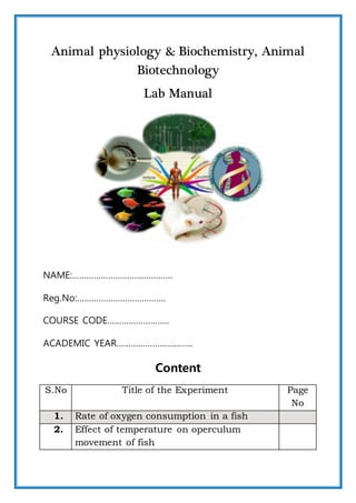
  • 1. Animal physiology & Biochemistry, Animal Biotechnology Lab Manual NAME:…………………………………… Reg.No:………………………………. COURSE CODE…………………….. ACADEMIC YEAR………………………….. Content S.No Title of the Experiment Page No 1. Rate of oxygen consumption in a fish 2. Effect of temperature on operculum movement of fish
  • 2. 2 3. Qualitative analysis of carbohydrate 4. Qualitative analysis of protein 5. Qualitative analysis of lipid 6. Demonstration of blood pressure using sphygmomanometer 7. Estimation of haemoglobin 8. Action of salivary amylase in relation to enzyme concentration 9. Action of salivary amylase in relation to temperature 10. Counting of different kinds of blood cells using haemocytometer 11. Isolation of genomic DNA from E. coli 12. Estimation of protein by Lowry’s Method Dos and Don’ts in the Lab Important Precautions to be taken  Do plan and prepare for every laboratory exercise before coming to lab. Always do some homework before carrying out experiments in the lab.
  • 3. 3  Keep work bench neat and clean before leaving the laboratory. Do wipe with detergents if necessary.  Dispose of biological materials such as microbes by sterilizing and in special containers for biological hazards. Do not throw other materials into these bags.  Tie back long hair to avoid contamination and fire hazard.  Be very careful with Bunsen burners. Always keep the burner at distance from the organic solvents. The burner must be turned off soon after the use.  Wash your hands before leaving the laboratory, even if you wore gloves, wash with disinfectant in running tap water before and after the work.  Be familiar with the chemicals in the laboratory and take maximum care in handling each chemical.  Keep chemicals back in position after use.  You must cut your nails regularly.  Tightly close all bottles and caps Don’ts  Don’t mishandle the chemical solutions, spirit lamp, UV light, instruments/apparatus or electricity.  Don‘t roam here and there in the laboratory without work or any aim.  Never misplace lab wares and other equipment.  Don‘t eat, drink, chew gum or apply cosmetics in the lab. Never use a lab microwave, laboratory fridge or freezer for food.
  • 5. 5 Ex. No: 1 Date: RATE OF OXYGEN CONSUMPTION IN A FISH BACKGROUND: Respiration by fish includes the uptake of oxygen from the environment at sites of gas exchange (typically the gills), the use of oxygen at mitochondria within individual cells, and the excretion of waste gases to the environment. Respiration provides oxygen for aerobic conversion of the energy contained in food to high-energy chemical bonds, such as those formed when adenosine diphosphate (ADP) is changed to adenosine triphosphate (ATP). The energy thus stored is then used to keep fish alive (maintenance) and to allow fish to move, digest food, grow, reproduce, and carry out all other energy-requiring functions. Measurements of respiration often can tell us how a fish is responding to environmental conditions and what its physiological state may be. Respirometry gives quantitative measures of how rapidly energy and oxygen are used. Respiratory data are important in the construction of bioenergetics models that can be used, for example, to calculate capacities for growth and reproduction. Respiration rates can be sensitive indicators of altered environmental conditions or physiological states, and thus can reveal much about a fish's recent and current activity, acclimation, and stress. AIM: To determine the rate of oxygen consumption by the given fish. PRINCIPLE OF TEST When manganous sulphate is added to the sample of water followed by alkaline iodide, manganous hydroxide is formed. This combines with
  • 6. 6 dissolved oxygen present in the water forming manganic hydroxide. This on acidification in the presence of concentrated sulphuric acid release certain amount of iodine which is equivalent to the amount of oxygen dissolved in the water. The amount of iodine released is determined by titrating against 0.01 N sodium thio sulphate solution using 1% starch as an indicator. The reactions are shown below: MnSO4 + 2KOH -------àMn(OH)2 + K2SO4 2 Mn(OH)2 +O2 ----à 2 MnO(OH)2 (Basic manganic oxide) MnO(OH)2 + H2SO4 ----àMnSO4 + 2H2O + [O] 2KI + H2SO4 + [O] -----à K2SO4 + H2O + I2 I2 + 2Na2S2O3 ------à 2NaI + Na2S4O6 MATERIALS REQUIRED:  100 ml beaker with siphon arrangement burette  Burette stand  Pipette  Conical flask  Reagent bottles  Thermometer  Balance REAGENT REQUIRED:  Manganese Sulphate  Alkaline iodide  Concentrated H2SO4  0.01 N Sodium thiosulphate  1% Starch METHOD:
  • 7. 7 A living tilapia fish is kept in container containing one litre of tap water. The same tap water is closed in a glass stopper control of bottle known volume (250 ml). A fine layer of kerosene is added to the surface of the experimental medium to prevent mixing with atmospheric air. The starting time of the experiment is noted. In this condition the fish is allowed to respire for half an hour in room temperature. The tap water in the control bottle is kept under control during the time of experiment, so that no change in the medium affect the control, after half an hour the water in the control bottle is siphoned out into the analyzing bottle. The volume of analyzing bottle is determined by Winkler’s method. The difference between the oxygen in control water and the experimental water gives the volume of oxygen consumed by the fish in half an hour. ESTIMATION OF OXYGEN IN THE WATER SAMPLE: A reagent bottle is taken and filled with water. The stopper is replaced without enclosing any air bubble. After removing the stopper, 1 ml of Manganous Sulphate and 1 ml of alkaline mixture were added. The stopper is replaced and the bottle is shaken well to ensure maximum chemical reaction and hence maximum absorption of oxygen. This is done for the uniform distribution by precipitation. Then the precipitate is allowed by settle down for few minutes. After this 1 ml of con H2SO4 is added to the solution and is shaken well till the precipitate is not seen in the bottle. The burette is rinsed with water and 0.1 N Sodium thiosulphate, filled with 0.1 N Sodium thiosulphate solution. Initial reading of the burette is noted. About 50 ml of solution from the bottle is transferred to a conical flask. To this 1 ml of 1% starch is added as an indicator. Now the color of the solution turns to dark blue. This solution is titrated against 0.01 Sodium thiosulphate and the titration is continued till the blue color disappeared. This is the achromic point and the final reading is noted. The volume of Sodium thiosulphate required for titrated is noted. The titration is repeated for concordant values. The difference between oxygen content of the control and of the experimental bottle gives the volume of oxygen consumed by the
  • 8. 8 fish in half an hour. Then the fish is removed from the experimental medium. Weight of the fish is taken. Oxygen consumed by the fish in one hour is calculated. The rate of oxygen consumption by the fish is calculated by using the formula. TABULATION: RATE OF OXYGEN CONSUMPTION IN A FISH S.No Volume of sample(ml) Burette reading Volume of Na2S2O3 Concordant value(ml) Indicator Weight of fishInitial (ml) Final (ml) CALCULATION: O2/ml/litre= CD* M*E*0.698*1000*Vt vs CD= volume of reagent bottle Volume of reagent bottle-volume of reagent used M= Molarity of sodium thiosulphate(0.01) E= Equivalent weight of oxygen 1000= To express litre Vt= Volume of sodium thiosulphate used in titration vs= Volume of sample titrated O2 in control water=………………………parts/ml/hr.
  • 9. 9 RATE OF OXYGEN CONSUMPTION IN THE EXPERI MENTAL WATER S.No Volume of sample(ml) Burette reading Volume of Na2S2O3 Concordant value(ml) Indicator Weight of fishInitial (ml) Final (ml) CALCULATION: O2/ml/litre= CD* M*E*0.698*1000*Vt vs CD= volume of reagent bottle Volume of reagent bottle-volume of reagent used M= Molarity of sodium thiosulphate(0.01) E= Equivalent weight of oxygen 1000= to express litre Vt= Volume of sodium thiosulphate used in titration vs= Volume of sample titrated O2 in experimental water=………………………parts/ml/hr.
  • 10. 10 Oxygen consumed by the fish in half an hour= 02 content in the tap water- O2 content in the experimental water Oxygen consumed by the fish in one hour=……………..*2 Rate of oxygen consumed by the fish=…..…………../weight of the fish =….…………..parts/ml/hr RESULT: Rate of oxygen consumption by the fish= …………………….parts/ml/hr. DISCUSSION: RVIEW QUESTIONS: 1. What is dissolved oxygen (DO)? 2. What are the approved methods for determination of DO? 3. What reagents are required for D.O. determinations using the modified Winkler titration method? 4. What interferences affect DO determinations by the modified Winkler titration procedure?
  • 11. 11 Ex. No: 2 Date: EFFECT OF TEMPERATURE ON OPERCULUM MOVEMENT OF FISH BACKGROUND Fish obtain their oxygen from water by ventilating their gills. These breathing apparatus are highly vascularized out pocketing of the body surface. The operculum, a protective flap, covers and protects the gills, which are soft and delicate. The gills are ventilated by pumping water across them in one direction. Both the mouth and operculum work together to pump the water through the mouth, into the gills, and then out to the sides of the body. Counting gill cover (operculum) movements is a way to calculate respiration rates in fish. An oxygen-carbon dioxide exchange occurs. Then the operculum opens to allow the carbon dioxide-rich water to exit. Fish are exothermic; their body temperature is the same as water temperature and changes directly with it. Many physiological processes, such as metabolic rate, heart rate, and enzyme activities occur faster as body temperature increases and slow down at colder body temperature. By counting gill cover movements, students are getting an idea of a fishes' response to an ecological change.
  • 12. 12 AIM: To determine the rate of operculum movement of a given fish at different temperature. MATERIALS  Fish  Crushed ice  Large beaker  Hot water  Glass stirring rod  Thermometer  Stop clock METHOD A living fish is taken gently without rubbing on the mucous membrane and placed into a plastic jar containing water at room temperature. The temperature of the water sample is recorded with the help of thermometer. After 5 minutes a fish is exposed to the sample water, the number of operculum movement per minute at this temperature is recorded with the help of stop watch. The observation is repeated 3 times until two
  • 13. 13 concordant values are obtained. After that hot is added to the water sample until the temperature of the medium is increased by 5˚c. Again the experiment is repeated and the number of gill movement per minute are calculated. This same experiment is repeated with different temperature like 30˚c, 35˚c, 40˚c. The temperature of sample water is decreased by the addition of ice water and the time taken at 25˚c 20˚c and 15˚c is noted. The rate of operculum movement (1/t) is calculated. TABULATION S.No Temperature Time taken for seconds Concordant value 1/t rate of activity FORMULA: Q10 is the factor by which the reaction rate increases when the temperature is raised by ten degrees. Q10=K2 (10)/K1 (t2-t1) Speed of rate in reciprocal=1/t Where k1, K2 represent the velocity constant of particular temperature t2& t1 respectively t1= Lower temperature t2= Higher temperature
  • 14. 14 K1= Rate of activity at low temperature. K2= Rate of activity at high temperature. RESULTS: Effects of temperature Q10 on operculum movement of fish Q10= Q102= DISCUSSION: REVIEW QUESTIONS: 1. Are fish warm-blooded or cold-blooded animals? 2. Do you think that all animals would be affected in this manner by temperature changes? Explain.
  • 15. 15 Ex. No: 3 Date: QUALITATIVE ANALYSIS OF CARBOHYDRATE BACKGROUND: Carbohydrates are polyhydroxy aldehydes and ketones or substances that hydrolyze to yield polyhydroxy aldehydes and ketones. Aldehydes (– CHO) and ketones (= CO) constitute the major groups in carbohydrates. Carbohydrates are mainly divided into monosaccharides, disaccharides and polysaccharides. The commonly occurring monosaccharides includes glucose, fructose, galactose, ribose, etc. The two monosaccharides combine together to form disaccharides which include sucrose, lactose and maltose. Starch and cellulose fall into the category of polysaccharides, which consist of many monosaccharide residues. AIM: To detect the presence of different group carbohydrate in the given solution. GENERAL MATERIALS: 0.1% of glucose, Test solution I. ANTHRONE TEST: PRINCIPLE:
  • 16. 16 Carbohydrates are dehydrated with concentrated H2SO4 to form “Furfural”, which condenses with anthrone to form a green color complex which can be measured by using colorimetrically at 620nm (or) by using a red filter. Anthrone react with dextrins, monosaccharides, disaccharides, polysaccharides, starch, gums and glycosides. But they yields of color where is to form carbohydrate to carbohydrate. REAGENTS:  0.2% anthrone in con H2SO4  Test tubes  Spirit lamp  2ml pipette. PROCEDURE:  Take 2 ml of sample solution in a test tube.  Add 1 ml of anthrone reagent and mix thoroughly OBSERVATION: A bluish green color develops in the test tube indicates the presence of sugar. II. FEHLING’S TEST: This is an important test to detect the presence of reducing sugars. Fehling’s solution A is copper sulphate solution and Fehling’s solution B is potassium sodium tartrate. On heating, carbohydrate reduces deep blue solution of copper (II) ions to red precipitate of insoluble copper oxide. MATERIALS:  Fehling’s reagent
  • 17. 17  Test tube  Holder  Spit lamp PROCEDURE:  Take 5 ml of fehling’s solution in a test tube.  Add 1ml of test solution to it.  Mix well and boil in water bath. OBSERVATION: The production of brownish red cuprous oxide precipitate indicates the presence of reducing sugar. III. BENDICT’S TEST: PRINCIPLE: The copper sulfate (CuSO4) present in Benedict's solution reacts with electrons from the aldehyde or ketone group of the reducing sugar. Reducing sugars are oxidized by the copper ion in solution to form a carboxylic acid and a reddish precipitate of copper (I) oxide MATERIALS:  Benedict’s reagent  Test tube  Test tube holder  Sprit lamb  Measuring cylinder PROCEDURE:
  • 18. 18  Take 5 ml of sucrose solution in a test tube  Add 2 ml of Benedict’s reagent  Boil for two minutes. OBSERVATION: A yellowish- red precipitate occurs within 2 to 5 minutes indicates the presence of monosaccharide. IV. IODINE TEST Iodine test is used to detect the presence of starch. Iodine is not much soluble in water so iodine solution is prepared by dissolving iodine in water in presence of potassium iodide. Iodine dissolved in an aqueous solution of potassium iodide reacts with starch to form a starch/iodine complex which gives characteristics blue black color to the reaction mixture. PROCEDURE:  Take 2 ml of sample solution in a test tube.  Add 0.5 ml of iodine solution.  Dilute the solution with distilled water. OBSERVATION Appearance of blue coloration indicates the presence of starch RESULT: Sample…………… indicates the presence of carbohydrates in the test solution. DISCUSSION: REVIEW QUESTIONS: 1. Write a test which helps to differentiate between a disaccharide and a monosaccharide? 2. If a sample being tested gives a positive result for Benedict’s test and Fehling's test, then what will the sample be?
  • 19. 19 Ex. No: 4 Date: QUALITATIVE ANALYSIS OF PROTEIN BACKGROUND: Proteins are large biological molecules composed of α-amino acids (Amino acid in which amino group is attached to α-carbon, which exist as zwitter ions and are crystalline in nature). They contain carbon, hydrogen, oxygen, nitrogen and sometimes phosphorous and sulphur. Protein is an important macronutrient essential for survival. They are constituent of calls and hence are present in all living bodies. 10-35% of calories should come from protein. Protein is found in meats, poultry, fish, meat substitutes, cheeses, milk etc. AIM: To detect the presence of protein in the given sample. BIURET TEST: PRINCIPLE: This test is used to detect the presence of peptide bond. When treated with copper sulphate solution in presence of alkali (NaOH or KOH), protein reacts with copper (II) ions to form a violet colored complex called biuret
  • 20. 20 MATERIALS Biuret reagent PROCEDURE:  Take 2 ml of sample solution in a test tube  Add 2 ml of Biuret reagent solution to it.  Mix well and observe the color. OBSERVATION: Development of violet color in 15 minutes indicates the presence of protein. XANTHO PROTEIN TEST: PRINCIPLE: Aromatic amino acids, such as Phenyl alanine, tyrosine and tryptophan, respond to this test. In the presence of concentrated nitric acid, the aromatic phenyl ring is nitrated to give yellow colored nitro-derivatives. At alkaline pH, the color changes to orange due to the ionization of the phenolic group. MATERIALS • Conc.HNO3 • Blank solution PROCEDURE:  Take 1 ml of sample solution in a test tube.  Add 0.5 ml of concentrated HNO3.
  • 21. 21  Observe the yellow color  Boil the tube in a water bath, cool and add few drops of NaOH solution. OBSERVATION: The yellow colored solution is turn into orange. RESULT: Sample………….. Indicates the presence of protein in the solution. DISCUSSION: REVIEW QUESTIONS: 1. All the amino acids with aromatic side chains give positive test to Xanthoprotein test- justify. 2. Define “activated benzene ring”. 3. Write the reaction(s) involved in Biuret’s Test.
  • 22. 22 Ex. No: 5 Date: QUALITATIVE ANALYSIS OF LIPID BACKGROUND: Lipids are one of the major constituents of foods, and are important in our diet for a number of reasons. They are a major source of energy and provide essential lipid nutrients. Nevertheless, over-consumption of certain lipid components can be detrimental to our health, e.g. cholesterol and saturated fats. In many foods the lipid component plays a major role in determining the overall physical characteristics, such as flavor, texture, mouth feel and appearance. For this reason, it is difficult to develop low-fat alternatives of many foods, because once the fat is removed some of the most important physical characteristics are lost. Finally, many fats are prone to lipid oxidation, which leads to the formation of off-flavors and potentially harmful products. Some of the most important properties of concern to the food analyst are: • Total lipid concentration • Type of lipids present • Physicochemical properties of lipids, e.g., crystallization, melting point, smoke point, rheology, density and color • Structural organization of lipids within a food.
  • 23. 23 AIM: To determine the presence of lipids in the given sample. GREASE SPOT TEST: PRINCIPLE: The working principle is that most grease or fat have a high boiling point. So, they are non-volatile. In room temperature, the spot of water can absorb enough heat from the air and evaporated. But the spot of grease can never absorb enough heat to evaporate. When the liquid is inside the sheet of paper, it diffracts light. So, light can pass from one side of the paper to another side. This gives the phenomenon of "translucent". When there is no liquid in the paper, there is no diffraction. So, light cannot pass it through. MATERIALS:  Coconut oil  Paper PROCEDURE A drop of oil is put over a piece of ordinary paper. OBSERVATION: Oil spreads and leaves on oily impression on the paper. The translucent spot indicates the presence of fat. TEST FOR SOLUBILITY: PRINCIPLE: The oil will float on water because of lesser specific gravity. MATERIALS:  Take 5 ml of water and chloroform in a separate test tube.  Add to it a few drops of sample solution and shake well. OBSERVATION:
  • 24. 24 The oil drops are completely dissolved chloroform. This indicates the presence of lipids in the given solution. EMULSIFICATION TEST: PRINCIPLE: When oil and water, which are immiscible, are shaken together, the oil is broken up into very tiny droplets which are dispersed in water. This is known as oil in water emulsion. The water molecule due to the high surface tensions has a tendency to come together and form a separate layer. This is why the oil and water emulsion is unstable in the presence of substances that lower the surface tension of water. Eg: Sodium carbonate, soap, bile salts etc. MATERIALS:  Coconut oil PROCEDURE:  Take 2 drops of sample solution in a test tube  Add 2 ml of water to it and shake vigorously. OBSERVATION: A milky white emulsion indicates the presence of lipid. RESULTS: Sample…………indicates the presence of lipids in the test solution. DISCUSSION: REVIEW QUESTIONS: 1. Give the classification of Lipids and write the biological importance. 2. Describe the structure of the triglycerides. 3. Define obesity. Name three diseases associated with obesity.
  • 25. 25 Ex. No:6 Date: DEMONSTRATION OF BLOOD PRESSURE USING SPHYGMOMANOMETER BACKGROUND Blood Pressure (BP) is the force or pressure which the blood exerts on the wall of blood vessels. When the left ventricle contracts and pushes blood into the aorta the pressure produced within the arterial system is called systolic blood pressure and in the complete cardiac diastole stage, the heart is resting following the ejection of blood, the pressure within the arteries is called diastolic blood pressure. In adult normal systolic BP ranges about 110-130 mm Hg and the diastolic BP ranges about 70-85 mm Hg in adult. A sphygmomanometer is a device that measures blood pressure. It is composes of an inflatable rubber cuff, which is wrapped around the arm. A measuring device indicates the cuff's pressure. A bulb inflates the cuff and a valve releases pressure. A stethoscope is used to listen to arterial blood flow sounds. Types There are two types of sphygmomanometers:  Manometric
  • 26. 26  Digital. A manometric sphygmomanometer is the old-fashioned blood pressure- measuring device. It is the more accurate of the two types of sphygmomanometers. A digital sphygmomanometer uses digital readouts. With this type of sphygmomanometer, the blood pressure reading appears on a small screen or is signaled in beeps. Parts of the Device A sphygmomanometer usually consists of a:  A rubber bag surrounded by a cuff.  A manometer (usually a mechanical gauge, sometimes electronic, rarely a mercury column).  An inflating bulb to elevate the pressure.  A deflating valve. WORKING PRINCIPLE: A sphygmomanometer consists of a pump, dial, cuff and a valve. To measure blood pressure, the cuff is wrapped around the arm and then inflated using the pump. The pressure applied by the cuff closes off the brachial artery so that no blood flows through. The pressure is then slowly released and sounds are detected using a stethoscope placed on the brachial artery. Blood pressure readings are given as a fraction of two numbers, 120/80 mm Hg for example. The first number is the systolic pressure; this
  • 27. 27 is the blood pressure when the ventricles of the heart are contracting. The number is recorded when a sound is heard for the first time as pressure in the cuff is slowly relaxed. The second number is the diastolic pressure, which is the blood pressure in between contractions when the heart is relaxed. This number is recorded when the thumping of blood squeezing through the narrowed brachial artery is no longer heard. It is important to note that multiple measurements of a person’s blood pressure must be made in order to gain an accurate reading. APPARATUS:  Sphygmomanometer  Stethoscope. METHODS OF USING SPHYGMOMANOMETER Open the adjusting screw at the proximal end of the pumping bulb. So that air is removed from the cuff and tubes. Then wrap the cuff around the upper arm just above the elbow joint and fix it with the help of attached hook in a manner that neither it is too tight nor too loose. While wrapping the cuff care should be taken that the tubes attached to it must face towards the lower end. Then tighten the adjusting screw and pump the air be pumping rubber bulb rapidly so that mercury is raised up to 110-120 mm Hg mask in the manometer. Observe the sound of arterial blood branchial artery with the help of a stethoscope. Again pump the air slowly and keep on observing the sound of arterial blood in the branchial artery with stethoscope to disappear. As and when sound is not felt, note the mercury level in the manometer.Pump a litter more air and again note the proper arterial sound to reappear. This would be the systolic pressure, now loose the screw, remove all air and open the cuff and keep the apparatus in a safe place. The difference in a pressure between systole and diastole is called the pulse pressure which is normally 30-35 mm hg. The lower limit systole pressure in a normal adult is estimated about 105 mm Hg and the upper limit about 150 mm Hg. In women the blood pressure is from 5 to 10 mm Hg less than in man.
  • 28. 28 REVIEW QUESTIONS: 1. What are the benefits of monitoring blood pressure? 2. What are the home remedies for controlling high blood pressure? 3. Why do we use mercury in a sphygmomanometer even though it is toxic to our body? Ex. No:7 Date: ESTIMATION OF HAEMOGLOBIN BACKGROUND: The apparatus consists of haemeter having haemoglobin pipette, glass stirrers and a dropper. Haemometer Housing: This is a chamber with 3 compartments and is open on oneside and closed behind with an opaque glass plate. The central compartment holds a Hn flux which can be inserted and removed on eitherside of this compartment 2 vertical tubes are present that are fixed. The Hb tube is graduated on one side with gm % one side and on the other side % alone.
  • 29. 29 Pipette and Stirrer: The pipette is known as sucking pipette. It is having a capillary pore and marker on the top as 20 micro litre. The blood sample is sucked upto this mark. The glass stirrer has a flattened end and is used for mixing blood and acid in the Hb-tube. AIM: To determine the percentage of haemoglobin in human blood sample. PRINCIPLE: To estimate the percentage of haemoglobin a define quality of Haemoglobin (Hb) is converted to acid haematin by addition of 0.1 N hydrochloric acid and resulting brown color is compared with standard brown glass reference blocks of a haemometer. MATERIAL REQUIRED:  Haldon’s haemometer  haemoglobin pipette  blood sample  Glass rod REAGENT:  N/10 Hcl METHOD: Haemoglobinometer has a graduated tube or dilution tube which may be round or square shaped and standard light brown glass plate rod. Usually haemometer with square meter or glass rod is used. The haemoglobin pipette has uniform diameter and 20 cubic meter marked on it. The graduated haemoglobin tube was sterilized and picked with sterilized needle up to 20mm3. Blood was poured into the haemoglobin tube already containing N/10 Hcl. They prevent the production air bubbles. The mixture is stirred well with the glass rod and the preparation is left for 10 minutes. The haemoglobin of the sample blood is converted into acid haematin. The mixture is diluted by N/10 Hcl is added drop into the haemometer till the
  • 30. 30 color matches with that by drop of standard brown color glass rod. The reading of haemoglobin tube showing percentage of haemoglobin is noted. DISCUSSION: REVIEW QUESTIONS: 1. Write the composition of blood and functions of blood. 2. Can a healthy diet and nutrition help keep optimal hemoglobin levels? Ex. No:8 Date: ACTION OF SALIVARY AMYLASE IN RELATION TO ENZYME CONCENTRATION BACKGROUND: Saliva mixes up with the food and helps its digestion. That is, the enzyme ptyalin or amylase present in human saliva hydrolyzes the big molecules of food into many molecules. For example starch into mono- saccnaricies maltose and glucose; proteins into amino acids and fats into fatty acids and glycerol. Thus saliva not only helps in digestion of food but convert it into energy generating substances. Further, enzymes and their activity are very sensitive to temperature and pH. Even a slight variation in these two factors, can disturb the action of enzymes. In other words, digestion of food by salivary amylase is also effected by pH and temperature and can be verified experimentally. For example, hydrolysis of starch can be verified by testing it with iodine solution. Starch forms blue colored complex with iodine. If no starch is present in a system it will not give blue color with iodine. AIM: To determine amylase activity of human saliva in relation to enzyme concentration. MATERIALS:
  • 31. 31  Test tubes  Stop watch  Thermometer  Pipette  Porcelain tile  Glass rod  Iodine PRINCIPLE: The enzyme salivary amylase is present in saliva and is responsible for the partial digestion by facilitating the formation of easy absorbable amylase also known as ptyalin. Amylase activity can be studied by time taken for the digestion of standard starch solution. When digestion is proceeding starch is hydrolyzed to dextrin and the color produced by iodine is blue. It slowly changes to reddish violet, brownish violet brown and finally the iodine color. This point is called acromic point and time taken for complete hydrolysis of known amount of starch by a known strength of enzyme is the measure of amylase activity. First product of the action of the enzyme amylase in the substrate. Starch is the formation of an intensive blue color in combination with summarized as follows.
  • 32. 32 PROCEDUR E: Mouth is washed thoroughly with water and about 5 ml of saliva is collected in a clean dry test tube. 1 ml of starch is taken in a test tube and a drop of iodine is added. The starch gives blue color. To this mixture 1ml of 25% saliva is added and the stop clock is switched on. The test tube is continuously shaken till the blue color is disappeared. At the time of disappearance of blue color (achromic point) the stop clock is switched off and the time taken is noted. The same procedure is repeated for various enzyme concentrations like 50%, 75% and 100%. Tabulate the results and draw a graph marking the enzyme concentration in “X” axis and the rate of activity in “Y” axis. Calculate the Q10 value. TABULATION: S. No Enzyme concentration Time taken in seconds Rate of activity(1/t)
  • 33. 33 RESULT: The rate of digestion of different concentration of enzymes  25%=  50%=  75%=  100%= DISCUSSION: REVIEW QUESTIONS: 1. What is an achromic point? 2. What are the functions of the salivary enzyme amylase during digestion? Ex. No:9 Date: ACTION OF SALIVARY AMYLASE IN RELATION TO TEMPERATURE BACKGROUND: All enzymes are proteinaceous in nature. At a lower temperature, the enzyme salivary amylase is deactivated and at the higher temperature, the enzyme is denaturated. Therefore, more time will be taken by an enzyme to digest the starch at lower and higher temperatures. Optimum temperature for the enzymatic activity of salivary amylase ranges from 32 °C to 37 °C. The optimum temperature means that the temperature at which the enzyme
  • 34. 34 shows the maximum activity. At this optimum temperature, the enzyme is most active and hence, takes less time to digest the starch. AIM: To determine amylase activity of human saliva in relation to temperature. MATERIALS:  Test tubes  Stop watch  Thermometer  Pipette  Porcelain tile  Glass rod  Iodine and starch PROCEDURE: 5ml of saliva is collected in a clean beaker. In a test tube 3 ml of 1% starch solution is taken. The test is immersed in water bath in a beaker. The temperature of water bath is noted. A drop of iodine is added in the test tube after adding 1ml of saliva. The color change is noted. The stop clock is
  • 35. 35 switched on at the time of introducing enzyme (saliva). At the achromic point, the stop watch is stopped and the time taken for the complete digestion is recorded. This indicates the stage in which all the starch is completely hydrolyzed by amylase at room temperature of 30˚c. Thus the experiment is repeated with different temperature (25 ˚c, 30 ˚c, and 35˚c). Tabulate the results and draw a graph marking temperature in “X” axis and the rate of activity in “Y” axis. Calculate the Q10 value. S. No Temperature Time taken in seconds Rate of activity(1/t)
  • 36. 36 Tabulation: FORMULA: Q10=K2/K1* 10/t2-t1 T= Time taken in seconds t2= Higher temperature t1= Lower temperature K2= Rate of activity at higher temperature K1= Rate of activity at lower temperature RESULTS: The rate of digestion at 25 ˚c= 30 ˚c= 35˚c=
  • 37. 37 Q10= DISCUSSION REVIEW QUESTIONS: 1. The experiment set up to study the effect of temperature on the activity of salivary amylase on starch is carried out at 10 °C. The solution mixture that contains amylase and starch keeps on giving blue color for iodine test about half an hour. What is the reason for this? 2. What is the optimum temperature for the enzymatic activity of salivary amylase?
  • 38. 38 Ex. No:10 Date: COUNTING OF DIFFERENT KINDS OF BLOOD CELLS USING HAEMOCYTOMETER AIM: To count the RBC and WBC of human blood sample. PRINCIPLE: Whole blood is diluted with a 3% acetic acid solution, which hemolyzes mature erythrocytes and facilitates leukocyte counting. The standard dilution for leukocyte counts is 1:20. This dilution is prepared using the leukocyte Unopette system. The dilution is mixed well and incubated to permit lysis of the erythrocytes. Following the incubation period, the dilution is mounted on a hemacytometer. The cells are allowed to settle and then are counted in specific areas of the hemacytometer chamber under the microscope. The number of leukocytes are calculated per µL (x 109/L) of blood. MATERIALS:  Haemocytometer  Pipette  Counting chamber  Compound microscope REAGENTS: Dilution fluid (Haem’s solution) and 15% acetic acid. PROCEDURE:
  • 39. 39 Finger tips and pins were sterilized carefully by 70% alcohol. When it becomes dry, finger is punctured to allow a free flow of blood the tip of the pipette is place over the blood and sucking up to 0.5 mark in the RBC pipette. The tip of the pipette is wiped out with a piece of clean cloth and is immediately placed in the dilution fluid and it is sucked until the mark shake the pipette well to ensure uniform suspension of cells mean while a coverslip is placed on the counting chamber.After discarding 3 to 4 drops of diluted blood. The blood from the pipette is placed on the raised center of the counting chamber at the edge of the coverslip. So that a drop is drawn under the coverslip by capillary action. After 5 minutes the counting chamber is placed under high power microscope and adjusted in such a way that squares can be seen clearly. Red blood corpuscles were counted for WBC another pipette having the white blood is taken. Similar to RBC counting of the blood cells the blood is drown up to 5 marks and diluted to 1.1 using 1.5% acetic acid as a dilution
  • 40. 40 fluid. The diluted blood is placed in the counting chamber focused in low power microscope. The WBC corpuscles were seen as blank spots and they were seen as black spots and they were counted in the four corners of the square. CALCULATION: RBC COUNTING Total RBC/mm3 = Number of RBC counted × Dilution factor × Depth factor No. of chambers counted WBC COUNTING Total WBC/mm3 = Number of WBC counted × dilution factor × depth factor No. of chambers counted RESULT: No. of RBC in one cubic mm………………………../mm3 No. of WBC in one cubic mm…………………/mm3 DISCUSSION: REVIEW QUESTIONS: 1. Which are the different dyes used to differentiate between viable and non-viable cells? 2. Explain the different cell counting techniques available other than Haemocytometer.
  • 42. 42 Ex. No: 11 Date: ISOLATION OF GENOMIC DNA FROM E. coli AIM: To isolate the genomic DNA from E .coli cells. PRINCIPLE: The isolation and purification of DNA from cells is one of the most common procedures in contemporary molecular biology and embodies a transition from cell biology to the molecular biology (from in vivo to in vitro). The isolation of DNA from bacteria is a relatively simple process. The organism to be used should be grown in a favorable medium at anoptimal temperature, and should be harvested in late log to early stationary phase for maximum yield. The genomic DNA isolation needs to separate total DNA from RNA, protein, lipid, etc. Initially the cell membranes must be disrupted in order to release the DNA in the extraction buffer. SDS (sodium dodecyl sulphate) is used to disrupt the cell membrane. Once cell is disrupted, the endogenous nucleases tend to cause extensive hydrolysis. Nucleases apparently present on human fingertips are notorious for causing spurious degradation of nucleic acids during purification. DNA can be protected from endogenous nucleases by chelating Mg2++ ions using EDTA. Mg2++ ion is considered as a necessary cofactor for action of most of the nucleases. Nucleoprotein interactions are disrupted with SDS, phenol or proteinase K. Proteinase enzyme is used to degrade the proteins in the disrupted cell soup. Phenol and chloroform are used to denature and separate proteins from DNA. Chloroform is also a protein denaturant, which stabilizes the rather unstable boundary between an aqueous phase and pure phenol layer. The denatured proteins form a layer at the interface between the aqueous and the organic phases which are removed by centrifugation. DNA released from disrupted cells is precipitated by cold absolute ethanol or isopropanol.
  • 43. 43 MATERIALS REQUIRED:  LB Broth, E. coli cells REAGENTS  TE buffer (pH 8.0)  10% SDS  Proteinase K  Phenol-chloroform mixture  5M Sodium Acetate (pH 5.2)  Isopropanol  70% ethanol  Autoclaved Distilled Water  Eppendorf tubes 2 ml  Micropipette & Microtips  Microfuge PROCEDURE: 1. 2 ml overnight culture is taken and the cells are harvested bycentrifugation for 10 minutes 2. About 500 μl of TE buffer is added to the cell pellet and the cells areresuspended in the buffer by gentle mixing. 3. 100 μl of 10% SDS and 5 μl of Proteinase K are added to the cells. 4. The above mixture is mixed well and incubated at 37º C for an hour inan incubator. 5. 1 ml of phenol-chloroform mixture is added to the contents, mixedwell by inverting and incubated at room temperature for 5 minutes. 6. The contents are centrifuged at 10,000 rpm for 10 minutes at 4º C. 7. The highly viscous jelly like supernatant is collected using cut tipsand is transferred to a fresh tube. 8. The process is repeated once again with phenol-chloroform mixtureand the supernatant is collected in a fresh tube. 9. 100 μl of 5M sodium acetate is added to the contents and is mixedgently.
  • 44. 44 10. 2 ml of isopropanol is added and mixed gently by inversion till white strands of DNA precipitates out. 11. The contents are centrifuged at 5,000 rpm for 10 minutes. 12. The supernatant is removed and 1ml 70% ethanol is added. 13. The above contents are centrifuged at 5,000 rpm for 10 minutes. 14. After air drying for 5 minutes 200 μl of TE buffer or distilled water isadded. 15. 10 μl of DNA sample is taken and is diluted to 1 or 2 ml with distilledwater. 16. The concentration of DNA is determined using a spectrophotometer at 260/280 nm. 17. The remaining samples are stored for further experiments. RESULTS: DISCUSSION: REVIEW QUESTIONS: 1. Can I use a microscope to see the DNA that I extract? 2. Why must we add the proteinase K enzymes to the mixture in order to extract the DNA? 3. Why does the DNA clump together?
  • 45. 45 Ex. No: 12 Date: ESTIMATION OF PROTEIN BY LOWRY’S METHOD AIM: To estimate the amount of protein in the given sample by Lowry’s method. PRINCIPLE: The method is based on both the Biuret reaction, where the peptide bonds of proteins react with copper under alkaline conditions producing Cu+, which reacts with the Folin reagent, and the Folin-Ciocalteau reaction, which is poorly understood but in essence phosphomolybdotungstate is reduced to heteropolymolybdenum blue by the copper-catalyzed oxidation of aromatic amino acids. The reactions result in a strong blue color, which depends partly on the tyrosine and tryptophan content. The Lowry method is sensitive to pH changes and therefore the pH of assay solution should be maintained at 10 - 10.5. . The Lowry method is sensitive to low concentrations of protein. REAGENTS: A. 2% Na2CO3 in 0.1 N NaOH B. 1% NaK Tartrate in H2O C. 0.5% CuSO4.5 H2O in H2O D. Reagent I: 48 ml of A, 1 ml of B, 1 ml C E. Reagent II- 1 part Folin-Phenol [2 N]: 1 part water. PROCEDURE: 1. Different dilutions of BSA solutions are prepared by mixing stock BSA solution (1 mg/ ml) and water in the test tube as given in the table. The
  • 46. 46 final volume in each of the test tubes is 5 ml. The BSA range is 0.1 to 0.5 mg/ ml. 2. From these different dilutions, pipette out 5.5 ml reagent I to different test tubes .Mix the solutions well. 3. This solution is incubated at room temperature for 10 mins. 4. Then add 0.5 ml of reagent II to each tube and incubate for 30 min. Zero the colorimeter with blank and take the optical density (measure the absorbance) at 660 nm. 5. Plot the absorbance against protein concentration to get a standard calibration curve. 6. Check the absorbance of unknown sample and determine the concentration of the unknown sample using the standard curve plotted above. Tabulation: S.No Vol. of BSA (ml) Conc of BSA (mg/ml) Vol. of Distilled water (ml) Vol. of reagent I (ml) Incubationfor10min Vol. of Reagent II (ml) Incubationfor30min OD At 660nm 1. 0.1 100 3.9 5.5 0.5 2. 0.2 200 3.8 5.5 0.5 3. 0.3 300 3.7 5.5 0.5 4. 0.4 400 3.6 5.5 0.5 5. 0.5 500 3.5 5.5 0.5 6. Unknown 100 3.9 5.5 0.5 7. Blank 0 4 5.5 0.5
  • 47. 47 Calculations: Result: The amount of protein present in the given sample was found to be ………… Review Questions: 1. Explain the principle behind lowry’s method? 2. Beer-Lamberts law. 3. Compare Lowry’s method with other methods of protein estimation.
  • 49. 49 Content 1. Glucose 2. Fructose 3. Glycogen 4. Sucrose 5. Amino acids 6. Cholesterol 7. Intestinal villi 8. Haemoglobin 9. ECG 10. Sphygmomanometer 11. Haemometer 12. Haemocytometer 13. Kymograph 14. Cardiac Muscle 15. Striated & Non striated muscle 16. Simple muscle Twitch 17. PBR 322 18. Plasmid 19. Lambda phage 20. Recombinant DNA 21. Gene cloning 22. Southern blotting 23. Stem cells
  • 50. 50 24. RFLP 25. Dolly 26. Transgenesis 27. Animal cloning
  • 51. 51 Glucose Glucose 1. The most common carbohydrate is glucose (C6H12O6). 2. Glucose is a monosaccharide, an aldohexose and a reducing sugar. 3. Glucose contains six carbon atoms and an aldehyde group. 4. Glucose exists as D-(+)-glucose and L-(-)-glucose. 5. The D-(+)-glucose form is used as fuel by your body and is also called dextrose. 6. L-(-)-glucose cannot be used by your body as a source of energy. 7. Glucose is a white, sweet-tasting powder, so it can be used as a sweetener. 8. Glucose is one of the products of photosynthesis in plants and some prokaryotes. 9. In animals and fungi, glucose is the result of the breakdown of glycogen but in plants the breakdown substrate is starch.
  • 52. 52 10.Glucose is found in the human bloodstream where it is referred to as "blood sugar". 11.The first step in the breakdown of glucose in all cells is glycolysis, producing pyruvate which is the starting point for all other processes in cellular respiration. 12.A major part of the use of the energy from glucose oxidation is the conversion of ADP to ATP, with the energy-rich molecule ATP being subsequently used as the energy currency of the cell. Fructose Fructose 1. Fructose (or levulose) is a simple sugar (monosaccharide) with the same chemical formula as glucose (C6H12O6) but a different atomic arrangement. 2. Sources of fructoseinclude honey, fruits, and some root vegetables.
  • 53. 53 3. Fructose is also known as Fruit sugar, levulose, D-fructofuranose, D-fructose, D-arabino-hexulose. 4. The D-fructose is the most predominant isomer in nature compared to L-fructose. 5. It is the most reactive and most water soluble sugar compared to other natural sugars. 6. Fructose is a 6-carbon keto sugar and also known as D- fructopyranose. 7. Fructose is not a product of photosynthesis. 8. Fructose is produced commercially from sugarcane, sugar beets, and corn. 9. Fructose is difficult to crystallize from an aqueous solution compared to glucose. 10.Fructose is mainly important for beverage industries and bakery products because it contributes to enhancing palatability and taste, and for browning or color development. Sucrose
  • 54. 54 Sucrose 1. Sucrose is a disaccharide (glucose + fructose) with the molecular formula C12H22O11. 2. Sucroseis a non- reducing sugar in which alpha glucose is linked to fructo furanoseby a glycosidic link. 3. Sucroseis fine and colorless. It is odor-free and is a crystalline powder with a sweet taste 4. It is best known for its role in human nutrition and is formed by plants but not by higher animals. 5. Sucroseis soluble in water with melting point 180° C. 6. Sucroseis heated above its melting point, it forms a brown substance known as caramel. 7. Hydrolysis of sucroseyields D-glucose and D-fructose Sucrose + water =hydrolysis=> Glucose + Fructose C12H22O11 + H2O =hydrolysis=> C6H12O6 + C6H12O6 8. Sucrose has the ability to dissolve in water and form crystals when heated properly. 9. Sucrose is too large to cross cell membranes. 10.So our body break down the large molecule in to glucose and fructose with an assistance of enzyme sucrose and the process called hydrolysis.
  • 55. 55 11. The bacterial metabolism of sucrose can cause abdominal cramping, bloating, constipation and diarrhea. Amino Acids Amino Acids 1. Amino acids are the building blocks of proteins, each one corresponding to one or more DNA codon. 2. The physical and chemical properties of a protein are determined by its constituent amino acids. 3. They consist of an amine group, a carboxylic acid group, a hydrogen and a group unique to each residue around a central carbon atom. 4. Amino acids are forming a peptide bond between the amine group of one amino acid and the carboxyl group of the next. 5. Amino acids are more soluble in water than they are in ether, dichloromethane, and other common organic solvents. 6. In a polypeptide chain, the end with the free amine group is called the N-terminus and the end with the free carboxylic acid the C-terminus.
  • 56. 56 7. They can be classified according to the core structural functional groups' locations as alpha- (α-), beta- (β-), gamma- (γ-) or delta- (δ-) amino acids. 8. Humans can synthesize about half of the amino acids needed to make proteins. Other amino acids, called the essential amino acids, must be provided in the diet. 9. Amino acids have high melting points, generally over 200 °C. 10.All natural amino acids are L‐amino acids. 11.Essential Amino Acids: Histidine, Isoleucine, Leucine, Lysine, Methionine, Phenylalanine, Threonine, Tryptophan, and Valine. 12.Nonessential Amino Acids: Alanine, Asparagine, Aspartic Acid, Glutamic Acid. 13.Conditional Amino Acids: Arginine (essential in children, not in adults), Cysteine, Glutamine, Glycine, Proline, Serine, and Tyrosine. 14.classification depends on the side chain structure, and experts recognize 5 types in this classification: Containing sulfur (Cysteine and Methionine) Neutral (Asparagine, Serine, Threonine, and Glutamine) Acidic (Glutamic acid and Aspartic acid) and basic (Arginine and Lysine) Alphatic (these include Leucine, Isoleucine, Glycine, Valine, and Alanine) Aromatic (these include Phenylalanine, Tryptophan, and Tyrosine) Cholesterol
  • 57. 57 Cholesterol 1. Cholesterol is a sterol and a lipid found in the cell membranes of all body tissues. 2. It is transported in the blood plasma of all animals. 3. Its molecular formula is C27H46O. 4. Its pure state it is a white, crystalline substance that is odourless and tasteless. 5. Cholesterol is present in higher concentrations in tissues or densely packed membranes, for example, the liver, spinal cord and brain. 6. Cholesterol is required to build and maintain cell membranes and it makes the membrane's fluidity, degree of viscosity and stable over wider temperature. 7. Cholesterol also serves as a precursor for the biosynthesis of steroid hormones and bile acids. 8. High levels of cholesterol in the bloodstream will cause atherosclerosis.
  • 58. 58 9. Cholesterol is insoluble in the blood; it must be attached to certain protein complexes called lipoproteins in order to be transported through the bloodstream. Glycogen 1. Glycogen is a homo polysaccharide. 2. Glycogen is a polymer of glucose residues in the main chains are connected by α (1→4) glycosidic bonds. 3. The branches are attached by α (1→6) glycosidic linkages. 4. It is a major reserve carbohydrates in animals. So it is called as animal starch. 5. It is a white powder, readily dissolves in water. 6. Glycogen is a large molecule produced in the liver and stored mainly in the liver and muscle cells. 7. Glycogen is the analogue of starch, a glucose polymer that functions as energy storage in plants.
  • 59. 59 8. It is a non-reducing sugar. It gives red color with iodine. The red color disappears on boiling and reappears on cooling. 9. The blood always contains 1% glucose. When it exceeds 1% the excess glucose is transported to the liver and is converted into glycogen. This process is called glycogenesis. 10.When blood sugar level is below 1% the liver glycogen is changed into blood glucose. This process is called glycogenolysis. 11.Muscle glycogen is utilized as the energy source during muscle contraction. Intestinal Villi Intestinal Villi
  • 60. 60 1. The wall of the small intestine contains millions of fingerlike projections called villi. 2. Their fingerlike shape greatly increases the surface area of the intestine and increase the overall rate of absorption. 3. The villi and intestinal walls are richly supplied with blood vessels and lymphatic vessels to carry away absorbed food substances. 4. Each villus transports nutrients to a network of capillaries and fine lymphatic vessels called lacteals close to its surface. 5. They have very thin walls, only one cell thick, this enables molecules to pass through easily. 6. The villi also have digestive enzymes on their surface. 7. Digested food passes into the villi through diffusion. 8. The villi are connected to blood vessels to allow nutrients to be carried away by the blood. 9. Villi also help the food to move along the digestive tract. 10.. Villus capillaries collect the simple sugars and amino acids into the bloodstream. Hemoglobin
  • 61. 61 Hemoglobin 1. Hemoglobin is the oxygen binding protein of red blood cells 2. It is a globular protein with quaternary structure. 3. A hemoglobin molecule is composed of a globin protein group and four heme groups. 4. Each RBC contains over 600 million hemoglobin molecules. 5. Each heam group associated with one Fe2+ atom. 6. Each hemoglobin molecule can bind 4 oxygen molecules. 7. The heme molecules give hemoglobin its red color. 8. Hemoglobin consists of four polypeptide subunits are 2 alpha chains and two beta chains. 9. Hemoglobin transports oxygen in the blood from the lungs to the rest of the body. 10.Hemoglobin develops in cells in the bone marrow that become red blood cells.
  • 62. 62 11.During phagocytosis globin protein is broken down to amino acids, which are recycled into new proteins and the heme is broken down into Iron, Biliverdin and Carbon Monoxide. 12.Hemoglobin plays an important role in the modulation of erythrocyte metabolism. Electrocardiogram Electrocardiogram 1. An electrocardiogram is abbreviated as EKG or ECG. 2. The electrocardiogram can measure the rate and rhythm of the heartbeat, as well as provide indirect evidence of blood flow to the heart muscle. 3. The ECG as a graph, plotting electrical activity on the vertical axis against time on the horizontal axis. 4. Standard ECG paper moves at 25 mm per second during real-time recording.
  • 63. 63 5. ECG paper is marked with a grid of small and large squares. Each small square represents 40 milliseconds (ms) in time along the horizontal axis and each larger square contains 5 small squares, thus representing 200 ms. 6. Standard paper speeds and square markings allow easy measurement of cardiac timing intervals. This enables calculation of heart rates and identification of abnormal electrical conduction within the heart 7. The first electrical signal on a normal ECG originates from the atria and is known as the P wave. 8. Depolarisation of the ventricles results in usually the largest part of the ECG signal and this is known as the QRS complex. 9. The Q wave is the first initial downward deflection 10.The R wave is then the next upward deflection. 11.The S wave is then the next downward deflection. 12.ST segment and the T wave electrical signal reflecting repolarisation of the myocardium. 13.Doctors use ECGs to detect and study many heart problems, such as heart attacks, arrhythmias and heart failure.
  • 64. 64 Sphygmomanometer Sphygmomanometer 1. A sphygmomanometer is a device that measures blood pressure. There are two types of sphygmomanometers:  Manometric  Digital. 2. A manometric sphygmomanometer is the old-fashioned blood pressure-measuring device. It is the more accurate of the two types of sphygmomanometers. 3. A digital sphygmomanometer uses digital readouts. With this type of sphygmomanometer, the blood pressure reading appears on a small screen or is signaled in beeps.
  • 65. 65 4. A sphygmomanometer usually consists of a:  A rubber bag surrounded by a cuff.  A manometer (usually a mechanical gauge, sometimes electronic, rarely a mercury column).  An inflating bulb to elevate the pressure.  A deflating valve. 5. To measure blood pressure, the cuff is wrapped around the arm and then inflated using the pump. The pressure applied by the cuff closes off the brachial artery so that no blood flows through. 6. The pressure is then slowly released and sounds are detected using a stethoscope placed on the brachial artery. 7. Sphygmomanometer is kept at the level of heart of the subject and cuff is wrapped around the upper arm just above the elbow. 8. The chest piece of the stethoscope is placed upon the brachial artery. 9. The other ends of the stethoscope are connected with two ears. 10.The bag of the cuff is filled by the pump up to 240 mm Hg. pressure. 11.The pressure inside the cuff is released slowly by losing the air with the air adjustment screw. 12.As the pressure is released sudden appearance and disappearance of a sound is heard and recorded. 13.Sudden onset of trapping sound is systolic blood pressure and the sudden disappearance of sound is diastolic pressure. 14.Blood pressure readings are given as a fraction of two numbers, 120/80 mm Hg.
  • 66. 66 Haemometer 1. Haemometer is an apparatus used for the estimation of blood haemoglobin. It is otherwise called as haemoglobinometer. 2. It consists of a chamber and a capillary pipette. The chamber contains three test tubes held against a black background. 3. The two lateral tubes are filled with standard acid haematin and are sealed. They are called comparison tubes. 4. The central tube is graduated and is called experimental tube. 5. The experimental tube is graduated and is called experimental tube. 6. The experimental tube is graduated from 2 to 22gm% on one side and form 10 to 140gm% on the other side. 7. This tube has a glass rod to stir the blood when it is mixed with Hcl. 8. The capillary pipette is called Pasteur pipette. It is marked with 20 cu.mm. It is connected to a sucking rubber tube. 9. There is a small bottle to take N/10 Hcl and cleaning brush.
  • 67. 67 10.To estimate haemoglobin the experimental tube is filled up to the level of 2 gm. with N/10 Hcl. 11.The finger is picked with the needle and blood is sucked into the capillary pipette up to the level of 20 cubic mm. the blood is poured into the experimental tube. 12.The blood is mixed with Hcl by sucking and releasing with the pipette several times. 13.Allow 5 minutes to complete the reaction. 14.The red color of blood changes to brown indicating the formation of acid haematin. 15.Now start diluting acid haematin by adding distilled water drop by drop with the help of a dropper till the brown color matches the standard solution color. 16.The result was observed in bright sunshine. 17.The amount of haemoglobin is represented in g%. It is the measure of the height of the solution in the experimental tube. 18.The normal level of haemoglobin is 12g%. Haemocytometer
  • 68. 68 Haemocytometer 1. The haemocytometer was originally invented by Louis-Charles Malassez to count blood cells. 2. A haemocytometer has two chambers areas separated by an H-shaped moat and each chamber has a microscopic grid etched on the glass surface. 3. The chambers are overlaid with a glass coverslip that rests on pillars exactly 0.1 mm above the chamber floor. 4. These two squares are identical and each has a total area of 9 mm2 (1 mm on each side). 5. These squares are divided into nine primary squares with an area of 1mm2 . 6. The four corner primary squares are used when counting leukocytes. 7. These 4 large corner squares contain 16 smaller secondary squares, each with an area of 0.04 mm2 .
  • 69. 69 8. All 25 secondary squares of the center primary square are used to count platelets. 9. The raised edges of the haemocytometer hold the coverslip 0.1 mm off the marked grid, giving each square a defined volume. 10.The distance between the bottom of the coverslip and the surface of the counting area of the chamber is 0.1 mm Kymograph 1. A kymograph is a device that graphically records changes in position over time. 2. This instrument was invented in the 1840s by German physiologist Carl Ludwig. 3. The kymograph has been used most commonly in the field of medicine to study various physiological and muscular processes, for example blood pressure, respiration and muscle contractions.
  • 70. 70 4. This instrument consists of a motor, a central rod, a rotating drum, a stand, a battery, a switch and an induction coil. 5. The muscle is suspended from the clamp of a stand. 6. The free end of the muscle is tied with a lever. The lever has a pointer which is directed to the rotating drum. 7. A battery supplies the electric current which acts as a stimulus. 8. The current passes through an induction coil. 9. A smoked paper is pasted on the rotating drum. 10.A clockwork or electric motor drives the drum, rotating it slowly. 11.The stylus traces a graphic record on a piece of paper wrapped around the drum. 12.The two terminals of the wires coming out from the induction coil are connected to the two ends of the muscle (ex. Frog thigh muscle). 13.When an electric shock is given, the muscle contracts and hence shortens. 14.As a result, the pointer moves up and down makes a lines on the smoked paper of the moving drum. 15.The graphic record generated by the kymograph instrument is commonly translated into a graph, showing changes in pressure or motion on the horizontal x-axis, and time elapsed on the vertical y- axis. Cardiac muscle
  • 71. 71 Cardiac muscle 1. Cardiac muscle (heart muscle) is involuntary striated muscle. 2. It is found in the walls and histological foundation of the heart. 3. Cardiac muscle fibres are essentially long, cylindrical cells with one nuclei. 4. The arrangement of actin and myosin is similar to skeletal striated muscle. 5. Intercalated disks are located between cardiac muscles cells. 6. The intercalated disks form tight junctions between the cells so that they cannot separate under the strain of pumping blood. 7. The cardiac muscles are autorhythmic because they contract independently of the nervous system and has an intrinsic rhythm. 8. The muscle fibres are uninucleate and do not have a sarcolemma. 9. The cytoplasm consists of a large number of mitochondria and numerous glycogen granules. 10.They have a stable resting membrane potential of -90 mV. It has a prolonged action potential.
  • 72. 72 11.Millions of cardiac muscle cells working together are easily able to pump all of the blood in the body through the heart in less than a minute. Striated Muscle Striated Muscle 1. Skeletal muscle is called "striated" because of its appearance consisting of light and dark bands visible using a light microscope. 2. Skeletal muscle, also called voluntary muscle in vertebrates. 3. A single skeletal muscle cell is long and approximately cylindrical in shape, with many nuclei located at the edges of the cell. 4. A skeletal muscle fiber is around 20-100 µm thick and up to 20 cm long. 5. It is a form of striated muscle tissue which is under the voluntary control of the somatic nervous system.
  • 73. 73 6. Most skeletal muscles are attached to bones by bundles of collagen fibres known as tendons. 7. They have number of glycogen granules and mitochondria. 8. The most striking feature of the striated muscle fibre is the presence of al-ternating dark and light transverse bands called stripes or striations. 9. The dark bands are called anisotropic or A bands. 10. Each A band has at its middle a light zone called the H zone or Henson's line. 11.Skeletal muscle fibres are bound together by connective tissue and communicate with nerves and blood vessels. 12.The lighter band is iso-tropic and is called the I band. 13.Each I band has at its centre a dark line called the membrane of Krause or Z line or Z band. 14. Myofilaments are composed of thick (myosin) and thin (actin) filaments, which interact to cause muscle contractions. Non-Striated muscle
  • 74. 74 Non-Striated muscle 1. Smooth muscle are also called non-striated or non-straight muscles. 2. The muscle fibres are made up of long narrow spindle shaped cells that are generally shorter than striated muscle cells. 3. They average a length of about 0.2 mm. 4. Each muscle fibre has a single nucleus in the central thick portion. 5. The sarcoplasm consists of a number of parallely arranged myofibrils. 6. The walls of blood vessels, the tubes of the digestive system, and the tubes of the reproductive systems are composed primarily of smooth muscle. 7. Contractions of smooth muscle move food through the digestive tracts and push blood through the blood vessels. 8. The muscle cells are not bound by a true sarcolemma. 9. Smooth muscle fibres may occur individually or in the form of bundles of sheets.
  • 75. 75 10.The muscle fibres of viscera are usually arranged in the form of sheets made up of many layers of fibres. 11.In villi of the small intestine the muscle fibres occur singly. 12.Smooth muscles undergo slow and rhythmic contraction regulated by the autonomous nervous system. So they are called involuntary muscles. Simple Muscle Twitch Simple Muscle Twitch 1. A muscle twitch is contraction of a muscle in response to a stimulus that causes an action potential in one or more muscle fibres. 2. A twitch can last for a few milliseconds or 100 milliseconds, depending on the muscle type.
  • 76. 76 3. The tension produced by a single twitch can be measured by a myogram, an instrument that measures the amount of tension produced over time. 4. A simple twitch can be divided into a latent period, a contraction phase, and a relaxation phase. 5. The latent period begins at stimulation and typically lasts about two milliseconds. 6. The time it takes the muscle to react to the stimulus is called the latent period. 7. The contraction period is the time where the muscle is actually contracting and tension rises to a peak. 8. The last period is when the muscle length returns back to normal and it continues for about another twenty-five milliseconds. Plasmid pBR322 Plasmid pBR322 1. Plasmid pBR322 is one of the first multipurpose cloning vectors constructed for use in Escherichia coli.
  • 77. 77 2. It is cloning vector used for reproducing the DNA fragment. 3. pBR322 was the first artificial plasmid created by Mexicans Bolivar and Rodriguez in 1977. 4. The name pBR denotes the p stands for "plasmid," and BR for "Bolivar" and "Rodriguez." 5. pBR322 is 4363 base pairs in length and contains a replicon region (source plasmid pMB1). 6. It has two antibiotic resistance genes like ampicillin (ampR) and tetracycline (tetR). 7. The gene for ampicillin resistance are Pst I, Pvu I, and Sca I. 8. It have the gene for tetracycline resistance are BamH I, BspM I, EcoR V, Nhe I, Nru I, Sal I, Sph I, and Xma III. 9. It contains the origin of replication of pMB1, and the rop gene, which encodes a restrictor of plasmid copy number. 10.It also contains the unique EcoRI restriction site GAATTC and splices the DNA between G and A from 5' to 3' position. Ti plasmid
  • 78. 78 Ti plasmid 1. Tumor inducing plasmid or Ti plasmid are double stranded circular DNA molecule present in Agrobacterium tumefaciens. 2. Ti plasmids are classified based on the type of opines (octopine and nopaline. ) they produce in the host cell during infection. 3. Almost all Ti plasmids have identical structure except in their sequence for opine metabolism. 4. The plasmid has 196 genes that code for 195 proteins. 5. Size of the plasmid is ~250 kbp and 206,479 nucleotides long, the GC content is 56% and 81% of the material is coding genes. 6. It has T DNA which is of 20kb and in addition it has several genes such as vir genes for virulence, ori gene for origin of replication, tra genes for transfer and genes for opine synthesis. 7. Vir region contains eight operons they are Vir A, Vir B, Vir C, Vir D, Vir E, Vir F, Vir G and Vir H which together span about 40kb of DNA. 8. Virulence genes are responsible for the transfer of T DNA into the host cell and integration of T DNA with host genome.
  • 79. 79 9. T DNA is region of Ti plasmids common to both octopine and nopaline plasmids. But generally T DNA segment of Octopine plasmids is shorter than in Nopaline plasmids. 10.Ti plasmids have a single repeated sequence at both ends of T DNA. This sequence is called as the bordered sequence. 11. T DNA encode for the production of growth hormones like Cytokinin and Auxin which is necessary for tumor induction. 12.Vir region is activated by the phenolic compounds, namely Acetosyringone and alpha hydroxy acetosyringone produces by plant wounds. 13.These bind to Vir A proteins activates Vir G gene by phosphorylation which in turn activates other genes. 14.Vir D proteins together with Vir D proteins participate in the formation of conjugal tube formation between the bacterial cell and plant tissue during the infection process. 15.In transferring gene of interest to Ti plasmid, an intermediate vector is used such as pBR 322. T DNA portion of the Ti plasmid is separated. T DNA is inserted into the pBR 322 vector which results in formation of ashuttle vector. This shuttle vector can replicate in E. coli and in Agrobacterium. 16.Ti plasmids serve as ideal vectors for plant genetic engineering as it has the capability of transferring gene of interest into the target site with a high efficiency. It is easy to screen recombinant cells as marker genes are present.
  • 80. 80 Lambda phage Lambda phage 1. Easter Lederberg discovered lambda phage in E.coli K12 strain in 1951. 2. Enterobacteria phage λ (lambda phage, coliphage λ) is a temperate bacteriophage that infects Escherichia coli. 3. It belongs to a Group I, dsDNA phages, Order-Caudovirales, Family- Siphoviridae, Genus- lambda like viruses and Species lambda phage. 4. A lambda phage has a head measuring around 50-60 nanometers in diameter and a flexible tail that is around 150 nanometers long.
  • 81. 81 5. The head functions as a capsid that contains its genome, which contains 48,490 base pairs of double-stranded linear DNA. 6. The λ genome contains of  an origin of replication  genes for head and tail proteins  enzymes for DNA replication  gene for lysis and lysogeny cycle  single-stranded protruding cohesive ends of 12 bases (5’GGGCGGCGACCT; the other end is complementary to it, i.e., CCCGCCGCTGGA5′). 7. The single-stranded parts are known as sticky sites and are also called the cos site, which encircles the DNA in the host cytoplasm. 8. They can have either lytic or lysogenic cycle, depending on the environment. 9. The lysogenic cycle to the lytic cycle transformation is mainly controlled by the proteins cI (also known as λ repressor) and Cro. 10. λ phages are commonly used in DNA cloning and it can be used to clone up to 20-25 kb DNA. Recombinant PBR 322 (rDNA)
  • 82. 82 1. One of the most notable plasmids, termed pBR322 after its developers Bolivar and Rodriguez (pBR) pBR322. 2. pBR322 has sequences derived originally from three different plasmids as follows:  The blaR gene from plasmid Rl, a typical antibiotic resistance plasmid found in natural populations of E. coli.  The tet gene is derived from plasmid R6-5, another antibiotic resistance plasmid.  The origin of replication from plasmid pMBl, which is a close relative of plasmid ColEl. 3. The useful features of this vector are as follows:  Plasmid pBR322 has been completely sequenced.
  • 83. 83  There are over 40 enzymes with unique cleavage sites on the pBR322 genome.  Small size (-4.4 kb) enables easy purification and manipulation.  Two selectable markers (bla and tet) permit easy selection of recombination DNA,  High copy number of 15 copies per cell, which  Can be amplified to up to 1,000 to 3,000 when protein synthesis is blocked, e.g., by applying chloramphenicol. Gene Cloning
  • 84. 84 Gene Cloning 1. It is technique used in genetic engineering that involves the identification, isolation and insertion of gene of interest into a vector such as a plasmid or bacteriophage to form a recombinant DNA molecule. 2. This technique is also used to produce large quantities of that desire gene fragment or product encoded by that gene. 3. The major steps involved in cloning a gene are:  Step1: Identification and isolation of gene of interest (from Genomic library, cDNA library, Chemical synthesis of gene).  Step II: joining of this gene into a suitable vector (construction of recombinant DNA Eg: pBR 322) I. In the process, restriction enzymes functions as scissors for cutting DNA molecules. II. Ligase enzyme is the joining enzyme that joins the vector DNA with gene of interest. III. The resulting DNA is called the recombinant DNA, chimera or recombinant vector.  Step III: Introduction of this vector into a suitable organism Introduction of recombinant vector into host cell is achieved by different gene transfer methods like Electroporation, Microinjection, Liposome mediated gene transfer etc.  Step VI: Selection of transformed recombinant cells with gene of interest
  • 85. 85 Test like antibiotic resistance, visible characters, assay for biological activity are used to isolate the gene of interest.  Step V: Multiplication or expression of the gene of interest. Southern Blotting Southern Blotting 1. A Southern blot is a method used in molecular biology for detection of a specific DNA sequence in DNA samples. 2. Southern blotting combines transfer of electrophoresis-separated DNA fragments to a filter membrane and subsequent fragment detection by probe hybridization. 3. The method is named after its inventor, the British biologist Edwin Southern. 4. Southern blot are used in gene discovery and mapping, evolution and development studies, diagnostics and forensics.
  • 86. 86 Method:  Restriction endonucleases are used to cut high-molecular-weight DNA strands into smaller fragments.  The DNA fragments are then electrophoresedon an agarose gel to separate them by size.  Once separated, the fragments are denatured from double-stranded to single-stranded form by treatment with sodium hydroxide (NaOH).  After the denaturation step a positively charged nylon filter and a stack of paper towels are placed atop the gel and fragments are pulled towards the positively charged nylon by capillary action.  The membrane is then baked in a vacuum or regular oven at 80 °C for 2 hours or exposed to ultraviolet radiation (nylon membrane) to permanently attach the transferredDNA to the membrane.  The membrane is then exposed to a hybridization probe solution.  Hybridization of the probe to a specific DNA fragment on the filter membrane indicates that this fragment contains DNA sequence that is complementary to the probe.  Excess probe is washed off by reducing NaCl concentration.  Finally, the radio-labelled fragments are exposed to X rays film by autoradiography.  The position of the fragments on the nylon sheet corresponds to black bands where X-rays have been absorbed.
  • 87. 87 STEM CELL 1. Stem cells are cells which can generate new copies of themselves, and can turn into the more specialized cells (e.g. red blood cells) that perform functions in the body. 2. There are different types of stem cells, a. Some can turn into any cell in the body called pluripotent stem cells. b. Some can only turn into certain cells in one type of tissue called tissue specific stem cells. 3. Adult and foetal stem cells are tissue specific, they only turn into certain types of specialized cells. 4. We all have adult stem cells in many of our tissues which repair and replenish damaged cells.
  • 88. 88 5. The cells isolated from the embryo give rise to one cell line which can produce an infinite number of cells. 6. Embryonic stem cells exist only briefly after fertilization. 7. Human embryonic stem cells are obtained from very early stage embryos, within 5-6 days of fertilization. 8. Most embryos used in stem cell research were initially created for use in IVF and are donated by parents. 9. All types of stem cell are used in basic research to understand how our bodies work and develop, and to understand disease. 10.Blood from the umbilical cord, which is rich in stem cells, can be stored indefinitely for use in later life. Cord blood can be donated to public banks, or private companies offer storage for use by the baby or his/her relatives in later life. Restriction Fragment Length Polymorphism (RFLP)
  • 89. 89  Restriction Fragment Length Polymorphism (RFLP) is a molecular method of genetic analysis  It allows individuals to be identified based on unique patterns of restriction enzyme cutting in specific regions of DNA. For RFLP, the steps are: 1. Separate white and red blood cells with a centrifuge. 2. Extract DNA nuclei from the white blood cells. 3. Cut DNA strand into fragments using a restriction enzyme. 4. Place fragments into one end of a agarose gel. 5. Use an electric current to sort the DNA segments by length. This process is called agarose gel electrophoresis.
  • 90. 90 6. Use a sheet of nitrocellulose or nylon to blot the DNA. 7. The sheet is stained so the different lengths of DNA bands are visible to the naked eye. 8. By treating the sheet with radiation, anautoradiograph is created. 9. This is an image on x-ray film left by the decay pattern of the radiation. 10.The autoradiograph, with its distinctive dark-colored parallel bands, is the DNA profile. Some of the applications for RFLP analysis include:  DNA fingerprinting. Forensic scientists may use RFLP analysis to identify suspects based on evidence samples collected at scenes of crimes.  Paternity.RFLP is also used in the determination of paternity or for tracing ancestry.  Genetic diversity. The technique can be used in studying evolution and migration of wildlife, studying breeding patterns in animal populations, and in the detection and diagnosis of certain diseases.  The technique of using RFLP detection of variation in genomes is a vital tool in genome mapping and genetic disease analysis. DOLLY
  • 91. 91 1. Cloning is a process by which a genetically identical individual organism is produced.
  • 92. 92 2. Dolly the sheep was the first mammal to be cloned, not from a cell taken from embryos, but from an adult cell. 3. Dolly was cloned by using a technique known as somatic cell nuclear transfer (SCNT). 4. For cloning the steps :  Taking an oocyte (egg cell) and removing its nucleus.  Then the nucleus of a somatic (body) cell, which contains the DNA to be cloned.  It is implanted into the egg cell from which the nucleus had been removed. 5. The cell used as the donor for the cloning of Dolly was taken from the mammary gland of a 6 years old Finn-Dorsetewe. 6. The unfertilized egg was taken from a Scottish Blackface ewe. 7. From 277 fertilized eggs, 29 early embryos developed. They were planted into 13 surrogate mothers. Only one pregnancy went full term resulting in the birth of Dolly on 5 July 1996. 8. Dolly was cloned at the Roslin Institute in Scotland by British embryologist Ian Wilmut 9. Dolly lived her entire life at the Roslin Institute in Edinburgh, Scotland. 10.Dolly was died on 14 February 2003 as she had developed a form of lung cancer called Jaagsiekte and severe arthritis. The disease was common in sheep kept indoors and Dolly had to be kept indoors for security reasons.
  • 93. 93 11.A Finn Dorset has a life expectancy of around 11 to 12 years but Dolly lived for only 6.5 years. 12.Cloning may prove beneficial in saving endangered and newly extinct species by resurrecting them from frozen tissue. TRANSGENISIS 1. Transgenesis is a genetic engineering processes that remove genetic material from one species of plant or animal and add it to a different species. 2. The most common method for producing transgenic animals is gene transfer by DNA microinjection, which involves the following steps:  DNA containing the desired transgene is identified and cloned (copied tens of thousands of times in bacteria) before insertion into the animal host.  The host animals (cows, pigs, or sheep) are induced to super ovulate and their eggs are collected.  The eggs are fertilized in a laboratory dish.  Using a fine, hollow needle, a solution of DNA containing the transgene is injected into the male pronucleus of the fertilized egg (the nucleus of the sperm cell that entered the egg) before it fuses with the female pronucleus.
  • 94. 94  The transgenic embryos are grown in cell culture and then implanted into the uterus of a surrogate mother, where they complete their development.  Screening is performed to determine which of the offspring have inherited the transgene. 3. Transgenic organisms have also been developed for commercial purposes. Examples of transgenic organisms with commercial value: 4. Golden rice: modified rice that produces beta-carotene, the precursor to vitamin A. 5. Goats modified to produce FDA-approved human antithrombin (ATryn), which is used to treat a rare blood clotting disorder in humans. 6. Goats have also been genetically modified to produce spider silk. 7. The main drawback of DNA microinjection is its low success rate: only between 1 and 4 percent of microinjected eggs result in the live birth of a sheep, goat, or cow containing the transgene, and about 80 to 90 percent of transgenic embryos die during early development. Animal Cloning
  • 95. 95 1. Cloning is defined as the deliberate production of genetically identical individuals. Each newly produced individual is a clone of the original. 2. Animal Cloning is done by somatic cell nuclear transfer (SCNT) method. 3. This procedure starts with the removal of the chromosomes from an egg to create an enucleated egg. 4. The chromosomes are replaced with a nucleus taken from a somatic (body) cell of the individual or embryo to be cloned. 5. This cell could be obtained directly from the individual, from cells grown in culture, or from frozen tissue. 6. They treat the reconstructed egg with chemicals or electricity to stimulate cell division. 7. If the egg divides normally and forms a blastocyst (a small clump of cells that forms after an egg is fertilized), scientists will transfer it into a surrogate mother to develop into a new animal.
  • 96. 96 8. The successful implantation of the blastocyst in a uterus can result in its further development, culminating sometimes in the birth of an animal. 9. This animal will be a clone of the individual that was the donor of the nucleus. 10.Its nuclear DNA has been inherited from only one genetic parent. Application of Animal Cloning:  Genetically engineered pigs or suitable breeds of pigs can be cloned for organ transplantation.  Population of endangered species of animals can be increased by cloning. Cloning can be of immense use in improving the pedigree of livestock. Superior breeds of animals can be multiplied by this technique.
  • 97. 97 REFERENCES:  https://cyhsanatomy1.wikispaces.com/What+is+a+Simple+Twitch%3 F  http://www.southsudanmedicaljournal.com/archive/may-2010/how- to-read-an-electrocardiogram-ecg.-part-one-basic-principles-of-the- ecg.-the-normal-ecg.html  http://geekymedics.com/understanding-an-ecg/  http://www.practicalclinicalskills.com/ecg-interpretation.aspx  https://www.nhlbi.nih.gov/health/health-topics/topics/ekg  http://www.emedicinehealth.com/electrocardiogram_ecg/page6_em. htm
  • 98. 98  http://www.interactive-biology.com/3955/function-and-synthesis-of- hemoglobin/  http://www.britannica.com/science/hemoglobin  http://www.fastbleep.com/biology-notes/40/116/1172  http://proteopedia.org/wiki/index.php/Hemoglobin  http://hannahhelpbiology.blogspot.in/2013/02/231-describe- structure-of-villus-and.html  http://www.ask.com/science/function-villi-9890a6c92b86d436  https://answers.yahoo.com/question/index?qid=20071216082414AA dPQDP https://www.boundless.com/physiology/textbooks/boundless- anatomy-and-physiology-textbook/digestive-system-23/absorption- 225/absorption-in-the-small-intestine-1105-808/  http://biology-thedigestivesystem.blogspot.in/2012/11/explain-how- structure-of-villus-is.html  http://www.livestrong.com/article/151766-chemical-properties-of- sucrose/  http://www.ehow.com/about_5454423_properties-sucrose.html  http://pediaa.com/difference-between-glucose-and-fructose/  http://www.newworldencyclopedia.org/entry/Fructose  http://www.chemicalformula.org/sucrose  http://chemistry.tutorvista.com/organic- chemistry/disaccharides.html  http://healthyliving.azcentral.com/digestion-absorption-sucrose- 17531.html
  • 99. 99  https://www.verywell.com/what-is-glycogen-2242008  https://www.rpi.edu/dept/bcbp/molbiochem/MBWeb/mb1/part2/glyc ogen.htm http://www.preservearticles.com/2012042131039/the- structure-of-the-smoothstriatedcardiac-muscle-tissue-with-a- diagram.html  http://www.innerbody.com/image_musc01/musc71.html  http://slidingfilament.webnode.com/cardiac-muscle/  http://www.preservearticles.com/2012042131039/the-structure-of- the-smoothstriatedcardiac-muscle-tissue-with-a-diagram.html  https://www.boundless.com/biology/textbooks/boundless-biology- textbook/the-musculoskeletal-system-38/muscle-contraction-and- locomotion-218/skeletal-muscle-fiber-structure-824-12067/  http://www.britannica.com/science/skeletal-muscle  http://www.ivyroses.com/HumanBody/Tissue/Tissue_Muscular- Tissue.php  https://cyhsanatomy1.wikispaces.com/What+is+a+Simple+Twitch%3F  http://www.southsudanmedicaljournal.com/archive/may-2010/how-to-read-an- electrocardiogram-ecg.-part-one-basic-principles-of-the-ecg.-the-normal-ecg.html  http://geekymedics.com/understanding-an-ecg/  http://www.practicalclinicalskills.com/ecg-interpretation.aspx  https://www.nhlbi.nih.gov/health/health-topics/topics/ekg  http://www.emedicinehealth.com/electrocardiogram_ecg/page6_em.htm  http://www.interactive-biology.com/3955/function-and-synthesis-of-hemoglobin/  http://www.britannica.com/science/hemoglobin  http://www.fastbleep.com/biology-notes/40/116/1172  http://proteopedia.org/wiki/index.php/Hemoglobin  http://hannahhelpbiology.blogspot.in/2013/02/231-describe-structure-of-villus-and.html  http://www.ask.com/science/function-villi-9890a6c92b86d436
  • 100. 100  https://answers.yahoo.com/question/index?qid=20071216082414AAdPQDPhttps://www.b oundless.com/physiology/textbooks/boundless-anatomy-and-physiology- textbook/digestive-system-23/absorption-225/absorption-in-the-small-intestine-1105- 808/  http://biology-thedigestivesystem.blogspot.in/2012/11/explain-how-structure-of-villus- is.html  http://www.livestrong.com/article/151766-chemical-properties-of-sucrose/  http://www.ehow.com/about_5454423_properties-sucrose.html  http://pediaa.com/difference-between-glucose-and-fructose/  http://www.newworldencyclopedia.org/entry/Fructose  http://www.chemicalformula.org/sucrose  http://chemistry.tutorvista.com/organic-chemistry/disaccharides.html  http://healthyliving.azcentral.com/digestion-absorption-sucrose-17531.html  https://www.verywell.com/what-is-glycogen-2242008  https://www.rpi.edu/dept/bcbp/molbiochem/MBWeb/mb1/part2/glycogen.htmhttp:// www.preservearticles.com/2012042131039/the-structure-of-the- smoothstriatedcardiac-muscle-tissue-with-a-diagram.html  http://www.innerbody.com/image_musc01/musc71.html  http://slidingfilament.webnode.com/cardiac-muscle/  http://www.preservearticles.com/2012042131039/the-structure-of-the- smoothstriatedcardiac-muscle-tissue-with-a-diagram.html  https://www.boundless.com/biology/textbooks/boundless-biology-textbook/the- musculoskeletal-system-38/muscle-contraction-and-locomotion-218/skeletal-muscle- fiber-structure-824-12067/  http://www.britannica.com/science/skeletal-muscle  http://www.ivyroses.com/HumanBody/Tissue/Tissue_Muscular-Tissue.php