Explanation of Major parts of human eye and iLasik reviews By Vipin Buckshey at Visual Aids Centre
•Download as PPT, PDF•
23 likes•1,353 views

Optometrist Vipin Buckshey is explaining about the major parts of human eye. He has also covered the functionality of major parts . Vipin Buckshey is the owner of Visual Aids Centre which is popular for iLasik surgeries .Read reviews about Visual Aids Centre and Vipin Buckshey in this slide.

Recommended
More Related Content
Viewers also liked
Recently uploaded
Recently uploaded (20)
Explanation of Major parts of human eye and iLasik reviews By Vipin Buckshey at Visual Aids Centre
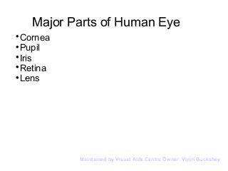
- 1. Major Parts of Human Eye Cornea Pupil Iris Retina Lens Maintained by Visual Aids Centre Owner: Vipin Buckshey
- 2. Cornea The cornea is the outer covering of the eye. This dome-shaped layer protects your eye from elements that could cause damage to the inner parts of the eye. There are several layers of the cornea, creating a tough layer that provides additional protection. The cornea also allows the eye to properly focus on light more effectively. Those who are having trouble focusing their eyes properly can have their corneas surgically reshaped to eliminate this problem. Maintained by Visual Aids Centre Owner: Vipin Buckshey
- 3. Pupil The pupil appears as a black dot in the middle of the eye. This black area is actually a hole that takes in light so the eye can focus on the objects in front of it. The pupil may appear to open (dilate) and close (constrict), but it is really the iris that is the prime mover; the pupil is merely the absence of iris. The pupil determines how much light is let into the eye. Both pupils are usually of equal size. If they are not, the condition is called anisocoria. Maintained by Visual Aids Centre Owner: Vipin Buckshey
- 4. Iris The iris is the area of the eye that contains the pigment which gives the eye its color. This area surrounds the pupil, and uses the dilator pupillae muscles to widen or close the pupil. This allows the eye to take in more or less light depending on how bright it is around you. If Light is too bright, the iris will shrink the pupil so that they eye can focus more effectively. Maintained by Visual Aids Centre Owner: Vipin Buckshey
- 5. Retina The light focuses by the lens will be transmitted onto the retina. This is made of rods and cones arranged in layers, which will transmit light into chemicals and electrical pulses. The retina is located in the back of the eye, and is connected to the optic nerves that will transmit the images the eye sees to the brain so they can be interpreted. The back of the retina, known as the macula, will help interpret the details of the object the eye is working to interpret. Maintained by Visual Aids Centre Owner: Vipin Buckshey
- 6. Lens The lens sits directly behind the pupil. This is a clear layer that focuses the light the pupil takes in. It is held in place by the ciliary muscles, which allow the lens to change shape depending on the amount of light that hits it so it can be properly focused. Maintained by Visual Aids Centre Owner: Vipin Buckshey
- 7. Lens The lens sits directly behind the pupil. This is a clear layer that focuses the light the pupil takes in. It is held in place by the ciliary muscles, which allow the lens to change shape depending on the amount of light that hits it so it can be properly focused. Maintained by Visual Aids Centre Owner: Vipin Buckshey