Central venous pressure (cvp)
•Download as DOC, PDF•
60 likes•133,943 views

Central Venous Pressure (CVP) monitoring

Recommended
More Related Content
What's hot
What's hot (20)
Viewers also liked
Similar to Central venous pressure (cvp)
Similar to Central venous pressure (cvp) (17)
Central venous pressure (cvp)
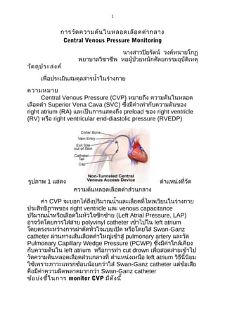
- 1. 1 การวัด ความดัน ในหลอดเลือ ดดำา กลาง Central Venous Pressure Monitoring วัต ถุป ระสงค์ นางสาวปิยรัตน์ วงค์หนายโกฏ พยาบาลวิชาชีพ หอผู้ป่วยหนักศัลยกรรมอุบัติเหตุ เพื่อประเมินสมดุลสารนำ้าในร่างกาย ความหมาย Central Venous Pressure (CVP) หมายถึง ความดันในหลอด เลือดดำา Superior Vena Cava (SVC) ซึ่งมีค่าเท่ากับความดันของ right atrium (RA) และเป็นการแสดงถึง preload ของ right ventricle (RV) หรือ right ventricular end-diastolic pressure (RVEDP) รูปภาพ 1 แสดง ความดันหลอดเลือดดำาส่วนกลาง ตำาแหน่งที่วัด ค่า CVP จะบอกได้ถึงปริมาณนำ้าและเลือดที่ไหลเวียนในร่างกาย ประสิทธิภาพของ right ventricle และ venous capacitance ปริมาณนำ้าหรือเลือดในหัวใจซีกซ้าย (Left Atrial Pressure, LAP) อาจวัดโดยการใส่สาย polyvinyl catheter เข้าไปใน left atrium โดยตรงระหว่างการผ่าตัดหัวใจแบบเปิด หรือโดยใส่ Swan-Ganz catheter ผ่านทางเส้นเลือดดำาใหญ่เข้าสู่ pulmonary artery และวัด Pulmonary Capillary Wedge Pressure (PCWP) ซึ่งมีค่าใกล้เคียง กับความดันใน left atrium หรือการทำา cut drown เพื่อสอดสายเข้าไป วัดความดันหลอดเลือดส่วนกลางที่ ตำาแหน่งเหนือ left atrium วิธีนี้นิยม ใช้เพราะภาวะแทรกซ้อนน้อยกว่าใส่ Swan-Ganz catheter แต่ขอเสีย ้ คือมีค่าความผิดพลาดมากกว่า Swan-Ganz catheter ข้อ บ่ง ชี้ใ นการ monitor CVP มีด ัง นี้
- 2. 2 1. ประเมิน preload ในผู้ป่วย hypovolemia/septic shock/valvular problems/congestive heart failure 2. ประเมินในรายสงสัย right ventricular dysfunction ที่สัมพันธ์ กับ severe lung disease, pulmonary hypertention,cardiac temponade 3. Major surgery ที่คาดว่าจะเสียเลือด > 1 blood volume ตำา แหน่ง เส้น เลือ ดที่ใ ช้ส ำา หรับ monitor CVP เช่น Basilic vein Brachial vein Cephalic vein Saphenous vein นอกจากนี้สามารถ monitor CVP ทาง catheter ซึ่งแทงผ่าน ผิวหนังเข้าไปในเส้นเลือดดำาใหญ่ ได้แก่ External/Internal jugular vein หรือทาง Subclavian vein catheter สาย catheter ซึ่งมักใช้ feeding tube No. 8 ยาว 100 cm. โดยปลาย สายจะอยู่ที่ Superior Vena Cava ก่อนเข้า right atrium ในกรณีที่ผู้ ป่วยใส่ Swan-Ganz catheter สามารถ monitor CVP ได้ทาง proximal line ซึ่งมีรูเปิดอยู่ใน right atrium ปัจ จัย ที่ส ่ง ผลต่อ การเปลี่ย นแปลงค่า ความดัน ในหลอดเลือ ดดำา ส่ว นกลาง(จันทรา พรหมน้อย;2555) ได้แ ก่ 1. ปริมาตรในหลอดเลือด (intravascularvolume) ส่งผลให้เกิดได้ทั้ง ภาวะนำ้าเกินและภาวะ ขาดนำ้าภาวะนำ้าเกินพบบ่อยในผู้ป่วยไตวาย (renalfailure) หรือมีภาวะ หัวใจล้มเหลว (heart failure) ทำาให้ค่าความดันในหลอดเลือดดำาส่วนกลางสูงขึ้นในทางตรงกันข้าม ภาวะขาดนำ้าหรือปริมาตรไม่เพียง พอ มีสาเหตุจากร่างกายสูญเสียเลือด หรือสูญเสียนำ้าร่างกายได้รับสาร นำ้าไม่เพียงพอ ทำาให้ค่าความดันใน หลอดเลือดดำาส่วนกลางลดลง (Scales, & Pilsworth,2008) นอกจากนี้ ยังมีสาเหตุจากการสูญเสียนำ้าใน ช่องว่างระหว่างเซลล์เนื่องจากหลอดเลือดสูญเสียความสามารถในการ ซึมผ่าน (increase capillary
- 3. 3 permeability) เช่น ภาวะช็อคจากการติดเชื้อในกระแสเลือด (septic shock) หรือมีระดับโปรตีนในเลือดตำ่า ทำาให้ความดันออนโคติก (oncotic pressure) ลดลง เช่น ผู้ป่วยที่มีปัญหาโรคตับ หรือ แผลไหม้ เป็นต้น 2. ความตึง ตัว ของหลอดเลือ ด (vasculartone) ได้แก่ การหดรัดตัว และการขยายตัวของหลอด เลือดดำาในภาวะหลอดเลือดดำาส่วนปลายขยายตัวทำาให้การไหลเวียน เลือดกลับสู่หัวใจลดลง ค่าความดัน ในหลอดเลือดดำาส่วนกลางจึงลดลง พบได้ในกลุ่มผู้ป่วยที่มีไข้สูง ติดเชื้อ ในกระแสเลือด มีภาวะช็อค จาก การแพ้ (anaphylactic shock) หรือได้รับยาขยายหลอดเลือด (vasodilating drug) ในทางตรงกันข้าม ขณะที่ภาวะหลอดเลือดดำาส่วนปลายหดตัวทำาให้ปริมาตรไหลเวียนเลือด กลับสู่หัวใจเพิ่มขึ้น ความดัน ในหลอดเลือดดำาส่วนกลางในระยะแรกจึงสูงขึ้น เช่นผู้ป่วยที่มีภาวะสูญ เสียเลือด หรือปริมาณนำา h ในร่างกายน้อย 3. ประสิท ธิภ าพการทำา งานของหัว ใจด้า นขวา (right heart function) เมื่อร่างกายอยู่ในภาวะที่ ประสิทธิภาพการทำางานของหัวใจด้านขวาลดลง ทำาให้หัวใจไม่ สามารถบีบเลือดไปยังหลอดเลือดแดง ปอดได้ เกิดภาวะเลือดคั่งค้างในหัวใจห้องบนขวาความดันในหัวใจห้อง บนขวาเพิ่มขึ้น ค่าความดันใน หลอดเลือดดำาส่วนกลางจึงสูงขึ้น 4. ความดัน ในทรวงอก (intrathoracic pressure) ในภาวะที่ความ ดันในทรวงอกสูงขึ้นจะส่ง ผลให้ค่าความดันหลอดเลือดดำาส่วนกลางสูงกว่าค่าปกติ เช่น กรณีผู้ ป่วยใส่เครื่องช่วยหายใจความดันบวก(PEEP) (Magder, 2006) วิธ ีก ารวัด CVP 1. บอกให้ผู้ป่วยทราบและล้างมือให้สะอาด 2. จัดท่าผู้ป่วยให้นอนหงายราบ (ผู้ป่วยบางรายมีข้อจำากัดในการนอน ราบหรืออาจหอบ เหนื่อยขณะที่นอนราบ จัดท่าศีรษะสูงได้ไม่เกิน 45 องศา และแขนขา ขณะที่วัดควรเหยียดตรง) 3. หาตำาแหน่งของ zero จุดตัดของ midaxillary line กับ fourth intercostal space และอาจขีดระดับไม่ว่าจะเป็น external jugular
- 4. 4 ,subclavian vein,cutdown ให้วัดที่ตำำแหน่ง zero หรือ phlebostatic axis รูปภำพ 2 แสดงตำำแหน่ง phlebostatic axis 4. หมุน three-way ให้ IV fluid ไหลเข้ำไปในสำย iv ด้ำนไม้บรรทัด โดยปิดด้ำนผู้ป่วยไว้ก่อน ควรให้ IV fluid อยู่ในสำย ในระดับเกือบเต็ม สำย หรือมำกกว่ำค่ำเดิม (ประมำณ 5 cm) จำกนั้นหมุนปิด three-way ด้ำนไม้บรรทัด 5. นำำไม้บรรทัดวำงทำบที่ผู้ป่วย โดยให้ตำำแหน่งของ zero หรือเลข ศูนย์ ซึ่งจุดที่วำงต้องอยู่ระดับเดียวกับ right atrium นั่นคือที่ตำำแหน่งจุด ตัดของ midaxillary line กับ fourth intercostal space 6. หมุน three-way เปิดเฉพำะด้ำนผู้ป่วยกับไม้บรรทัด ปิดด้ำน IV (กรณีที่มี three-way หลำยอัน ให้ปรับเฉพำะอันที่อยู่ติดกับสำย cut down หรืออันที่มีไม้บรรทัด) รูปภำพที่ 3 แสดงวิธีกำรวัดควำมดันหลอดเลือดดส่วนกลำง
- 5. 5 7. กำรอ่ำนค่ำ CVP ที่ work ดี จะต้อง fluctuate หรือมีกำรเต้นขึ้นลง ของระดับนำ้ำในสำยที่ไม้บรรทัดตำมจังหวะกำรหำยใจ (หำกพบว่ำเต้น ขึ้นลงตำมชีพจร แสดงว่ำปลำยสำย CVP อยู่ลึกเกินไปลงเข้ำไปถึงใน หัวใจ) ให้อ่ำนค่ำเมื่อเริ่มคงที่ โดยอ่ำนค่ำช่วงหำยใจออกสุด (end of expiration) เนื่องจำกควำมดันในช่องทรวงอกจะใกล้เคียงกับควำมดัน บรรยำกำศ *กรณีที่ผู้ป่วยใส่เครื่องช่วยหำยใจ และสำมำรถหำยใจเองได้ ไม่มีหอบ เหนื่อย ขณะอ่ำนค่ำให้ปลดเครื่องช่วยหำยใจ เนื่องจำกเครื่องช่วย หำยใจจะทำำให้ได้ค่ำ CVP สูงกว่ำค่ำจริง *กรณีที่มีกำรใส่ PEEP จะทำำให้ค่ำ CVP สูงกว่ำค่ำจริงมำกขึ้น เนื่องจำกควำมดันในช่องทรวงอกมำก แต่ในกำรวัด CVP ผู้ป่วยที่ on PEEP โดยเฉพำะที่ค่ำ PEEP > 5 cmH2O จะวัด CVP โดยไม่ปลด เครื่องช่วยหำยใจ ทั้งนี้เนื่องจำกผู้ป่วยเหล่ำนี้มักมีภำวะของ hypoxia และกำรปลดเครื่องบ่อยจะมีผลให้ประสิทธิภำพในกำรถ่ำงถุงลมปอดลด ลง ดังนั้นในกำรอ่ำนค่ำ CVP ทุกครั้ง ควรบันทึกไว้ด้วยว่ำวัดขณะใส่เครื่อง ช่วยหำยใจหรือปลดเครื่อง นอนศีรษะสูงกี่องศำ 9. เมื่ออ่ำนค่ำ CVP เสร็จแล้ว ให้หมุน three-way อยู่ในลักษณะเดิม คือ ปิดด้ำนไม้บรรทัด 10. ตรวจสอบควำมเรียบร้อยอีกครั้ง โดยเฉพำะกำรหมุน three-way, rate IV fluid และข้อต่อต่ำงๆ ไม่ให้หลวมหรือหลุด 11. จัดท่ำผู้ป่วยให้เหมือนเดิมหรือตำมควำมเหมำะสม กำรแปลค่ำ CVP - ใช้ pressure transducer ซึ่งจะมีหน่วยเป็น millimeters of mercury (mmHg) - ใช้ water manometer หรือใช้ไม้บรรทัดที่มีสำยยำง (extension tube) ซึ่งใช้บ่อยบนคลินิก จะมีหน่วยเป็น centimeters of water (cmH2O) หมำยเหตุ: 1 cmH2O=1.36 mmHg ,1mmHg.= 0.76 cmH2O ค่ำ CVP ปกติ อำจอยู่ในช่วง 6-12 cmH2O ทั้งนี้มักใช้ค่ำ CVP ในกำรเปรียบเทียบกำรเปลี่ยนแปลงจำกกำรรักษำในผู้ป่วยรำยนั้นๆ มำกกว่ำ ค่ำ CVP ตำ่ำ หมำยถึง ปริมำณนำ้ำและเลือดในร่ำงกำยลดลง ค่ำ CVP สูงขึ้นมัก หมำยถึงปริมำณนำ้ำและเลือดในร่ำงกำย มำกกว่ำปกติ ที่สำำคัญในกำรแปลค่ำ CVP จะต้องดูอำกำรและอำกำร แสดงอื่นร่วมด้วย เช่น blood pressure, heart rate, urine output, urine specific gravity, intake/output, conscious, ฟังปอดได้ยินเสียง
- 6. 6 ผิดปกติ อำกำรหอบเหนื่อย ควำมตึงตัว ควำมอุ่น เย็น ชื้นของผิวหนัง เป็นต้น ค่ำ CVP สูง และตำ่ำ พบได้ใ นหลำยๆ สำเหตุ ดัง นี้ สำเหตุท ี่ท ำำ ให้ CVP สูง Elevated vascular volume Increased cardiac output (hyperdynamic cardiac function) Depressed cardiac function (RV infarct, RV failure) Cardiac tamponade Constrictive pericarditis Pulmonary hypertension Chronic left ventricular failure สำเหตุท ี่ท ำำ ให้ CVP ตำ่ำ Reduced vascular volume Decreased mean systemic pressure (e.g., as in late shock state) Venodilation (drug induced) ภำ วะ แท รก ซ้อ น ทั้ง จำ กขั้น ต อ น ก ำ ร ใ ส่ส ำ ย CVP แล ะ กำ รวัด มี ดัง นี้ 1. Hemothorax 2. Pneumothorax 3. Nerve injury 4. Arterial puncture 5. Thoracic duct perforation
- 7. 7 6. Arrhythmias 7. Systemic or local infection 8. Perforation or erosion of vascular structure 9. Thrombosis 10. Air embolism Central Venous Pressure Monitoring อุป กรณ์ set IV ,0.9NSS 100ml ,three way,extention ,transducer ,แป้น transducer, syring วิธ ีก าร 1.ขั้น ต่อ อุป กรณ์ ต่อ set iv เข้ากับ ขวดนำ้าเกลือ 0.9 NSS 100 ml. แล้วต่อสาย IV เข้ากับตัว transducer แล้วต่อสาย extension เข้ากับ transducer ต่อแป้นสำาหรับวาง transducerg เข้ากับเสานำ้าเกลือ โดย
- 8. 8 ตัวแป้นต้องอยู่ในตำาแหน่ง phlebostatic axis คือ midaxillary line กับ fourth intercostal space ดังภาพ Set IV Extension จากนั้นเปิดนำ้าเกลือเพื่อไล่ air ที่อยู่ใน set ทั้งหมด เสร็จแล้วต่อสาย extension เข้ากับสาย cutdown หรือ subclavian vein โดย subclavian ต่อเข้ากับสาย สีนำ้าตาล หรือ proximal lumen 2.ต่อ monitor ต่อสาย cable เข้าที่จอ monitor สาย 3.การต่อ monitor
- 9. 9 เมื่อต่อสาย cable หน้าจอจะปรากฏดัง ภาพ เลือกชนิด cable ที่ตอเข้าไปเป็น CVP ่ โดย เลือกไปที่ label เลือก CVP ถึ ง ขั้ น ต อ น calibration เ มื่ อ ว า ง ตำา แหน่ ง transcuder ที่ จุ ด phlebostatic axis แ ล้ ว ห มุ น three way ข อ ง transducer ด้ า นผู้ ป่ ว ย แล้ ว เปิ ด จุ ก ออก “close patient open to air”ดังภาพ กด zero cal รอเครื่อง calibrate ให้ cvp = 0 mmHg เสร็ จ แล้ ว ก็ ห มุ น three way มาด้ า นจุ ก three way ตามเดิมและปิดจุก หมายเหตุ ควรทำา การ flush สาย CVP ทุก เวร เพื่อ ป้อ งกัน การอุด ตัน ของสาย แ ล ะ cribrate เ มื่ อ wave CVP เ มื่ อ มี การ Overdamping การอ่า นค่า CVP wave
- 10. 10 จากรูป เป็น ปกติข อง CVP A wave - occurs after the P wave of the ECG complex during the PR interval. It reflects the increased atrial pressure that occurs with atrial contraction. Note that the A wave will be absent in patients who do not have a distinct atrial contraction, such as those with atrial fibrillation. Since the CVP value should be a reflection of the Right Ventricular End-Diastolic Pressure, the CVP reading is taken at the last half of the A wave at the midpoint of the X descent. Calculate the CVP by averaging the pressure measured at the peak of the A wave and at the subsequent trough. due to atrial contraction. Absent in atrial fibrillation. Enlarged in tricuspid stenosis, pulmonary stenosis and pulmonary hypertension. The C wave - occurs at the end of the QRS complex at the beginning of the ST segment on the ECG tracing. It reflects closure of the tricuspid valve between the right atrium and right
- 11. 11 ventricle and the slight bulging of the tricuspid valve during ventricular contraction. The C wave is not always visualized. X descent - due to atrial relaxation. The V wave occurs at the end of the T wave on the ECG tracing. It reflects the increased pressure during passive atrial filling. The Y descent occurs prior to the P wave on the ECG tracing. It reflects the opening of the tricuspid valve and the passive flow of blood from the right atrium into the right ventricle prior to atrial contraction. Canon waves - large waves not corresponding to a, v or c waves. Due to complete heart block or junctional arrhythmias. ตัว อย่า ง 1 Measure CVP here Inspiration Expiration FIGURE 3 FIGURE เอกสารอ้า งอิง
- 12. 12 AnaesthesiaUK. The central venous pressure trace. สืบค้นเมื่อ 12 ธันวาคม 2556 จาก http://www.frca.co.uk/article.aspx?articleid=100036. นุชนารถ บุญจึงมงคล . Advanced Hemodynamic monitoring . สืบค้นเมื่อ 12 ธันวาคม 2556 จาก http://www.google.co.th/url? sa=t&rct=j&q=advanced%20hemodynamic%20monitorin g%20&source= web&cd=1&cad=rja&ved=0CD0QFjAA&url=http%3A%2F %2Fthaists.or g%2Fnews_files%2Fnews_file _385.pdf&ei=CkDHUI3GL8TorQfCgYHIDA&usg=AFQ jCNFZOuIrMeFDs5z0KlgpVQhx0eAbYA&bvm= bv.1354675689,d.bmk.