Compartment syndrome
•Download as PPTX, PDF•
83 likes•44,744 views

Seminar presentation by 4th year medical student of Lincoln University College, supervised by HRPZ Orthopedic's specialist. Reference were from reliable medical websites and also from texttbook; Apley and Solomon's Concise System of Orthopaedics and Trauma, 4th Ed.

Recommended
More Related Content
What's hot
What's hot (20)
Similar to Compartment syndrome
Similar to Compartment syndrome (20)
More from yuyuricci
More from yuyuricci (20)
Recently uploaded
Recently uploaded (20)
Compartment syndrome
- 1. a u d i a f f a n h i d a y u l i y a n a h u s n a s a t i s h COMPARTMENT SYNDROME
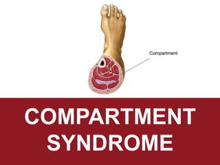
- 2. An elevation of interstitial pressure in a closed osteo-fascial compartment that result in microvascular compromise It is a TRUE orthopedics emergency. WHAT IS COMPARTMENT ? A c l o s e d a r e a o f m u s c l e g r o u p , n e r v e s a n d b l o o d v e s s e l s s u r r o u n d e d b y f a s c i a Muscle are arranged in different compartments and surrounded by one fascia, this arrangement is called osteofascial compartment. Normal compartment pressure: 5 – 15 mmHg Intacompartmental pressure rises to 35 – 40 mmHg DEFINITION
- 4. ANATOMY COMPARTMENT OF THE FOREARM Volar Dorsal Lateral (mobile ward) Most commanly affected Includes : 1. Flexor carpi radialis 2. Flexor carpi ulnaris 3. Flexor digitorum superficialis and profundus 4. Pronator teres 5. Pronator quadratus 6. Median nerve 7. Ulnar nerve Muscle : 1. Ext. carpi ulnaris 2. Ext. digiti minimi 3. Ext. digitorum 4. Supinator Rarely involved Muscles : 1. Brachioradialis 2. Ext. carpi radialis longus 3. Ext. carpi radialis brevis
- 5. ANATOMY COMPARTMENT OF THE HAND There are 10 compartments in the hands, which include 1. Hypothenar 2. Thenar 3. Adductor pollicis 4. Dorsal interosseous – 4 5. Volar(palmar) interosseous – 3
- 6. ANATOMY COMPARTMENT OF THE THIGHS
- 7. ANATOMY COMPARTMENT OF THE THIGHS Anterior Posterior Adductor 1. Quadriceps a) Rectus femoris b) Vastus lateralis c) Vastus intermedius d) Vastus medialis 2. Sartorius 1. Hamstrings a) Semitendinosus b) Semimembranous c) Biceps femoris (long and short) 1. Adductors a) Adductor longus b) Adductor brevis c) Adductor magnus d) Gracilis Femoral nerve Sciatic nerve Obturator nerve
- 8. ANATOMY COMPARTMENT OF THE LEGS
- 9. ANATOMY COMPARTMENT OF THE LEGS Anterior Lateral Deep posterior Superficial posterior Function Dorsiflexion of foot and ankle Plantarflexion and eversion of foot Plantarflexion and inversion of foot Mainly plantarflexion of foot and ankle Muscles 1.Tibialis anterior 2.Ext hallucis longus 3.Ext digitorum longus 4.Peroneus tertius 1.Peroneus longus 2.Peroneus brevis Only affect superficial peroneal nerve 1.Tibialis posterior 2.Flx digitorum longus 3.Flx hallucis longus 1. Gastrocnemius 2. Soleus 3. Plantaris
- 10. Any condition that reduces the volume of a compartment or increases the fluid content of the compartment can lead to an acute compartment syndrome. Increased Fluid Content Decreased Compartment Size 1. Fracture of the bones (mainly tibia and femur fracture) 2. Trauma 3. Burns 4. Hemorrhage 5. Vascular disruption/puncture 6. Snake venom 1. Burns 2. Cast 3. Bandage AETIOLOGY
- 11. AETIOLOGY
- 12. Acute Compartment Syndrome Chronic Compartment Syndrome Medical emergency Caused by severe injury The classic sign of acute compartment syndrome is pain, especially when the muscle within the compartment is stretched. The pain is more intense than what would be expected from the injury itself. Using or stretching the involved muscles increases the pain. There may also be tingling or burning sensations (paresthesias) in the skin. The muscle may feel tight or full. Numbness or paralysis are late signs of compartment syndrome. They usually indicate permanent tissue injury. Known as exertional compartment syndrome Not a medical emergency Most often caused by athletic exertion Chronic compartment syndrome causes pain or cramping during exercise. This pain subsides when activity stops. It most often occurs in the leg. Symptoms may also include: Numbness Difficulty moving the foot Visible muscle bulging TYPES
- 13. TISSUE SURVIVAL RATE 3-4 HOURS Reversible changes 6 HOUR Variable damage 8 HOUR Irreversible changes MUSCLE 2 HOUR LOOSE NERVE CONDUCTION 4 HOUR NEUROPRAXIA 8 HOUR IRREVERSIBLE CHANGES NERVE Delayed diagnosis will cause: Permanent sensory and motor deficit Contractures Infections & amputations
- 14. PATHOPHYSIOLOGY
- 15. PATHOPHYSIOLOGY
- 16. PATHOPHYSIOLOGY
- 17. PATHOPHYSIOLOGY In the setting of acute extremity compartment syndrome regardless of mechanism, the compartment pressure increases, causing hypoxia of tissues. There is normally an equilibrium between venous outflow and arterial inflow. When there is an increase in compartmental pressure, there is a reduction in venous outflow. This causes a build-up of venous pressure and an increased venous capillary pressure. If the intra-compartmental pressure becomes greater than arterial pressure, a decrease in arterial inflow will also occur. The decrease of venous outflow and arterial inflow result in decreased oxygenation of tissues causing ischemia. If the deficit of oxygenation becomes great enough then, irreversible necrosis may occur.
- 18. DIAGNOSIS HISTORY AND PHYSICAL paralysis pain pallor paraesthesia pulselessness poikilothermia Inability to regulate body temperate Loss of muscle function Abnormal sensation; tingling / numbness Of the skin But is usually intact Pain with passive stretch
- 19. DIAGNOSIS DIFFERENTIAL DIAGNOSIS 1. Cellulitis 2. DVT and thrombophlebitis 3. Gas gangrene 4. Necrotizing fasciitis 5. Peripheral vascular injuries
- 20. DIAGNOSIS EVALUATION A diagnosis of acute compartment syndrome is not always obvious. Therefore, serial examinations and frequent assessments are necessary to make such a diagnosis. Assess tenderness in the muscle compartment Evaluate motor and neurologic function Radiographs should be obtained if a fracture is suspected In an appropriate setting, measurement of intra-compartmental pressure using a solid- state transducer intra-compartmental catheter (STIC) can aid in a diagnosis. This device allows monitoring up to 16 hours. The normal pressure within the compartment is between 0 mmHg to 8 mmHg (i.e., less than 10 mmHg). An intra-compartmental pressure greater than 30 mmHg indicates compartment syndrome
- 21. DIAGNOSIS INVESTIGATIONS - LABORATORY 1. Full blood count 2. Coagulation profile 3. Creatinine phosphokinase 4. Urine myoglobin 5. X-ray 6. Ultrasound
- 22. ACUTE COMPARTMENT SYNDROME (ACS) Acute compartment syndrome is a surgical emergency, so a prompt diagnosis and treatment are important for an optimal outcome. Once the diagnosis is confirmed, immediate emergent surgical fasciotomy is important, since surgical fasciotomy can potentially reduce intra-compartmental pressure. MANAGEMENT 15 mmHg to 20 mmHg Serial compartment pressure measurement 20mmHg to 30 mmHg Surgical consultation and admit Greater than 30 mmHg Surgical fasciotomy Provide supplemental oxygen. Remove any restrictive casts, dressings or bandages to relieve pressure. Keep the extremity at the level of the heart to prevent hypo-perfusion. Prevent hypotension and provide blood pressure support in patients with hypotension.
- 23. ACUTE COMPARTMENT SYNDROME (ACS) Urgent decompression is necessary to reduce the increased compartment pressure. The ideal timeframe for fasciotomy is within six hours of injury to ensure full recovery; it is not recommended after 36 hours following the injury. When tissue pressure remains elevated for that much time, irreversible damage may occur, and fasciotomy may not be beneficial in this situation. After a fasciotomy is performed and swelling dissipates, a skin graft is most commonly used for closure. Patients must be closely monitored for complications, such as infection, acute renal failure, and rhabdomyolysis. If necrosis occurs before fasciotomy is performed, there is a high likelihood of infection which may require amputation. If infection occurs, debridement is necessary to prevent the systemic spread or other complications. Renal failure may also occur. MANAGEMENT
- 24. FASCIOTOMY Fasciotomy or fasciectomy is a surgical procedure where the fascia is cut to relieve tension or pressure commonly to treat the resulting loss of circulation to an area of tissue or muscle. Fasciotomy is a limb-saving procedure when used to treat acute compartment syndrome. It is also sometimes used to treat chronic compartment stress syndrome. The procedure has a very high rate of success, with the most common problem being accidental damage to a nearby nerve. Complications can also involve the formation of scar tissue after the operation. A thickening of the surgical scars can result in the loss of mobility of the joint involved. This can be addressed through occupational or physical therapy. MANAGEMENT
- 25. EMERGENCY FASCIOTOMY 1. Dual medial-lateral incision approach Two 15-18cm vertical incisions separated by 8cm skin bridge 1. Anterolateral incision 2. Posteromedial incision Post-operative Dressing changes followed by delayed primary closure or skin grafting at 3-7 days post decompression MANAGEMENT
- 26. EMERGENCY FASCIOTOMY 2. Single lateral incision approach Single lateral incision from head of fibula to ankle along line of fibula MANAGEMENT
- 29. COMPLICATION OF FASCIOTOMIES MANAGEMENT 1. Altered sensation within margin of wounds (77%) 2. Dry, scaly skin (40%) 3. Pruritis (33%) 4. Iatrogenic neurovascular injury 5. Swollen limb 6. Tethered scars 7. Recurrent ulceration 8. Pain related to wound
- 30. CHRONIC COMPARTMENT SYNDROME Chronic compartment syndrome in the lower leg can be treated conservatively or surgically. Conservative treatment includes rest, anti- inflammatories, and physical therapy. Elevation of the affected limb in patients with compartment syndrome is contraindicated, as this leads to decreased vascular perfusion of the affected region. Ideally, the affected limb should be positioned at the level of the heart. If symptoms persist after conservative treatment or if an individual does not wish to cease engaging in the physical activities which bring on symptoms, compartment syndrome can be treated by fasciotomy. Surgery is the most effective treatment for compartment syndrome. This decompression will relieve the pressure on the venules and lymphatic vessels, and will increase blood flow throughout the muscle. Left untreated, chronic compartment syndrome can develop into the acute syndrome and lead to permanent muscle and nerve damage. MANAGEMENT
- 31. Permanent nerve damage Permanent muscle damage and reduced function of affected limb Rhabdomyolysis which will lead to acute renal failure Volkmann ischemic contracture (infarcted muscle replaced by inelastic fibrous tissue) DIVC Neurologic deficit Infection Amputation COMPLICATIONS
Editor's Notes
- Ddx for chronic compartment syndrome: vascular problems – more common (hard to differentiate)
- 1. Pain 2. Swelling 3. Measure how tensed is the swelling. ACS is diagnosed clinically.
- Cut the skin including the fascia