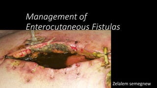
3. Enterocutaneous Fistulas
• Introduction
Fistula is defined as an abnormal communication between two
epithelialized surfaces.
Enteric fistulas are classified as
enterocutaneous or enteroatmospheric (exposed) fistulas
Enterocutaneous fistula
is a communication between the bowel lumen and the skin
An enteroatmospheric or exposed fistula
refers to drainage of intestinal contents from an open
abdomen with no overlying soft tissue
5. Classification
Site
( anatomy)
Small bowel
(65%)
Colon (30%)
Stomach/oe
sophagus
(rare)
Output
( physiology)
Low (<200
mL/24 hr)
Moderate
(200 – 500
mL/24 hr)
High (>500
mL/24 hr)
Complexity
Simple
Complex– long, multiple, associated
abscess, other organ involvement (e.g.
bladder, vagina
6. Clinical Presentation
• Recognition- 7 to 10 days following the procedure.
• Slow course with fevers and a prolonged ileus
• Development of wound erythema and, finally, drainage of enteric
contents through the wound spontaneously or following opening of
the skin over the wound
• Fluid and electrolyte imbalances(low sodium, potassium, phosphate
and magnesium)
• Malnutrition
• Sepsis
7. Management
• The goals of therapy for patients with enterocutaneous fistulas are
• to correct metabolic and nutritional deficits,
• close the fistula, and
• re-establish continuity of the gastrointestinal tract.
• The expected treatment course can be divided into five overlapping,
but sequential phase
9. Phase 1: Recognition and stabilization (24-48hrs)
• A . Identification
• B. Resuscitation (crystalloid, colloid, blood)
• C. Control of sepsis
• with antibiotics, percutaneous drainage of abscess under guidance or open drainage
• D. Nutritional support
• E. Control of fistula drainage
• F. Skin care to prevent excoriation
10. Phase 1…………….
• An immediate re-laparotomy is warranted when:
• There is evidence of clinical peritonitis.
• There is “ SIRS/sepsis” with proven or suspected intraperitoneal
abscesses
• Abdominal compartment syndrome exists.
• Somebody you do not trust performed the primary operation
• you never know what the findings will be
11. Phase 2: Investigation (7- 10 days)
• Investigation:
• A.) Fistulagram
• - Length of the tract
• Extent of the bowel wall disruption
• Location of the fistula
• Presence of a distal obstruction
• B.) CT scan, US
• C.) Intestinal contrast studies
12. Phase 3: Decision (7-10 d to 4-6 wk )
• Decision
• Assess likelihood of non operative closure
• Plan therapeutic course
• Decide optimal surgical timing
14. • M ore than 90% of small intestinal fistula which closed spontaneously, did so
within a month.
• Spontaneous closure rates dropped to less than 10% after 2 months and none
after 3 months.
• Factors possibly responsible for failure of spontaneous closure are:
a. Foreign Body
b. Radiation
c. Inflammation/ infection
d. Epithelialization [F-R-I-E-N-D-S]
e. Neoplasm
f. Distal intestinal obstruction
g. Steroids.
15. Average Time for closure
• Varies with anatomical location;
• Esophageal- 15-25 days
• Duodenal- 20-30 days
• Colonic - 30- 40 days
• Small Bowel- 40-60 days
16. Phase 4: Definitive therapy
• Surgical Indications
• Patients with an enterocutaneous fistula (ECF) with adverse factors may
require earlier surgical intervention
• surgical intervention should be undertaken after a 4- to 6-week trial of
conservative therapy, if no signs of spontaneous closure exist.
• Surgical procedures in patients with adverse factors can include
• draining an abscess,
• creating stomas by exteriorizing the bowel,
• or creating controlled fistulas.
• When feasible, resection of the fistula with restoration of GI continuity is performed.
• In patients with no associated adverse factors usually wait for about 3-4
months before surgical therapy
17. Phase 4: Definitive ……..…….
• Preoperative
• patients should be stable and free from sources of sepsis
• antibiotic prophylaxis and parenteral nutrition should be supplemented to
achieve good results
• Intraoperative
• Incision
• always enter the abdomen through a fresh incision
• Excision and restoration of bowel continuity
• the entire bowel from the ligament of Treitz to the rectum is made free of all adhesions
• the fistulous site is dissected free from the surrounding structures and a complete
excision is done
• do restoration of bowel continuity using a 2-layered anastomosis, employing interrupted,
nonabsorbable suture
18. Phase 4: Definitive ……..…….
• Treatment of abscess or diseased bowel
• If an abscess or diseased bowel segments are seen, then drainage of the abscess or
resection of the diseased bowel is performed
• If the patient is sick and cannot withstand a resectional procedure, then
exteriorization of the bowel via ileostomy or colostomy is carried out
• If anastomosis is performed close to a duodenojejunal flexure, then adequate
decompression by gastrostomy and feeding jejunostomy are carried out
• When it is not possible, fistula area bypass, Roux-en-Y drainage, serosa
patch technique is used.
• Duodenal fistula is better managed by bypass using gastrojejunostomy and
vagotomy without intervening the fistula
19. Phase 5 . Healing
• Continue nutrition support
• Antibiotic cover is needed if the operation is performed in the
presence of sepsis
• Patients who develop spontaneous fistula due to disease need
appropriate therapy during follow-up
• Physical and emotional rehabilitation
Most fistulas are the result of bowel injury during surgery, a leak from a bowel anastomosis, or erosion of mesh into adjacent bowel
Colonic: It can be due to postappendicectomy, postcolonic surgeries. It is common after emergency surgeries, and surgeries in unprepared bowel
Physiological—based on quantity of daily output:
High output—> 500 ml/day—
usually small bowel; 50% mortality; less chance of spontaneous closure.
• Moderate output—200-500 ml/day—
colonic and small bowel mixed.
• Low output—< 200 ml/day—
colonic; mortality is 15%; more chance of spontaneous closure
The first step of management is the resuscitation and stabilization of the patient.
> Needs to be accomplished within the first 24 to 48 hours of management.
> Initial efforts directed towards intravenous fluid resuscitation, control of infection,
protection of surrounding skin & measuring and replacing ongoing losses
Enterocutaneous fistulae are usually associated with hypokalemia and metabolic acidosis, which require correction
Somatostatin analogue Octreotide, at doses of 100 – 250 mcg TDS reduces
fistula output by 40 – 60% by the end of 24 hrs
Nutritional support should be initiated slowly after correction of fluid, electrolyte, and vitamin deficits
.
A fistulogram is performed by injecting a water soluble contrast agent into the cutaneous opening.
Following stabilization of the patient and maturation of the fistula tract, the anatomy of the fistula should be investigated radiographically. A fistulogram should be performed
Investigations are done to assess fistula and its causes. It is done in 7-10 days of fistula formation.
♦Fistulogram using water soluble contrast, CT fistulogramto see the pathological anatomy of fistula—site, number, length, status of bowel, distal obstruction, presence of abscess cavity.
♦Biochemical analysis(electrolytes, haematocrit and albumin) and renal, hepatic, respiratory, cardiac status should be assessed carefully
The expected time period for spontaneous closure, if it is to occur at all, varies with the anatomic location of the fistula. Fistulas from the esophagus and duodenum are expected to heal in two to four weeks. Colonic fistulas may heal in 30 to 40 days. Small bowel fistulas may take at least 40 to 60 days.